Home Blog Page 3

Integrative Skin Lesion Treatment and Patient Experience

Integrative Skin Lesion Treatment and Patient Experience

Integrative Skin Lesion Treatment and Pain Control

Abstract: An Integrative Approach to Cutaneous Lesion Management

In this educational overview, I will walk you through the procedural steps and clinical reasoning for excising a dysplastic nevus, a type of atypical mole. This procedure highlights the precision required to ensure complete removal while minimizing discomfort. We achieve this goal using a technique called a field block for local anesthesia. Furthermore, I will explain the physiological basis for this method and how it allows for a virtually painless patient experience. I will also place this procedure within the broader framework of our practice at Injury Medical Clinic. In addition, I’ll discuss how our multidisciplinary team, with crucial medical oversight from our Medical Director, Dr. Maria Guadalupe Cardenas, MD, collaborates to provide comprehensive, integrative care. This model combines chiropractic, functional medicine, rehabilitation, and conventional medical expertise. As a result, it addresses not just isolated symptoms but the patient’s overall health and well-being.

Integrative Skin Lesion Treatment and Patient Experience


Introduction: A Collaborative Model for Patient-Centered Care

As a clinician with diverse certifications spanning chiropractic (DC), advanced practice nursing (APRN, FNP-BC), and functional medicine (CFMP, IFMCP), my guiding philosophy has always been to view the patient as a whole. At Injury Medical Clinic, this philosophy is the cornerstone of our practice. Additionally, we have cultivated a unique, multidisciplinary environment where different fields of healthcare converge to provide superior patient outcomes.

A key element of our success is our collaboration with Dr. Maria Guadalupe Cardenas, MD, a highly respected, board-certified internist with over 40 years of experience. As our Medical Director and Collaborative Physician, Dr. Cardenas provides essential medical oversight to ensure our protocols meet the highest standards of care. Therefore, this integrative model allows us to blend the strengths of various disciplines:

  • Chiropractic Care (Dr. Alex Jimenez): Focused on spinal health, nervous system function, and musculoskeletal integrity.
  • Medical Oversight (Dr. Maria Cardenas): Provides diagnostic expertise, manages medical conditions, and ensures all treatments are safe and appropriate from an internal medicine perspective.
  • Functional Medicine: A systems-based approach to uncover and address the root causes of chronic illness.
  • Rehabilitation and Personal Injury Care: Specialized protocols to restore function and facilitate recovery after an injury.

This team-based approach allows us to manage a wide spectrum of health issues. We address both complex chronic conditions and straightforward procedures like the one I will detail today.

Preparing for the Excision: Pre-Procedural Planning

This morning, on September 1, 2026, I am preparing to see a 62-year-old gentleman who requires a re-excision of a skin lesion on his back. Approximately a month ago, a shave biopsy of this lesion revealed a dysplastic nevus with moderate atypia. A dysplastic nevus is an atypical mole that has abnormal cells and a higher potential to develop into melanoma, a serious form of skin cancer. The pathology report’s finding of “moderate atypia” signals a significant level of cellular abnormality. This finding necessitates a follow-up procedure to ensure all atypical cells are removed.

Our goal is to perform a complete excision with clear margins. This means removing the original biopsy scar along with a small, precise border of healthy tissue. Importantly, this “safety margin” is crucial to prevent recurrence and ensure the lesion is fully eradicated.

Mapping the Surgical Site

My first step is meticulous preparation of the surgical site. After cleansing the area with alcohol, I use a sterile surgical marker and a template to plan the excision.

  • Defining the Margins: The original lesion was approximately 5 x 6 millimeters. Based on current guidelines for a dysplastic nevus with moderate atypia, I plan to secure about a five-millimeter margin around the original lesion site, which equates to about two millimeters around the visible scar.
  • Creating the Ellipse: I draw an elliptical shape around the scar. This shape is not arbitrary; it’s designed to align with the natural skin tension lines (Langer’s lines). By orienting the excision this way, we can close the wound with minimal tension, which leads to better healing and a less noticeable scar.

Mastering Anesthesia: The Pain-Free Field Block Technique

Patient comfort is paramount. For a procedure like this, simply injecting anesthetic directly into the lesion site can be painful and less effective. Instead, I use a field block, a technique that creates an “anesthetic fence” around the entire surgical area. This approach numbs the nerves before they reach the target tissue, producing profound, widespread anesthesia.

The Physiological Rationale

The goal of a field block is to anesthetize the cutaneous nerves that supply sensation to the skin in a specific region. Instead of a single deep injection, we perform a series of interconnected, superficial injections in the subcutaneous tissue encircling the planned excision site. The anesthetic I am using is 1% lidocaine with epinephrine.

  • Lidocaine: This is the anesthetic agent. It works by blocking sodium channels in nerve cell membranes. Nerve impulses (including pain signals) are electrical signals that depend on sodium ion flow. By blocking these channels, lidocaine prevents the nerve from depolarizing and sending a pain signal to the brain.
  • Epinephrine: This is a vasoconstrictor, meaning it narrows the blood vessels in the area. Its inclusion is strategic for two reasons:
    1. Prolongs Anesthesia: By reducing blood flow, it slows the rate at which the lidocaine is carried away from the injection site, extending the duration of the numbness.
    2. Reduces Bleeding: The vasoconstriction also minimizes bleeding during excision, giving me a clearer view of the surgical field and allowing more precise work.

Executing the Block: A Step-by-Step Guide

  1. Initial Numbing Spray: To make the first needle stick completely painlessly, I use a vapocoolant spray (ethyl chloride). It rapidly cools the skin, creating an immediate, temporary numbing effect. As I asked the patient, “Is that cold?” he confirmed it was, and when I inserted the needle, he reported feeling nothing. This small step significantly enhances the patient experience.
  2. Creating the Subcutaneous Reservoir: I insert the needle at one corner of my marked ellipse and inject the lidocaine solution into the deeper subcutaneous tissue beneath the entire lesion. This creates a foundational layer of anesthesia, numbing the larger nerve branches that feed the area.
  3. The Intradermal Fence: This is the most critical part of the field block. Using the same entry point, I redirect the long, thin needle just under the skin’s surface (intradermally). I advance it along one side of the drawn ellipse and slowly inject the anesthetic as I withdraw the needle. This creates a visible wheal, or raised line, of anesthetic fluid. I then pivot the needle at the entry point and repeat the process on the other half of that side.
  4. Completing the Perimeter: I then move to the opposite corner of the ellipse and repeat the process, creating two more lines of intradermal anesthetic that connect with the first set. This completes the “fence,” ensuring the entire elliptical area is enclosed within a wall of numbness.

By the end of this process, the patient confirmed he felt no pain whatsoever. This is the power of a well-executed field block. It transforms what could be an uncomfortable procedure into a pain-free experience.

The Role of Integrative Chiropractic Care in Overall Wellness

While a skin lesion excision may seem purely medical, it’s an opportunity to consider the patient’s holistic health. In our practice, even when a patient comes in for a specific medical or minor surgical procedure, we see it through an integrative lens.

From a chiropractic and functional medicine perspective, skin health often mirrors internal health. Chronic inflammation, poor gut health, hormonal imbalances, and nutritional deficiencies can all manifest in the skin. Additionally, as a chiropractor, I am trained to assess the neuro-musculoskeletal system and understand that nerve function is critical for regulating all bodily processes, including tissue repair and immune surveillance.

If this patient were also seeing me for chiropractic care, my focus would be on:

  • Optimizing Nervous System Function: Through chiropractic adjustments, we can reduce nerve interference along the spine. Proper nerve supply is essential for healthy cellular communication, immune function, and the body’s ability to heal efficiently post-procedure.
  • Supporting the Body’s Healing Response: We would discuss anti-inflammatory nutrition, stress management techniques, and targeted supplementation (like Zinc and Vitamin C) to support wound healing and bolster the immune system.
  • Addressing Systemic Inflammation: A dysplastic nevus, while localized, can be influenced by systemic factors. A functional medicine workup could explore inflammatory markers, gut health, and toxin exposure to reduce the body’s overall inflammatory load, potentially lowering the risk for other health issues down the line.

This is the essence of our integrative model: we manage the immediate concern with precision and care. At the same time, we use it as a gateway to improve the patient’s foundational health and resilience. The collaboration between Dr. Cardenas and me ensures this comprehensive approach is both holistic and medically sound. Once the anesthesia has taken full effect, we are ready to proceed with the excision. We do so confident that the patient is comfortable and that we have a clear, well-prepared surgical field.
Unlocking the Secrets of Inflammation: Integrative Medicine Approach | El Paso, Tx (2023)

References

  • Kouba, D. J., LoPiccolo, M. C., & Alam, M. (2012). Guidelines for the use of local anesthesia in office-based dermatologic surgery. Journal of the American Academy of Dermatology, 66(3), 477-493. https://doi.org/10.1016/j.jaad.2011.08.012
  • Robinson, J. K., Hanke, C. W., Siegel, D. M., & Fratila, A. (Eds.). (2018). Surgery of the skin: Procedural dermatology (3rd ed.). Elsevier.
  • Tzarnas, S., & Jelinek, J. E. (2017). Local anesthetics. In StatPearls. StatPearls Publishing. Available from: https://www.ncbi.nlm.nih.gov/books/NBK534859/

Functional Medicine Benefits for Skin Health & Inflammation

Learn how functional medicine approaches can improve skin health by reducing inflammation and enhancing overall wellness.

Abstract

As a Doctor of Chiropractic with advanced certifications as a Nurse Practitioner and in Functional Medicine, I provide comprehensive, evidence-based care. In this educational post, I will guide you through diagnosing and managing a common clinical presentation: an inflamed skin lesion. We will explore the case of a middle-aged gentleman who presented with a painful, inflamed lesion on his hip, which we clinically identified as a probable seborrheic keratosis. This post details the step-by-step procedure for a shave skin biopsy, a crucial diagnostic tool for confirming the nature of such lesions. I will explain the physiological reasoning behind each step, from the local anesthesia technique using lidocaine with epinephrine to the shave biopsy itself and subsequent hemostasis with aluminum chloride.

Furthermore, I will discuss the importance of an integrative care model in managing patient health comprehensively. At our practice, Injury Medical Clinic, I work alongside Dr. Maria Guadalupe Cardenas, MD, our esteemed Medical Director and an internist with over 40 years of experience. This collaborative framework lets us merge the principles of chiropractic care, functional medicine, internal medicine, and rehabilitation to address not just the symptom—like a skin lesion—but the underlying systemic factors that may affect a patient’s overall health and well-being. We will delve into how this multidisciplinary approach ensures patient safety, provides a holistic perspective, and optimizes outcomes by treating the whole person.

Our Integrative Care Philosophy at Injury Medical Clinic

At Injury Medical Clinic, our mission is to provide patient-centered care that transcends traditional disciplinary boundaries. We have cultivated a unique clinical environment where multiple healthcare philosophies converge to offer a truly holistic treatment experience. This model is built on the collaborative relationship between me, Dr. Alex Jimenez (DC, APRN, FNP-BC, CFMP, IFMCP, ATN, CCST), and our Medical Director and Collaborative Physician, Dr. Maria Guadalupe Cardenas, MD (Board Certified in Internal Medicine).

Dr. Cardenas brings a wealth of knowledge from her more than four decades as an internist (NPI #1164426749, Texas MD License #J2933). Her role is fundamental to our practice. As Medical Director, she provides essential medical oversight, ensuring all our protocols and treatments meet the highest standards of medical safety and efficacy. This type of multidisciplinary setup is increasingly recognized as a best-practice model, particularly in integrative and injury care clinics. It allows a Doctor of Chiropractic (DC) like myself, with advanced training as a Family Nurse Practitioner (FNP-BC) and in Functional Medicine, to work in concert with an experienced Medical Doctor (MD).

This synergy allows our team to offer a broad spectrum of services under one roof:

  • Chiropractic Care: I focus on the body’s biomechanics, addressing musculoskeletal issues, nerve function, and spinal health. This is the foundation of our physical medicine approach.
  • Medical Oversight (Internal Medicine): Cardenas provides comprehensive medical evaluations, manages systemic health conditions, and offers crucial diagnostic insights to ensure we don’t overlook underlying medical issues.
  • Functional Medicine: We delve deep into the root causes of dysfunction, using advanced diagnostic testing to understand a patient’s unique biochemistry, genetics, and lifestyle factors.
  • Personal Injury and Rehabilitation: Our team is equipped to manage the complex needs of patients recovering from injuries, integrating physical therapies, rehabilitation exercises, and pain management strategies.

When a patient presents with an issue like a skin lesion, this integrated model becomes invaluable. While I may perform the initial assessment and procedure within my scope as an FNP, Dr. Cardenas’s oversight ensures a comprehensive differential diagnosis and follow-up plan. This collaboration ensures we treat not just a localized symptom but the patient’s entire physiological landscape, from musculoskeletal alignment to metabolic health.

A Clinical Scenario: The Inflamed Seborrheic Keratosis

A few weeks ago, a middle-aged gentleman came to our clinic with a specific concern. He pointed to the lateral aspect of his right hip, where a skin lesion had become increasingly noticeable over the past four weeks. It wasn’t just its appearance that troubled him; the lesion was now causing inflammation and pain.

On clinical examination, the lesion showed the classic features of an inflamed seborrheic keratosis. Seborrheic keratoses are very common, non-cancerous (benign) skin growths that people tend to develop as they age. They often appear as brown, black, or light tan growths on the face, chest, shoulders, or back. They have a waxy, scaly, and slightly elevated appearance, sometimes described as looking “pasted on” the skin.

While harmless, they can become a problem when irritated. This can happen from friction, like clothing rubbing against them, or for other unknown reasons. When irritated, they can become inflamed, red, itchy, and painful—exactly what our patient was experiencing. Although my clinical judgment strongly pointed toward a benign diagnosis, a core principle of evidence-based medicine is never to assume. The gold standard for a definitive diagnosis is a histopathological examination of the tissue. Therefore, the clear course of action was to perform a shave skin biopsy. This procedure would not only provide a tissue sample for the pathologist but would also serve a therapeutic purpose by removing the irritated lesion.

The Shave Biopsy Procedure: A Step-by-Step Explanation

The goal of a shave biopsy is to remove the outermost layers of the skin (the epidermis and a portion of the dermis) that contain the lesion. It is a minimally invasive, efficient, and highly effective technique for both diagnosing and treating raised skin lesions. I’ll walk you through the exact steps we took for this patient, explaining the “why” behind each action.

1. Preparation and Anesthesia: Ensuring Patient Comfort and Procedural Precision

The first and most critical step in any minor surgical procedure is ensuring the patient is comfortable and the area is properly prepared.

  • Cleansing the Area: I began by thoroughly cleaning the skin over and around the lesion with an alcohol pad. This standard aseptic technique reduces the microbial load on the skin’s surface, minimizing the risk of post-procedural infection.
  • Topical Anesthetic Spray: To mitigate the initial pain of the needle stick, I used a topical vasoconstrictor spray called Pain Ease® Mist. This spray rapidly cools the skin, creating a temporary numbing effect. The mechanism is based on the Gate Control Theory of Pain. The intense cold sensation travels along large-diameter A-beta nerve fibers, which are faster than the smaller A-delta and C fibers that transmit pain signals. The “gate” for pain signals in the spinal cord is effectively “closed” by the overwhelming cold sensation, making the subsequent needle insertion much more tolerable for the patient.
  • Local Anesthetic Injection: The cornerstone of local anesthesia for this procedure is injecting a local anesthetic agent. I used 1% lidocaine with epinephrine, injecting approximately 1 mL into the subcutaneous tissue. Let’s break down why this specific combination is so effective:
    • Lidocaine: This is an amide-type local anesthetic. Its primary function is to block nerve conduction, thereby preventing pain signals from reaching the brain. It achieves this by reversibly binding to and inactivating voltage-gated sodium channels within the nerve cell membrane. When these channels are blocked, the influx of sodium ions required to generate and propagate an action potential is prevented. No action potential means no pain signal transmission.
    • Epinephrine: This is a vasoconstrictor. Its inclusion serves two vital purposes. First, by constricting local blood vessels, it reduces blood flow at the injection site. This slows the rate at which lidocaine is absorbed into the systemic circulation, prolonging the anesthetic effect and keeping the area numb longer. Second, reduced blood flow provides hemostasis, minimizing bleeding during the biopsy procedure. This creates a clearer surgical field for me and reduces the total amount of bleeding the patient experiences.

I used a very fine 30-gauge, half-inch needle to minimize tissue trauma and discomfort. I quickly inserted it into the subcutaneous tissue just beneath the lesion and injected the anesthetic. The patient confirmed he felt no pain.

2. Creating an Intradermal Wheal: Elevating the Lesion for an Optimal Biopsy

After the initial subcutaneous injection, I let the anesthetic sit for a moment to take effect. Then, without fully removing the needle, I redirected it. This technique avoids a second painful needle puncture. I carefully advanced the needle horizontally, positioning it in the intradermal space directly underneath the lesion.

As I slowly injected the remaining lidocaine solution into this superficial layer, a visible, pale, and firm raised area of skin began to form. This is known as an intradermal wheal. The formation of this wheal is a crucial part of the technique for several reasons:

  1. Anesthetizes the Superficial Nerves: The dermis is rich with sensory nerve endings. Injecting the anesthetic directly into this layer ensures that the most superficial, and therefore most sensitive, part of the skin is completely numb. I asked the patient if he felt a stinging sensation, which is common as the fluid expands the dermal tissue, but he reported none.
  2. Elevates the Lesion: This is the most brilliant mechanical advantage of the intradermal wheal. By injecting fluid directly beneath the lesion, we use hydraulic pressure to lift the entire lesion up and away from the deeper subcutaneous tissues. It essentially “presents” the lesion to me on a platter.
  3. Creates a Firm Plane for Shaving: The wheal makes the skin taut and firm. This provides a stable, flat surface, allowing for a smooth, controlled, and even shave. It prevents the blade from skipping or digging too deep, ensuring that I can remove the lesion at the desired depth with a single, fluid motion.

You could see the wheal developing, and with it, the seborrheic keratosis was elevated perfectly, ready for removal.

3. The Shave Biopsy: Precise and Efficient Removal

With the lesion anesthetized and elevated, it was time to shave. Using a sterile, flexible razor blade (or a specialized tool like a DermaBlade), I began the incision about two millimeters lateral to the visible edge of the lesion. This margin ensures that the entire lesion is captured within the specimen, which is important for the pathologist to make an accurate diagnosis.

I held the blade parallel to the skin surface and used a gentle, horizontal, sawing motion to pass it through the base of the lesion, staying within the plane of the intradermal wheal. The goal is to be deep enough to remove the entire lesion but shallow enough to avoid significant scarring. The anesthetic wheal acts as a perfect guide for this depth.

I continued the motion through to the other side of the lesion. In one clean pass, I removed the entire lesion, leaving a smooth, shallow dermal wound. I immediately placed the specimen in a container with formalin, a preservative that fixes the tissue for transport to the pathology lab. On the patient’s skin, I saw no residual lesional tissue.

4. Hemostasis: Achieving a Bloodless Field

Even with epinephrine’s vasoconstrictive effects, a superficial dermal wound will ooze blood. Prompt, effective hemostasis (cessation of bleeding) is essential for patient comfort, infection prevention, and proper wound healing.

For shave biopsies, the hemostatic agent of choice is often chemical. In this case, I used aluminum chloride. I saturated a sterile gauze pad with a 20% aluminum chloride solution (a common concentration for this purpose). I applied it directly to the wound bed with firm pressure for about a minute.

Aluminum chloride works as a potent chemical styptic agent. Its mechanism of action is twofold:

  • It causes superficial tissue necrosis by denaturing proteins, which effectively seals the cut ends of the small capillaries in the dermis.
  • It interacts with blood proteins, particularly fibrinogen, to form a mechanical plug that occludes the bleeding vessels.

The effect was immediate and impressive. As soon as I removed the gauze, the bleeding stopped completely. The wound bed was clean and dry, ready for dressing.

5. Final Steps: Dressing and Patient Instructions

The final step was to apply a sterile dressing. I typically use a small amount of antibiotic ointment (like bacitracin or mupirocin) to provide a moist healing environment and an extra layer of infection protection, followed by a simple adhesive bandage.

I then provided the patient with clear aftercare instructions:

  • Keep the area clean and dry for the first 24 hours.
  • After 24 hours, he could gently clean the area daily with soap and water.
  • Apply a thin layer of antibiotic ointment and a new bandage daily until the wound has fully epithelialized (formed a new layer of skin), which usually takes 7-10 days.
  • We scheduled a follow-up appointment in two weeks to discuss the pathology report and check on the wound’s healing.

The Healing Diet: Combat Inflammation, Embrace Wellness- Video

The Healing Diet: Combat Inflammation, Embrace Wellness | El Paso, Tx (2023)

The Role of Integrative Chiropractic Care in Overall Wellness

Now, you might be wondering: what does a skin biopsy have to do with chiropractic care? This is where the beauty of an integrative and functional medicine approach comes into full view. At Injury Medical Clinic, my role as a Doctor of Chiropractic goes beyond spinal adjustments. My advanced training allows me to see the body as an interconnected system, where one issue can reflect a deeper, systemic imbalance.

While a seborrheic keratosis is a benign skin condition, skin health often mirrors internal health. Conditions like inflammation, oxidative stress, and hormonal imbalances can manifest on the skin. Here’s how our integrative model connects these dots:

Chiropractic Care and Neuro-Endocrine-Immune Function

The nervous system is the body’s master controller, regulating everything from muscle contraction to immune responses and hormone secretion. Chiropractic care, at its core, focuses on optimizing nervous system function by correcting vertebral subluxations—spinal misalignments that can interfere with nerve signaling.

  • Systemic Inflammation: Chronic pain and inflammation, whether from a skin lesion or a musculoskeletal issue, activate the body’s stress response system, primarily the Hypothalamic-Pituitary-Adrenal (HPA) axis. Persistent activation can dysregulate cortisol, the body’s main stress hormone. This can, in turn, promote systemic inflammation, which can exacerbate skin conditions and impair wound healing. Some research shows chiropractic adjustments can modulate the autonomic nervous system and influence the HPA axis, potentially helping downregulate systemic inflammation. By addressing musculoskeletal stress through chiropractic care, we can help calm the nervous system and, in turn, reduce the body’s overall inflammatory burden.
  • Immune System Modulation: The nervous system and the immune system are intricately linked in a field of study called psychoneuroimmunology. Nerve fibers innervate lymphoid organs like the spleen and lymph nodes. By improving neuromuscular function, chiropractic care may help support a balanced immune response, which is crucial for fighting potential skin infections and supporting proper wound healing.

Functional Medicine: Looking Deeper

As a Certified Functional Medicine Practitioner (CFMP, IFMCP), my goal is to investigate the “why” behind a patient’s symptoms. In the context of skin health, this means looking at:

  • Gut Health: The gut is a major regulator of inflammation and immune function. A condition known as “leaky gut” or increased intestinal permeability can allow inflammatory molecules and undigested food particles to enter the bloodstream, triggering systemic inflammation that can manifest in the skin (the “gut-skin axis”).
  • Nutrient Deficiencies: Proper wound healing is highly dependent on an adequate supply of key nutrients, including Vitamin C (for collagen synthesis), Zinc (for cell proliferation and immune function), Vitamin A (for epithelialization), and protein (the building blocks for new tissue). A functional medicine workup can identify and correct these deficiencies through targeted nutrition and supplementation.
  • Oxidative Stress: The process of inflammation generates free radicals, which cause oxidative stress and damage to cells. We can assess a patient’s antioxidant status and recommend a diet rich in colorful fruits and vegetables, along with specific supplements like glutathione or N-acetylcysteine, to help quench this oxidative stress and support cellular repair.

Putting It All Together for the Patient

For the gentleman with the hip lesion, our integrative plan extends beyond the simple biopsy.

  1. Immediate Care: The shave biopsy, overseen medically by Dr. Cardenas, addresses the immediate pain and inflammation.
  2. Chiropractic Assessment: I would also perform a full spinal and biomechanical assessment. Does he have a pelvic obliquity or sacroiliac joint dysfunction causing gait changes that lead to his clothing rubbing on that specific spot on his hip? Addressing this underlying biomechanical issue could prevent future irritation of other skin lesions.
  3. Functional Medicine Investigation: We would discuss his diet, stress levels, and overall health. If he shows other signs of systemic inflammation (e.g., joint pain, fatigue, digestive issues), we might recommend advanced testing to look at inflammatory markers, gut health, or nutrient levels.
  4. Rehabilitation and Lifestyle: Our plan would incorporate recommendations for an anti-inflammatory diet, stress management techniques, and appropriate physical activity to support his overall health, promote efficient wound healing, and reduce his risk of future inflammatory issues.

This comprehensive approach, guided by the collaborative expertise of Dr. Cardenas and me, ensures that we are not just removing a benign skin growth. We are using this clinical encounter as an opportunity to assess and optimize the patient’s entire well-being, embodying the true spirit of integrative medicine. The pathology report will give us a definitive diagnosis, and our holistic plan will support the patient’s journey toward lasting health.

References

Clinical Observations and Further Reading

For those interested in exploring the clinical application of these principles in more detail, I invite you to review my professional insights and case studies, which reflect my extensive experience in chiropractic, functional medicine, and advanced nursing practice.

SEO Tags: shave biopsy, seborrheic keratosis, integrative medicine, chiropractic care, functional medicine, Dr. Alex Jimenez, Dr. Maria Cardenas, El Paso TX, skin lesion, local anesthesia, lidocaine with epinephrine, intradermal wheal, aluminum chloride, hemostasis, inflammation, multidisciplinary care, internal medicine, personal injury, rehabilitation, holistic health, Injury Medical Clinic, wound healing

Percutaneous Tenotomy for Chronic Tendinopathy Approaches

Percutaneous Tenotomy for Chronic Tendinopathy Approaches

Percutaneous Tenotomy for Chronic Tendinopathy

As an advanced practice provider with a deep commitment to integrative and functional medicine, I am constantly exploring the latest evidence-based treatments to help my patients overcome chronic pain and regain function. My goal is to bridge gaps between medical disciplines, bringing together the best of chiropractic, functional medicine, and conventional medical care to create comprehensive, patient-centered solutions.

Today, I want to share exciting developments in treating chronic tendinopathy, drawing on the pioneering work of leading researchers like Dr. Brian O’Connor. Specifically, we’ll explore an innovative, minimally invasive procedure known as percutaneous tenotomy, a technique that is transforming how we manage conditions like tennis elbow, plantar fasciitis, and Achilles tendinopathy.

I will discuss how this procedure works, its applications, and how we integrate it into a broader, multidisciplinary care model at Injury Medical Clinic. This model combines my expertise in chiropractic and functional medicine with the invaluable medical oversight of Dr. Maria Guadalupe Cardenas, MD, our clinic’s Medical Director.

Percutaneous Tenotomy for Chronic Tendinopathy Approaches

Our Collaborative Care Model: Integrating Disciplines for Optimal Outcomes

At Injury Medical Clinic, we have cultivated a unique, multidisciplinary environment designed to provide the most effective and comprehensive care possible. A cornerstone of our practice is the collaborative relationship between me and Dr. Maria Guadalupe Cardenas, MD. Dr. Cardenas is a highly respected, board-certified internist with over 40 years of experience. As our Medical Director and Collaborative Physician, she provides essential medical oversight to ensure our treatment plans are safe, effective, and grounded in sound medical principles. Her NPI is #1164426749, and her Texas MD License is #J2933.

This partnership allows us to offer a truly integrative approach. While I focus on the biomechanical and neuromusculoskeletal aspects of health through chiropractic adjustments, functional movement, and rehabilitation, Dr. Cardenas provides the medical framework. This synergy is particularly vital when we incorporate advanced procedures like percutaneous tenotomy. Together, we manage patient care from diagnosis and treatment planning to post-procedure rehabilitation, ensuring we address all aspects of a patient’s health. Our services include:

  • Chiropractic Care: Focused on restoring spinal alignment, improving nervous system function, and correcting biomechanical imbalances.
  • Medical Oversight: Led by Dr. Cardenas to ensure patient safety and appropriate medical management.
  • Functional Medicine: Investigating the root causes of dysfunction, from metabolic issues to nutritional deficiencies.
  • Personal Injury and Rehabilitation: Specialized programs for recovery from accidents and injuries.
  • Advanced Regenerative Procedures: Incorporating cutting-edge, evidence-based treatments for chronic conditions.

Understanding Chronic Tendinopathy: When Rest and Ice Aren’t Enough

Many of my patients come to me frustrated after months or even years of battling chronic tendon pain. They’ve tried rest, ice, anti-inflammatory medications, and traditional physical therapy, yet the pain persists. This is because chronic tendinopathy is not simply an issue of inflammation; it’s a degenerative process.

The condition, more accurately termed tendinosis, involves the breakdown of the collagen fibers that make up the tendon. Over time, the tissue becomes disorganized, weak, and filled with dysfunctional, scar-like material. This pathological tissue lacks a healthy blood supply, which severely limits its ability to heal on its own. This is why passive treatments often fail. To achieve lasting relief, we must actively remove the diseased tissue and stimulate a new, robust healing response. That’s where percutaneous tenotomy comes in.

Percutaneous Tenotomy: A Minimally Invasive Solution for Chronic Tendon Pain

Percutaneous tenotomy is a revolutionary, FDA-approved procedure that uses ultrasound guidance to precisely target and remove damaged tendon tissue. The term “percutaneous” means “through the skin,” highlighting its minimally invasive nature. Unlike traditional open surgery, which requires large incisions and lengthy recovery times, this procedure is performed through a tiny entry point, often in an office setting.

As Dr. O’Connor shows in his research, this technique allows providers to “debride,” or clean out, the specific area of tendinopathy with incredible accuracy. This is a significant advance, especially for primary care and specialty clinics like ours, as it provides a powerful tool to address the root cause of chronic tendon pain without resorting to major surgery.

Current approved indications for percutaneous tenotomy include:

  • Extensor Carpi Radialis Brevis Tendinopathy (Tennis Elbow)
  • Flexor Tendinopathy (Golfer’s Elbow)
  • Patellar Tendinopathy (Jumper’s Knee)
  • Achilles Tendinopathy
  • Plantar Fasciopathy (Plantar Fasciitis)

The Technology Behind the Technique: Tenex and TenJet

Two primary systems perform percutaneous tenotomy, both designed to remove pathologic tissue while preserving healthy structures.

  1. Tenex Health TX™: This system is based on phacoemulsification, the same technology used in modern cataract surgery.
    • It utilizes a specialized handpiece with an 18-gauge double-lumen needle.
    • The needle tip vibrates at an ultrasonic frequency, emulsifying (liquefying) the targeted degenerative tissue.
    • At the same time, a second lumen within the needle suctions the emulsified debris away, leaving the healthy tendon intact.
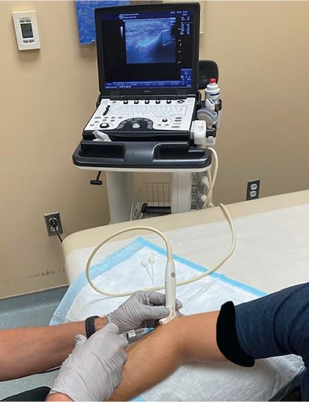
    • The entire process is performed under real-time ultrasound guidance, allowing for unparalleled precision. The provider can see the needle tip and the diseased tissue on the screen, ensuring only the damaged area is treated.
  2. TenJet™: This system offers an alternative method for tissue removal.
    • It uses a high-velocity, pressurized stream of saline to create a “cutting” effect.
    • This powerful water jet essentially dissects and removes the diseased tissue, which is then aspirated away through the same instrument.
    • Like Tenex, TenJet is also performed under ultrasound guidance to ensure targeted and safe debridement.

In our practice, we have found the Tenex system to be exceptionally effective. A foot pedal controls the procedure, allowing precise, hands-on control while you visualize the anatomy on the ultrasound monitor.

The Percutaneous Tenotomy Procedure: A Step-by-Step Look

Let’s walk through what a patient can expect during a percutaneous tenotomy procedure for tennis elbow, a very common application.

  1. Preparation and Sterilization: The procedure is conducted under strict sterile conditions. The treatment area is cleaned, and a local anesthetic is administered to numb the skin and the tissue surrounding the tendon. The ultrasound probe itself is covered in a sterile sheath.
  2. Ultrasound Visualization: Using high-resolution ultrasound, I identify the exact location of the tendinopathy. The diseased tissue often appears darker (hypoechoic), thickened, and disorganized on the ultrasound image, sometimes with tiny calcifications.
  3. Needle Insertion: A tiny incision is made, just large enough for the Tenex needle. Using the ultrasound image, the needle is advanced directly to the degenerative tissue. We typically use an “in-plane” approach, meaning we can see the entire length of the needle on the screen as it moves toward the target.
  4. Debridement: Once the needle is in position, the device is activated. The ultrasonic energy is applied, and the provider gently moves the needle tip back and forth through the diseased tissue. As Dr. O’Connor notes, this motion is similar to dry needling or a fenestration procedure, but with the added benefit of removing the debrided tissue at the same time. The entire “cutting time” is remarkably short, typically lasting between 30 seconds and two minutes. Patients may hear the device, but they should not feel pain because of the local anesthetic.
  5. Completion: After debridement, the needle is removed, and a small adhesive bandage is placed over the tiny incision. No stitches are required.

The Role of Integrative Chiropractic Care in Recovery

The procedure itself is only the beginning of the healing journey. Removing the diseased tissue creates an opportunity for the body to heal correctly, but it needs the right support. This is where our integrative model shines. My clinical observations consistently show that a structured post-procedure rehabilitation program is crucial for optimal outcomes.

Our post-tenotomy protocol is multifaceted and personalized, integrating chiropractic principles to restore function and prevent recurrence.

  • Initial Healing Phase (First 1-2 weeks): The focus is on protecting the healing tissue. We may recommend a brief period of modified activity. The body initiates an acute inflammatory healing response, which is a positive and necessary step. This is the body sending growth factors, stem cells, and blood supply to the area to build new, healthy collagen.
  • Restoring Mobility and Biomechanics: As a chiropractor, my primary goal is to ensure the joints and surrounding tissues are moving correctly. For tennis elbow, I would assess and adjust the wrist, elbow, shoulder, and even the cervical and thoracic spine. Misalignments in these areas can create abnormal loading patterns on the elbow, contributing to the initial injury. Correcting them is essential for long-term success.
  • Soft Tissue Mobilization: We use techniques like Graston Technique® or Active Release Technique (ART®) on the surrounding muscles to break down adhesions, improve blood flow, and reduce muscular tension that may be pulling on the newly repaired tendon.
  • Eccentric Loading Program: Research has conclusively shown that eccentric exercises are paramount for tendon remodeling. These exercises involve lengthening the muscle-tendon unit under load. For tennis elbow, this would involve slowly lowering a light weight. This controlled stress stimulates the fibroblasts (tendon cells) to lay down new, strong, and properly aligned collagen fibers.
  • Functional and Nutritional Support: As a Certified Functional Medicine Practitioner, I also look at systemic factors. We may recommend nutritional strategies to support collagen synthesis, such as adequate protein intake and supplementation with Vitamin C, zinc, and other cofactors. Managing systemic inflammation through diet can also create a better healing environment.

Conclusion

By combining the precision of percutaneous tenotomy with the holistic, biomechanical approach of chiropractic care and the oversight of internal medicine, we can guide the body through a complete and robust healing process. We are not just removing the pain; we are restoring function, rebuilding tissue, and resolving the underlying issues that led to the problem in the first place. This is the future of musculoskeletal care—integrated, evidence-based, and profoundly effective.

The Natural Way to Heal: Non-Surgical Chiropractic Care for Pain Relief | El Paso, Tx (2023)

References

O’Connor, B. (Presenter). (n.d.). Advanced Techniques: Percutaneous Tenotomy [Video].

Integrative Medicine: The Best Approaches for Menopause With HRT

Find out how HRT combined with integrative medicine can empower your journey through menopause with informed choices.

Abstract

As a clinician with dual credentials in chiropractic (DC) and advanced practice nursing (APRN), holding certifications in functional and integrative medicine, I, Dr. Alex Jimenez, have dedicated my career to understanding the intricate web of human physiology. In this educational post, I will explore the complex biological shifts that occur during perimenopause and menopause, explaining why simply administering Hormone Replacement Therapy (HRT) often fails to resolve the full spectrum of symptoms. We will explore the latest findings from leading researchers, moving beyond the simplistic view of hormonal decline to uncover three core biological problems: persistent inflammation and oxidative stress, mitochondrial dysfunction and localized insulin resistance, and critical NAD+ depletion. I will explain how these physiological disruptions lead to common menopausal symptoms like hot flashes, brain fog, weight gain, and mood instability. This post will also highlight the integrative approach we take at Injury Medical Clinic PA, where Dr. Maria Guadalupe Cardenas, MD, provides medical oversight that complements my chiropractic and functional medicine expertise. Together, we offer a multidisciplinary framework that addresses these underlying biological issues through targeted nutritional support, lifestyle modifications, and advanced chiropractic care, providing a comprehensive strategy to restore metabolic health and overall well-being.

Our Integrative Approach: A Partnership for Holistic Health

At Injury Medical Clinic PA, we have fostered a unique, collaborative clinical environment. I am Dr. Alex Jimenez, and my practice is founded on the belief that true healing requires a multifaceted approach. To that end, I am honored to work alongside Dr. Maria Guadalupe Cardenas, MD, our Medical Director and Collaborative Physician. Dr. Cardenas is a highly respected, board-certified internist with over 40 years of invaluable experience. Her NPI number is 1164426749, and she is licensed in Texas under license number J2933.

This multidisciplinary partnership is the cornerstone of our integrative model. Dr. Cardenas provides essential medical direction and oversight, ensuring that all our patient care plans meet the highest standards of medical safety and efficacy. My role involves integrating chiropractic care, functional medicine diagnostics, nutritional science, and personalized rehabilitation protocols. This synergy allows us to address health issues from multiple angles. For example, a patient presenting with chronic pain and fatigue may receive chiropractic adjustments to improve neuromusculoskeletal function, while Dr. Cardenas oversees any necessary medical evaluations or prescriptions. At the same time, we use functional medicine to investigate underlying issues like nutrient deficiencies or inflammation, which I then address through targeted lifestyle and supplementation plans. This collaborative structure is particularly effective for complex conditions, including personal injury recovery and the systemic challenges of menopause, as it ensures that both the structural and metabolic aspects of health are expertly managed.

Beyond Hormones: The Unseen Biological Challenges of Menopause

For years, the conventional understanding of menopause has been centered almost exclusively on the decline of estrogen and progesterone. The logical, albeit simplistic, solution was Hormone Replacement Therapy (HRT). The idea was straightforward: replace the missing hormones, and the symptoms will vanish. However, as a clinician who has worked with countless women navigating this transition, I can tell you that the reality is far more complex. Many of my patients come to me frustrated, reporting that even with HRT, they still “feel like crap.” They continue to struggle with hot flashes, persistent fatigue, brain fog, and unexplained weight gain.

This is because HRT, while often a valuable and necessary part of a comprehensive plan, only addresses one piece of a much larger biological puzzle. It handles the hormones, but it does not correct the underlying biology that has been disrupted. To truly understand and effectively manage menopausal symptoms, we must look deeper, into the cellular and metabolic shifts that are taking place. Based on groundbreaking research from leading institutions, we can identify three primary biological problems that HRT alone does not resolve. These hidden hurdles prevent so many women from feeling their best.

1. The Fire of Inflammation: When Estrogen Can’t Do Its Job

The first, and perhaps most significant, biological hurdle is persistent systemic inflammation and oxidative stress. Menopause is inherently an inflammatory state. As ovarian function declines, the body loses the potent anti-inflammatory effects of estrogen and progesterone. This hormonal shift often raises inflammatory markers, most notably C-reactive protein (CRP).

The Role of CRP and Oxidative Stress

C-reactive protein (CRP) is a substance produced by the liver in response to inflammation. In the context of menopause, elevated CRP is a sign that the body is in a state of chronic, low-grade inflammatory alert. This “inflammaging” is compounded by oxidative stress, an imbalance between the production of damaging free radicals and the body’s ability to counteract them with antioxidants. Think of oxidative stress as biological rust that damages cells, proteins, and DNA.

How Inflammation Hijacks HRT

Here is the critical point: you can introduce estrogen into the body via HRT, but if the cellular environment is inflamed, the hormone cannot function effectively. Let’s trace the pathway. When you apply transdermal estradiol, it enters the bloodstream and travels to target cells, such as those lining the blood vessels (the endothelium). Estrogen is a powerful vasodilator, meaning it signals the blood vessels to relax and widen. This action is crucial for maintaining cardiovascular health, delivering oxygen and nutrients throughout the body, and, importantly, regulating body temperature.

The estrogen molecule binds to its specific receptors on the endothelial cells, initiating a signaling cascade that should result in vasodilation. However, high levels of inflammation and oxidative stress throw a wrench in the works. The inflammatory molecules interfere with this signaling pathway. They essentially “deafen” the cell to estrogen’s message.

So, what happens?

  • The estrogen successfully docks with its receptor.
  • The cell receives the signal to dilate the blood vessel.
  • But the pervasive inflammation prevents the downstream cellular machinery from executing the command properly.

The result? The blood vessels remain constricted and dysfunctional despite adequate estrogen. This is precisely why many women on HRT continue to experience debilitating hot flashes. A hot flash is, at its core, a symptom of thermoregulatory collapse originating in the hypothalamus, but it shows up peripherally as chaotic vasodilation and vasoconstriction. If inflammation prevents blood vessels from responding properly to hormonal and neurological signals, hot flashes persist. It’s an incredibly frustrating scenario for patients: you’re taking the hormone, but you’re still suffering from one of the very symptoms it’s supposed to fix.

The Chiropractic and Functional Medicine Solution to Inflammation

This is where an integrative approach becomes indispensable. We cannot simply add hormones to an inflamed system and expect optimal results. We must actively work to quench the fire of inflammation.

  • Nutritional Intervention: We start by identifying and removing pro-inflammatory dietary triggers, such as processed foods, refined sugars, and industrial seed oils. We then introduce an anti-inflammatory eating plan rich in omega-3 fatty acids (from fish and flaxseeds), polyphenols (from colorful fruits, vegetables, and green tea), and other antioxidant-rich foods.
  • Targeted Supplementation: Based on functional testing, we may recommend specific anti-inflammatory and antioxidant supplements like high-potency curcumin, resveratrol, N-acetylcysteine (NAC), and a quality fish oil. These compounds help lower CRP and reduce oxidative stress at the cellular level.
  • Chiropractic Care and the Nervous System: Chronic inflammation is not just a chemical state; it is deeply intertwined with the nervous system. The autonomic nervous system (ANS), which controls our “fight or flight” (sympathetic) and “rest and digest” (parasympathetic) responses, plays a key role in regulating the immune system and inflammation. Misalignments in the spine, particularly in the upper cervical and thoracic regions, can create nerve interference that pushes the ANS toward a state of sympathetic dominance. This “fight or flight” state promotes the release of pro-inflammatory cytokines.
    • My Clinical Observation: I have consistently observed in my practice that patients suffering from chronic inflammatory conditions often present with significant spinal subluxations and poor heart rate variability (HRV), a key indicator of ANS dysfunction. Through specific chiropractic adjustments, we can restore proper spinal mechanics, reduce nerve irritation, and help rebalance the autonomic nervous system. By promoting a shift toward a more parasympathetic state, chiropractic care can help down-regulate the body’s inflammatory response, creating a more favorable biological environment for hormones to work effectively.

By addressing inflammation directly, we are not just managing a symptom; we are treating a root cause of HRT’s failure and restoring the body’s ability to respond to its own internal signals.

2. The Energy Crisis: Mitochondrial Dysfunction and Localized Insulin Resistance

The second major biological hurdle is a profound energy crisis happening at the cellular level. This crisis has two interconnected components: the breakdown of our cellular power plants (the mitochondria) and the development of localized insulin resistance. This explains the pervasive fatigue, brain fog, and stubborn weight gain that plague so many women during menopause.

Mitochondria: The Powerhouses of Our Cells

To understand this problem, we must first appreciate the role of the mitochondrion. Every cell in your body contains hundreds or even thousands of these tiny organelles. Their primary job is to take the fuel we consume—glucose (from carbohydrates) and fatty acids (from fats)—and convert it into adenosine triphosphate (ATP).

ATP is the universal energy currency of the body.

Every single biological process requires ATP. Think about it:

  • Muscle Contraction: Requires ATP.
  • Nerve Impulse Transmission: Requires ATP.
  • Synthesis of Neurotransmitters (like serotonin and dopamine): Requires ATP.
  • Cellular Repair and Regeneration: Requires ATP.
  • Maintaining Body Temperature (Thermoregulation): Requires ATP.

If your mitochondria don’t produce enough ATP, your entire system begins to sputter and fail. Metabolism slows down, cognitive function declines, and mood regulation becomes unstable. You feel exhausted, not because you are lazy or haven’t slept enough, but because your cells are literally running out of power.

The Double Whammy: Insulin Resistance in Key Tissues

During menopause, a peculiar and damaging phenomenon occurs. The body develops a localized insulin resistance, specifically in the brain (particularly the hypothalamus) and in metabolic tissues like muscle.

Insulin is the “key” that unlocks the cell door, allowing glucose to enter and be used for fuel. When a cell becomes insulin resistant, it’s like the lock on the door has become rusty. The key (insulin) is there, but it can’t open the door effectively.

Here’s the devastating consequence:

  1. Glucose Gets Trapped Outside: Cells in your brain and muscles can’t take in the glucose circulating in your bloodstream. This means the mitochondria inside those cells are starved of their primary fuel source. They are sitting in a sea of glucose but can’t use it.
  2. The Body Switches to “Fat Storage” Mode: Because the body thinks its cells are starving (even though there’s plenty of glucose in the blood), it goes into a panic mode. The liver converts the excess, unused glucose into triglycerides. Furthermore, the body cannot efficiently access the fat stored in adipocytes (fat cells) to burn for energy. Instead, every excess calorie is aggressively stored as visceral fat—the dangerous, inflammatory fat that accumulates around the organs in the abdominal cavity.

This is why menopausal weight gain is so stubborn and often settles around the midsection. It is not a simple caloric problem. You can be eating the same number of calories you always have, or even fewer, and still gain weight. The issue is metabolic. Your body has lost its ability to burn fuel efficiently and has become a hyper-efficient fat-storage machine.

This dysfunction explains so many hallmark symptoms:

  • Overwhelming Fatigue: Your cells have no ATP. The feeling of being “metabolically brittle” is real. You are fundamentally energy-deficient.
  • Depression, Anxiety, and Irritability: The prefrontal cortex is the part of your brain responsible for executive functions: mood regulation, impulse control, emotional stability, and rational thought. It is one of the body’s most energy-demanding regions. When the neurons in the prefrontal cortex are starved of ATP, they cannot function properly. They cannot synthesize or regulate key neurotransmitters like serotonin, dopamine, and GABA.
    • This is why I get so frustrated when I hear women being dismissed as “just hormonal.” It’s not a personality flaw. Your biology becomes neurochemically unstable because the cells that regulate your mood lack the fundamental energy to do their job.
    • This also explains why antidepressants often fail during menopause. Taking an SSRI to increase serotonin is like trying to get more water from a faucet when the main water line to the house is broken. You are treating a symptom (low serotonin), but the root problem is that the neurons don’t have the ATP to synthesize serotonin in the first place.

The Impact of HRT on this Energy Crisis

Hormone Replacement Therapy does not, by itself, rebuild mitochondria or fix localized insulin resistance. In fact, HRT can sometimes exacerbate an underlying problem. By speeding up metabolic processes and tissue repair, HRT can increase the demand for certain micronutrients. If these nutrients are already in short supply (which they often are), HRT can deplete mineral stores like magnesium and B vitamins even faster, further stressing an already compromised metabolic system.

The Integrative Strategy for Restoring Cellular Energy

To solve this energy crisis, we must focus on restoring mitochondrial health and improving insulin sensitivity.

  • Dietary Adjustments for Insulin Sensitivity: We implement a nutritional plan low in refined carbohydrates and sugars to rest insulin receptors. We emphasize high-quality proteins, healthy fats, and fiber-rich vegetables. This dietary pattern helps lower circulating glucose and insulin levels, allowing cells to regain sensitivity gradually.
  • Mitochondrial Support Nutrients: We use targeted supplementation to provide the raw materials mitochondria need to function and repair themselves. This “mito cocktail” often includes:
    • Coenzyme Q10 (CoQ10): A critical component of the electron transport chain, where ATP is generated.
    • Acetyl-L-Carnitine: Helps shuttle fatty acids into the mitochondria to be burned for fuel.
    • Alpha-Lipoic Acid: A powerful antioxidant that protects mitochondria from oxidative damage and improves insulin sensitivity.
    • B Vitamins: Essential cofactors for hundreds of enzymatic reactions in energy metabolism.
  • Exercise as Metabolic Medicine: The right kind of exercise is crucial. We focus on a combination of:
    • High-Intensity Interval Training (HIIT): Short bursts of intense effort have been shown to stimulate mitochondrial biogenesis (the creation of new mitochondria).
    • Resistance Training: Building muscle mass is one of the most powerful ways to combat insulin resistance. Muscle tissue is a major consumer of glucose, and more muscle means more places for glucose to go, taking the burden off the pancreas.
  • Chiropractic Care for Metabolic Regulation: The nervous system’s connection to metabolic health is profound. The hypothalamus, the brain region where localized insulin resistance is a key problem, is directly influenced by the autonomic nervous system.
    • My Clinical Observation: I have seen patients whose blood sugar regulation improves following a course of chiropractic care. By reducing stress on the nervous system through spinal adjustments, we can help modulate the hypothalamic-pituitary-adrenal (HPA) axis. An overactive HPA axis (chronic stress) leads to high cortisol levels, which directly promotes insulin resistance and visceral fat storage. By calming this axis, chiropractic adjustments can contribute to a more stable metabolic environment, supporting the dietary and nutritional interventions we implement.

3. The Power Outage: Catastrophic NAD+ Decline

The third biological problem is perhaps the most recently understood, yet it ties everything together. It involves the catastrophic decline of a molecule called Nicotinamide Adenine Dinucleotide, or NAD+.

A landmark 2021 study published in Nature Cell Biology provided definitive proof of what many in the functional medicine field had long suspected: NAD+ levels decline catastrophically during the menopausal transition.

What is NAD+ and Why is it So Important?

If ATP is the energy currency of the cell, NAD+ is the power supply that allows the mitochondrial factory to run. NAD+ is a coenzyme found in every cell of your body, and it is central to metabolism. It plays two main roles:

  1. Energy Production: NAD+ is essential for converting the food we eat into ATP. It acts as an electron shuttle, carrying high-energy electrons from the breakdown of glucose and fatty acids to the electron transport chain inside the mitochondria, where ATP is ultimately produced. Without sufficient NAD+, this entire energy production line grinds to a halt.
  2. Cellular Repair and Longevity: NAD+ is the required fuel for a class of proteins called sirtuins. Sirtuins are often called the “guardians of the genome.” They handle many critical functions, including DNA repair, reducing inflammation, and regulating cellular aging. When NAD+ levels are low, sirtuin activity plummets, leaving cells vulnerable to damage and accelerating aging.

So, the decline of NAD+ during menopause creates a perfect storm: the power supply to the mitochondria is cut, and the cell’s ability to repair itself is crippled.

The NAD+-Driven Consequences of Menopause

This dramatic drop in NAD+ is not a subtle change; it has direct, observable consequences that anchor many of the most challenging menopausal symptoms directly to dying and dysfunctional mitochondria.

  • Hot Flashes: As discussed, the hypothalamus is the body’s thermostat. It is an extremely energy-hungry part of the brain. When NAD+ levels fall, the mitochondria in the hypothalamic neurons starve. They can no longer produce the ATP needed to maintain stable thermoregulation. The result is a thermoregulatory collapse, leading to the chaotic signaling that causes hot flashes and night sweats.
  • Brain Fog and Cognitive Decline: The brain consumes about 20% of the body’s total energy, despite being only 2% of its weight. Low NAD+ means low ATP in the brain, leading to slower neural processing, difficulty recalling memories, and that pervasive feeling of mental “fog.”
  • Weight Gain: With low NAD+, metabolic pathways that burn fat for energy are severely impaired. The body defaults to storing energy as fat because it lacks the coenzymes needed for efficient energy conversion.
  • No Libido: The production of sex hormones and the neurological pathways for arousal are all energy-dependent processes. Low NAD+ contributes to a decline in both the physiological and psychological aspects of libido.
  • Depression, Anxiety, and Neurotransmitter Depletion: This brings us back to the energy-starved prefrontal cortex. Neurons cannot synthesize adequate amounts of serotonin, dopamine, and GABA without a steady supply of ATP, and they can’t make ATP without NAD+. The brain’s entire neurochemical balance hinges on this one critical molecule.

The decline of NAD+ is the unifying theory that explains why the mitochondria fail and why the energy crisis is so profound during menopause. Period.

The Integrative Approach to Boosting NAD+

Fortunately, we are not helpless in the face of declining NAD+. We can take proactive steps to support and replenish our NAD+ levels.

  • NAD+ Precursors: The body can synthesize NAD+ from certain precursors. We often recommend supplementation with Nicotinamide Mononucleotide (NMN) or Nicotinamide Riboside (NR). These molecules are the direct building blocks that the body uses to create new NAD+. Supplementing with them can help to elevate NAD+ levels systemically.
  • Lifestyle Factors: Certain lifestyle habits can preserve NAD+ levels:
    • Caloric Restriction and Intermittent Fasting: These practices have been shown to increase NAD+ levels and stimulate sirtuin activity.
    • Regular Exercise: Both endurance and resistance training can boost NAD+ in muscle and other tissues.
  • Sirtuin Activators: Compounds like resveratrol (found in grapes) and pterostilbene (found in blueberries) can help to activate the sirtuins, making them more efficient even with existing NAD+ levels.
  • Chiropractic Neurology and NAD+: The brain’s demand for NAD+ is immense. Any factor that increases metabolic stress on the brain can accelerate its depletion.
    • My Clinical Observation: Patients with chronic pain, poor posture, or unresolved injuries from accidents often live in a state of constant, low-level neurological stress. This “nociceptive barrage”—a continuous stream of pain signals to the brain—is metabolically expensive. It forces the brain to expend significant energy (and thus, NAD+) to process these signals. By using chiropractic adjustments to reduce pain, improve posture, and restore normal biomechanics, we can lessen this metabolic burden on the nervous system. This frees up valuable NAD+ and ATP, which can then be reallocated to other critical functions like mood regulation, cognition, and cellular repair. It’s a way of optimizing the body’s energy budget from a neurological perspective.

Aligned & Empowered: Chiropractic Conversations on Women’s Health-Video

Aligned & Empowered: Chiropractic Conversations on Women’s Health | El Paso, Tx (2020)

The Playbook: An Integrated Strategy for Hormonal and Metabolic Balance

Now that we understand the three biological hurdles—inflammation, the cellular energy crisis, and NAD+ depletion—we can create a comprehensive and logical plan that addresses these root causes alongside hormonal balancing. HRT is a tool, but it must be used intelligently within a supportive biological framework.

Here is a practical, evidence-based playbook for navigating this transition.

Step 1: Establish a Stable Hormonal Foundation

Before we can fine-tune the underlying biology, we need to stop the hormonal chaos. The most common mistake I see is patients and practitioners constantly tweaking HRT doses. This creates more problems than it solves.

The Fourteen-Day Rule

A fundamental principle of endocrinology is that hormones take about two weeks (fourteen days) to reach a steady state in the tissues. This means that after you start or change a dose, it takes fourteen full days for your body’s cells to adapt fully and for you to experience the true effect of that dose.

If you keep adjusting your dose every three or four days based on how you feel, you are riding a hormonal roller coaster. You will create surges and dips that your body interprets as instability, which can actually trigger more hot flashes and mood swings than you started with.

The Protocol:

  1. Be Patient: Once you and your provider have decided on a starting dose, commit to it for at least fourteen days. Do not make any changes during this period unless you experience a severe adverse reaction.
  2. Wait and Observe: Keep a simple symptom journal. Note the frequency and intensity of hot flashes, your sleep quality, mood, and energy levels.
  3. Adjust Methodically: After fourteen days, evaluate your progress. If symptoms persist, make a small, methodical adjustment. Then, you wait another fourteen days.

Standard Dosing Regimen

While every woman is unique, a common and effective starting point is:

  • Transdermal Estradiol: Applied once a day (e.g., a patch or gel). This method bypasses the liver, which is generally safer and provides more stable blood levels than oral estrogen.
  • Oral Progesterone: Taken at bedtime only. Progesterone has a natural sedative effect that can significantly improve sleep quality. It also serves to protect the uterine lining in women who still have a uterus.
  • DHEA: Taken in the morning. DHEA is a precursor hormone that can convert to estrogen and testosterone. It can help with energy, libido, and mood.
    • Caution: If you become irritable, anxious, or develop oily skin or acne, your DHEA dose may be too high. Cut the dose in half and reassess.

The Goal: Zero Flashes

The primary initial goal of HRT is to reduce hot flash frequency. Once your flash frequency hits zero, or is reduced to a minimal, non-disruptive level, stop messing with the doses. You have found your steady state. Chasing a specific number on a lab report while you feel good is counterproductive.

Troubleshooting:

  • Chest Tenderness: This is a common sign that the estrogen dose is too high. First, lower the estrogen dose slightly. If it persists, we investigate methylation pathways, as poor methylation (often related to B6, B12, and folate status) can impair the body’s ability to clear estrogen metabolites.
  • Feeling Jittery or Anxious: This is often a sign of too much DHEA. It can also indicate an underlying magnesium or taurine deficiency. These nutrients are critical for calming the nervous system, and starting HRT can increase the body’s demand for them.

Step 2: Get Your Labs—The Right Way

Lab testing is essential, but it must be timed correctly.

  1. Get Baseline Labs Today: Before you start any new protocol, get a comprehensive baseline panel. Include a full hormone panel (Estradiol, Progesterone, DHEA-S, Testosterone, FSH), inflammatory markers (hs-CRP), a complete metabolic panel, and key nutrients.
  2. Retest in Six Weeks: Get your labs drawn again six weeks after starting the full playbook (HRT and nutritional support). This gives your body enough time to reach a new equilibrium.

Interpreting Estradiol:

  • If your estradiol level is over 100 pg/mL, you are likely taking too much. This increases the risk of side effects like breast tenderness and is unnecessary for symptom control.
  • If your estradiol level is under 30 pg/mL, your dose is likely too low to be effective. A gentle increase is warranted.
  • The Sweet Spot: Many women feel their best with an estradiol level between 50-90 pg/mL, but the ultimate guide should be your symptoms, not the number.

Step 3: Implement the Non-Negotiable Foundational Nutrients

Regardless of what else you do, certain micronutrients are the essential raw materials required for your hormonal and metabolic systems to function. Their need is amplified during menopause. I consider these non-negotiable. Every woman navigating this transition should ensure she gets enough of these, either through diet or supplementation.

  • Magnesium: The “relaxation mineral.” It supports over 300 enzymatic reactions, including ATP production, nerve function, and blood sugar control. It helps with sleep, anxiety, and muscle cramps.
  • Potassium: Works with sodium to maintain fluid balance and is critical for nerve transmission and muscle contraction. Low potassium can contribute to fatigue and blood pressure issues.
  • Vitamin D3: Technically a hormone, it is essential for bone health, immune function, and mood. Most people are deficient.
  • Vitamin K2 (as MK-7): Works in synergy with Vitamin D3. D3 helps you absorb calcium, but K2 directs that calcium into the bones and teeth and keeps it out of the arteries and soft tissues, where it can cause calcification.
  • Boron: A trace mineral that plays a surprisingly important role in hormone metabolism. It helps to increase free testosterone and estradiol levels and is also important for bone health.
  • Zinc: Crucial for immune function, DNA synthesis, and the proper function of hundreds of enzymes. It also supports thyroid health and sex hormone production.
  • Taurine: An amino acid that has a stabilizing effect on cell membranes and is particularly important for calming the nervous system, supporting heart health, and aiding in the formation of bile salts for fat digestion.
  • Methylated B Vitamins: B6, B12, and Folate in their active, methylated forms are critical for energy production, neurotransmitter synthesis, and, importantly, the detoxification of estrogen metabolites through the methylation pathway.

These nutrients form the foundation for all other therapies. Comprehensive research playbooks detail specific dosages and forms, but their inclusion is universal. By supplying the body with these essential building blocks, you are empowering it to heal itself from the inside out.

Conclusion: A New Paradigm for Menopause Care

The journey through menopause is not a simple story of hormonal decline. It is a complex metabolic transition characterized by chronic inflammation, a cellular energy crisis driven by mitochondrial dysfunction, and a catastrophic drop in the vital molecule NAD+. Simply replacing hormones without addressing these fundamental biological problems is like repainting a house with a crumbling foundation. It may look better for a short time, but the underlying issues will inevitably surface.

At Injury Medical Clinic, our collaborative model, uniting the medical oversight of Dr. Maria Cardenas with my expertise in chiropractic and functional medicine, allows us to offer a truly holistic and effective paradigm for menopause care. We use HRT as a strategic tool to establish a stable hormonal baseline. Then, we get to the real work: quenching inflammation, rebuilding mitochondria, restoring insulin sensitivity, boosting NAD+ levels, and ensuring the body has all the nutritional cofactors it needs to thrive.

Through specific chiropractic adjustments, we address the often-overlooked neurological component, calm the stress response, reduce the metabolic burden of chronic pain, and optimize the nervous system’s ability to regulate the body’s internal environment. This integrated approach doesn’t just mask symptoms; it rebuilds health from the cell up. It transforms the menopausal transition from a period of decline and suffering into an opportunity for profound renewal and empowerment. You are not hormonal; your biology is crying out for the right support. By listening to those signals and responding with a comprehensive, evidence-based strategy, you can reclaim your energy, your clarity, and your vitality.

References

  • Covarrubias, A. J., Kale, A., Perrone, R., López-Domínguez, J. A., Pisco, A. O., Kasler, H. G., … & Verdin, E. (2021). Senescent cells promote tissue NAD+ decline during aging via the activation of CD38+ macrophages. Nature Metabolism, 3(9), 1265–1283. [https://www.nature.com/articles/s42255-021-00449-9](https://www.nature.com/articles/s42255-021-00449-9)
  • Ghavami, S., Yeganeh, B., St-Pierre, J., & Halayko, A. J. (2019). Autophagy, mitochondrial health, and the treatment of diseases. In Mitochondrial Regulation (pp. 41-62). Humana Press, Cham. (Note: This is a general reference for mitochondrial health concepts, providing a broader scientific basis for the discussion.)
  • Mauvais-Jarvis, F. (2015). Role of sex hormones in the development and progression of type 2 diabetes. Physiological Reviews, 95(3), 885-911. (Note: This reference supports the link between sex hormones, like estrogen, and metabolic function, including insulin sensitivity.)
  • Ruiz-Nunez, B., Pruimboom, L., Dijck-Brouwer, D. A. J., & Muskiet, F. A. J. (2013). Lifestyle and nutritional imbalances associated with Western diseases: causes and consequences of chronic low-grade inflammation. Journal of Nutritional Biochemistry, 24(7), 1183-1201. (Note: This reference provides a comprehensive overview of how diet and lifestyle contribute to the state of chronic low-grade inflammation discussed in the text.)
  • Yankner, B. A., Lu, T., & Lo, T. (2008). The aging brain, sirtuins, and NAD+. Cell, 134(3), 380-382. (Note: This seminal review discusses the critical roles of NAD+ and sirtuins in brain aging and neurodegeneration, underpinning the concepts of brain fog and cognitive changes.)

SEO Tags

menopause, perimenopause, HRT, hormone replacement therapy, Dr. Alex Jimenez, Dr. Maria Cardenas, integrative chiropractic, functional medicine, El Paso, Injury Medical Clinic, mitochondrial dysfunction, NAD+, inflammation, CRP, oxidative stress, insulin resistance, menopause weight gain, brain fog, hot flashes, fatigue, depression, anxiety, DHEA, estradiol, progesterone, magnesium, vitamin D3, K2, boron, taurine, methylated B vitamins, chiropractic adjustment, autonomic nervous system, clinical nutrition, anti-inflammatory diet, sirtuins

Frozen Shoulder Treatment With Hydroplasty Recovery Guide

Frozen Shoulder Treatment With Hydroplasty Recovery Guide

Frozen Shoulder Treatment With Hydroplasty Explained

Abstract

This educational post explores hydroplasty, also known as hydrodistension, a highly effective, minimally invasive procedure for managing adhesive capsulitis, or frozen shoulder. From my perspective as an integrative practitioner, I will guide you through the physiological rationale behind this technique and detail the step-by-step process as demonstrated by leading experts. We will examine the anatomical targets, the specific medications used, and how ultrasound guidance ensures precision and safety.

I will also explain how we incorporate this advanced procedure into a multidisciplinary framework at our clinic, where integrative chiropractic care plays a crucial role in post-procedural rehabilitation and long-term recovery. This article aims to provide a comprehensive understanding of hydroplasty, from the initial nerve block to the therapeutic capsular distension, and its place in a modern, evidence-based treatment plan for restoring shoulder function.

Frozen Shoulder Treatment With Hydroplasty Recovery Guide


Pain Solutions

As a practitioner with a diverse background in chiropractic, functional medicine, and advanced practice nursing, I am constantly seeking the most effective, evidence-based solutions for my patients. At Injury Medical Clinic PA, my mission is to integrate the best of multiple disciplines to create a synergistic healing environment. A cornerstone of this approach is our multidisciplinary team, where I work alongside our Medical Director and Collaborative Physician, Dr. Maria Guadalupe Cardenas, MD. With over 40 years of experience as a board-certified internist, Dr. Cardenas provides essential medical oversight, allowing us to offer a comprehensive spectrum of care that bridges traditional medicine, chiropractic, and rehabilitative therapies.

Our collaborative model is particularly vital when addressing complex conditions like adhesive capsulitis, commonly known as frozen shoulder. This painful and debilitating condition involves the thickening and tightening of the glenohumeral joint capsule, severely restricting movement. While chiropractic adjustments and physical therapy are foundational to our approach, some cases require a more direct intervention to break the cycle of inflammation and adhesion. Today, I want to share insights into one such advanced procedure: hydroplasty, or hydrodistension.

I’ll present the latest findings and techniques from leading researchers in the field, showcasing their work to show how modern, evidence-based methods are transforming patient outcomes. We will look at a procedure performed by Dr. Paul Taylor, a primary care sports medicine physician with OrthoVirginia, to understand the intricacies of this powerful intervention.

Understanding Hydroplasty for Frozen Shoulder

Hydroplasty is a procedure almost exclusively used to treat adhesive capsulitis. The core principle is straightforward yet profound: inject a large volume of fluid to mechanically stretch and break the restrictive adhesions within the shoulder joint capsule. This process, known as distension, creates space, reduces stiffness, and significantly improves range of motion, facilitating effective rehabilitation.

To visualize what we’re targeting, look at this ultrasound image of the posterior (back) shoulder.

  • Superior is at the top, and Inferior is at the bottom.
  • Medial is towards the body’s midline, and Lateral is away from it.
  • You can see the layers of tissue: the outer deltoid muscle, the deeper infraspinatus muscle, and then the crucial posterior capsule of the glenohumeral joint.
  • Beneath the capsule lies the humeral head (the “ball” of the ball-and-socket joint).

The goal of hydroplasty is to precisely inject fluid into the space between the humeral head and the capsule, forcing the capsule to expand and release.

The Three-Step Procedural Protocol

The hydroplasty procedure is a meticulous, multi-step process designed for maximum efficacy and patient comfort. The technique combines a specific anesthetic, anti-inflammatory medication, and saline, all delivered under precise ultrasound guidance.

Step 1: The Suprascapular Nerve Block

Before addressing the joint capsule directly, the first step is to manage pain. The suprascapular nerve provides the majority of sensation to the shoulder joint. By performing a nerve block at the spinoglenoid notch—a small anatomical passageway just medial to the glenoid (the “socket”)—we can achieve significant short- and intermediate-term pain relief.

  • Why is this done? Numbing this nerve accomplishes two things:
    1. It makes the subsequent intra-articular injection far more comfortable for the patient.
    2. It provides a window of pain relief that allows the patient to engage more effectively in post-procedure rehabilitation without being limited by severe pain.
  • The Injection: Use a 5 mL syringe containing 1% lidocaine (a fast-acting anesthetic). The needle is guided via ultrasound to the spinoglenoid notch, and the anesthetic is delivered around the nerve.

Step 2: Numbing the Joint Capsule Pathway

Next, a local anesthetic is administered directly into the path the main injection will take. This desensitizes the deeper tissues and the joint capsule itself.

  • The Cocktail: A small amount of 1% lidocaine is mixed with sodium bicarbonate.
  • Why use sodium bicarbonate? Lidocaine is naturally acidic, which can cause a stinging sensation upon injection. Sodium bicarbonate acts as a buffer, neutralizing the solution’s pH to match the body’s tissues. This simple addition significantly reduces injection discomfort.
  • The Procedure: Using a thin, 25-gauge needle, the buffered lidocaine is injected into the posterior glenohumeral joint space, right under the capsule. Ultrasound imaging confirms the needle is in the correct location—above the humeral head and beneath the posterior capsule.

Step 3: The Therapeutic Distension

This is the main event. A large volume of fluid is injected to stretch and ultimately rupture the thickened, adhered joint capsule.

  • The Injectate: A 51 mL cocktail is prepared. Formulation includes:
    • 10 mL of 1% lidocaine: For immediate pain control.
    • 10 mL of 0.5% ropivacaine: A longer-acting anesthetic to provide extended pain relief post-procedure.
    • 30 mL of sterile saline: Provides the primary volume needed to distend the capsule.
    • 1 mL of Kenalog (40 mg): A potent corticosteroid to powerfully reduce inflammation within the joint, helping to prevent the adhesions from reforming.
  • The Procedure: An 18-gauge needle is guided into the same intra-articular space. Because of the large volume and pressure required, this is often a two-person job. The physician guides the needle, while a medical assistant pushes the fluid from the syringe.
  • The Visual Confirmation: Under ultrasound, the process is remarkable. As the fluid is injected, you can see the posterior capsule lifting away from the humeral head. The space visibly expands as it fills. Then, suddenly, you see the capsule deflate. This is the “break”—the moment the adhesions tear and the capsule is released. This visible deflation signals a successful procedure.

The Role of Integrative Chiropractic Care in Recovery

Hydroplasty is a powerful “reset” for frozen shoulder, but it is not the end of treatment. In fact, it’s the beginning of a critical window for recovery. This is where our integrative model at Injury Medical Clinic PA truly shines.

Following a successful hydroplasty, the patient has a unique opportunity. The adhesions have been broken, the corticosteroid is controlling inflammation, and the long-acting anesthetic is managing pain. Now is the time for active rehabilitation to restore function and prevent the scar tissue from returning.

Our approach, overseen by Dr. Cardenas and led by my chiropractic and rehabilitation team, includes:

  • Gentle Mobilization: Immediately following the procedure, we begin gentle, passive, and active-assisted range-of-motion exercises. The goal isn’t to be aggressive, but to guide the shoulder through its newly regained movement patterns.
  • Chiropractic Adjustments: We focus on the entire kinetic chain. A frozen shoulder often leads to compensatory patterns in the neck (cervical spine), mid-back (thoracic spine), and shoulder blade (scapula). Chiropractic adjustments to these areas help restore proper biomechanics, reduce muscle tension, and ensure the nervous system functions optimally to support healing.
  • Myofascial Release and Soft Tissue Work: Techniques like trigger point therapy and active release target the surrounding muscles (deltoid, infraspinatus, and rotator cuff) to break up muscular adhesions and reduce protective spasm.
  • Personalized Rehabilitation Programs: We design a specific exercise program for each patient to progressively strengthen the rotator cuff and scapular stabilizing muscles. This is crucial for creating dynamic stability around the joint and preventing re-injury.
  • Functional Medicine Principles: We may also assess for underlying systemic inflammation through blood work and nutritional counseling. Chronic inflammation can drive conditions like adhesive capsulitis, and addressing it at a systemic level supports long-term joint health.

Conclusion

This combined approach ensures that we capitalize on the mechanical benefits of hydroplasty. The procedure creates the opportunity for movement, and our integrated chiropractic and rehabilitative care teaches the body how to use that movement correctly and sustainably. By managing the entire patient—from the joint capsule to the spine to their overall inflammatory status—we can achieve lasting relief and a full return to function.

Chiropractic: The Secret to Unlocking Mobility | El Paso, Tx (2023)

References

This article draws upon established medical procedures and principles. Further reading on adhesive capsulitis, hydrodistension, and related anatomy can be found in sources such as:

  • Macnab, I., & Rathbun, J. B. (1970). Macnab’s Elbow and Shoulder Pain. Williams & Wilkins.
  • Neviaser, J. S. (1945). Adhesive capsulitis of the shoulder: a study of the pathological findings in periarthritis of the shoulder. The Journal of Bone and Joint Surgery. American Volume, 27(2), 211–222.
  • Ryan, V., Brown, H., Minns Lowe, C. J., & Lewis, J. S. (2016). The pathophysiology associated with primary (idiopathic) frozen shoulder: A systematic review. BMC Musculoskeletal Disorders, 17(1), 340. https://doi.org/10.1186/s12891-016-1190-9

Chiropractic Rehabilitation Methods for Systemic Inflammation

Unlock the potential of chiropractic rehabilitation to address systemic inflammation and support your body’s natural recovery mechanisms.

Abstract

In this educational post, I, Dr. Alex Jimenez, DC, APRN, FNP-BC, CFMP, IFMCP, ATN, CCST, present a comprehensive, evidence-based exploration of systemic inflammation, its immunologic drivers, and its cross-system effects in metabolism, cardiovascular health, neurodegeneration, and autoimmunity-like phenomena. I explain how dysregulated innate immunity—especially sustained M1 macrophage bias and microglial activation—propagates multi-organ dysfunction, why inflammaging is a unifying mechanism for chronic disease, and how regulatory immune pathways and macrophage polarization relate to repair. I differentiate upstream and downstream targets, discuss the role and limits of medications, and outline integrative strategies spanning lifestyle, functional medicine, and integrative chiropractic care, including mechanical load management, neuro-musculoskeletal rehabilitation, and autonomic balancing. I also introduce our multidisciplinary model at Injury Medical Clinic PA (Mission Plaza Injury Medical Clinic) in El Paso, Texas, where Medical Director Dr. Maria Guadalupe Cardenas, MD (Board Certified in Internal Medicine; NPI #1164426749; Texas MD License #J2933) provides medical oversight alongside my chiropractic and functional practice, enabling coordinated care for personal injuries, chronic pain, and systemic inflammation. We review current research and provide practical protocols, clinical observations from my practice, and patient-centered frameworks for addressing the root causes of inflammation while restoring resilient function.

Note on scope: This post synthesizes current evidence up to the publication date and provides educational insights for patients and clinicians. It is not a substitute for individualized medical advice.

Understanding Systemic Inflammation: Why the Immune System Must Turn Off to Heal

  • Key terms: systemic inflammation, innate immunity, macrophages, M1/M2 polarization, TNF-alpha, IL-6, IL-1β, IL-10, TGF-β.
  • Clinical message: When the immune system cannot downshift out of a pro-inflammatory gear, the inflammatory program generalizes across tissues—adipose, liver, kidney, endothelium, and especially the brain—producing multi-system symptoms that seem unrelated but share a single upstream dysregulation.

I have long taught patients that acute inflammation is an essential phase of healing, but in chronic situations the immune system can become stuck in a self-amplifying loop. At the cellular level, macrophages, which are first responders of innate immunity, operate along a functional continuum:

  • M1 macrophages: Pro-inflammatory defenders producing TNF-α, IL-6, IL-1β, and reactive oxygen species (ROS). Think “burn it down” to eliminate perceived threats.
  • M2 macrophages: Pro-resolving cells secreting IL-10, TGF-β, and deploying enzymes like arginase-1 to facilitate debris clearance and tissue repair—”clean it up.”

The body’s health depends on the seamless progression from M1-driven containment to M2-driven resolution. When the switch fails and the immune system remains skewed toward M1, inflammation persists in many organs simultaneously. Patients then experience a constellation of issues—from metabolic dysfunction to vascular problems and cognitive changes—not because of a dozen separate diseases but because a shared inflammatory phenotype is playing out in diverse tissues.

The clinical implications are profound. Elevated IL-6 and TNF-alpha are not mere lab anomalies; they have been associated with adverse outcomes across conditions and are independently linked with mortality risk in large cohorts (see references). For our patients, this means that reducing systemic inflammatory tone is not only about symptom relief; it is about modifying long-term risk trajectories.

The Physiology of Macrophage Polarization: From Threat Response to Tissue Resolution

  • Key concepts: pattern recognition receptors (PRRs), TLR signaling, NF-κB, JAK-STAT, metabolic reprogramming, immunometabolism.
  • Why this matters: Targeting upstream triggers and intracellular signaling can restore balance between defense and resolution.

Macrophage polarization is both environmentally instructed and metabolically encoded:

  • Signals that drive M1:
    • Pathogen-associated molecular patterns (PAMPs) via Toll-like receptors (TLRs) activate NF-κB, promoting cytokine gene transcription.
    • Damage-associated molecular patterns (DAMPs) from injured tissues maintain vigilance even without infection.
    • Cytokine milieus rich in IFN-γ and TNF-α amplify M1 programming.
    • Metabolic reprogramming shifts toward glycolysis, fueling rapid pro-inflammatory outputs.
  • Signals that encourage M2:
    • IL-4, IL-13, and downstream STAT6 activation support reparative programs.
    • IL-10 and TGF-β dampen inflammatory transcriptional programs.
    • Metabolic reliance shifts toward oxidative phosphorylation and fatty acid oxidation, favoring sustained repair.

Chronic exposure to metabolic stressors—hyperglycemia, lipotoxicity, oxidative stress—keeps macrophages in surveillance mode. Environmental toxins, persistent low-grade infections, disrupted sleep, and psychosocial stress upregulate sympathetic tone and cortisol dysregulation, further biasing inflammatory pathways. Our integrative approach targets these drivers, aiming to re-establish the natural choreography from M1 containment to M2 resolution.

Inflammaging: The Unifying Thread in Chronic Disease Risk

  • Key terms: inflammaging, senescence-associated secretory phenotype (SASP), mitochondrial dysfunction, gut permeability, metaflammation.
  • Clinical takeaway: Low-grade, chronic inflammation links metabolic disease, cardiovascular disease, neurodegeneration, and more.

In mid-life and beyond, many patients carry a baseline of low-grade systemic inflammation—often called inflammaging. It is fueled by:

  • Cellular senescence: Senescent cells excrete pro-inflammatory mediators (SASP), sustaining chronic immune activation.
  • Mitochondrial stress: Damaged mitochondria release signals that mimic infection, keeping innate immune sensors engaged.
  • Metabolic dysregulation: Insulin resistance, adipose tissue inflammation, and altered lipid metabolism contribute to a chronic inflammatory tone.
  • Barrier dysfunction: Increased intestinal permeability and microbiome shifts feed immune activation.
  • Environmental factors: Sleep restriction, altered circadian rhythms, psychosocial stress, pollutants, and inactivity amplify immune stress.

Clinically, this unified phenomenon manifests as higher risk for type 2 diabetes, atherosclerosis, heart failure, NAFLD/NASH, osteoporosis, and neurodegenerative disorders. The lesson is not that all diseases are identical, but that chronic inflammatory signaling and impaired resolution act as a common “soil” in which many pathologies grow.

Neuroinflammation and Microglia: When the Brain’s Immune Cells Don’t Stand Down

  • Key terms: microglia, synaptic pruning, BDNF, neuroplasticity, ROS and cytokines, neuroimmune crosstalk.
  • Clinical message: Many neurologic and psychiatric symptoms share a core of immune-driven synaptic dysfunction.

The brain has its own macrophage lineage: microglia, which sculpt synapses and surveil neural integrity. Under chronic activation:

  • Microglia release TNF-alpha, IL-1β, IL-6, and ROS, compromising synapses.
  • They suppress BDNF, blunting neuroplasticity and learning.
  • Cross-talk with astrocytes and neurons reprograms networks toward hyperexcitability or hypoconnectivity, depending on region.

In patients, this can present as brain fog, slowed processing, memory lapses, mood alteration, sleep disturbance, and, in long arcs, contributions to degenerative conditions. The experience is inconsistent: some days better, others worse—mirroring immune cyclicity, sleep quality, glycemic control, and inflammatory triggers.

Our rehabilitative strategies explicitly target neuroimmune modulation: optimizing sleep architecture, stabilizing glycemic variability, structured aerobic and resistance training, vagal tone activation, cognitive-motor dual-tasking, and careful, titrated exposure to mechanical and sensory inputs that challenge—without overwhelming—the nervous system.

T Cell Education and the Thymus: Guardrails of Immune Tolerance

  • Key terms: thymic selection, positive/negative selection, central tolerance, T regulatory cells (Tregs).
  • Clinical message: T cell education is central to preventing inappropriate immune attacks on self.

The thymus functions as the training academy for T cells:

  • Positive selection ensures T cells recognize self-MHC.
  • Negative selection culls T cells that bind too strongly to self-antigens.
  • T regulatory cells (Tregs) emerge to police the periphery, preventing excessive responses and auto-reactivity.

With age, thymic involution and chronic inflammation impair selection processes and Treg function, increasing the risk of immune misdirection. Whether we label outcomes “autoimmune” or “immune dysregulation,” the practical reality is a breakdown in regulation. Our approach is to improve the terrain: reduce inflammatory load, support Treg competency through nutrition and lifestyle factors, address latent infections or dysbiosis, and correct micronutrient deficits needed for immune balance.

Upstream vs. Downstream: Medications, Biologics, and Regulatory Strategies

  • Key terms: upstream regulation, downstream symptom control, biologics, statins, metformin, SSRIs.
  • Clinical message: Many drugs target downstream consequences of inflammation; upstream strategies aim to restore regulatory balance.

Modern pharmacology provides valuable tools for symptom control and risk modification. However, chronic diseases often progress because the upstream drivers remain active. Examples:

  • Statins: Reduce LDL-C and exert some anti-inflammatory effects; vital in secondary prevention for many patients. Yet without addressing sleep, diet, visceral adiposity, and fitness, inflammatory drivers persist.
  • Metformin: Improves glycemic control and may modestly reduce inflammatory tone; still, diet quality, muscle mass, and circadian feeding patterns are major determinants.
  • SSRIs: Support mood and neuroplasticity; but neuroinflammation, sleep disruption, and physical inactivity may undermine gains unless addressed.

In our practice, we integrate medications judiciously under Dr. Cardenas’ medical direction while addressing upstream immune regulation through lifestyle, functional nutrition, rehabilitation, and stress physiology. This dual strategy honors both evidence-based pharmacology and the biology of resolution.

Our Multidisciplinary Model in El Paso: Integrative Chiropractic Care With Medical Oversight

  • Clinic: Injury Medical Clinic PA (Mission Plaza Injury Medical Clinic), El Paso, Texas.
  • Medical Director: Dr. Maria Guadalupe Cardenas, MD (Board Certified in Internal Medicine; NPI #1164426749; Texas MD License #J2933).
  • Clinical Lead: Dr. Alex Jimenez, DC, APRN, FNP-BC, CFMP, IFMCP, ATN, CCST.

We operate as a multidisciplinary integrative clinic, a model common in injury and functional care:

  • Cardenas provides medical direction and ensures diagnostics, pharmacologic options, and comorbidity management align with Internal Medicine standards.
  • I provide integrative chiropractic and functional medicine care—emphasizing neuromusculoskeletal integrity, load management, movement retraining, autonomic balance, and personalized nutrition.
  • Our team integrates rehabilitation, personal injury care, diagnostic imaging, and case coordination for a coherent plan of care.

This structure allows us to match the right tool to the right problem—leveraging medical interventions when needed while systematically addressing lifestyle and biomechanical contributors to inflammation.

Unlocking the Secrets of Inflammation: Integrative Medicine Approach- Video

Unlocking the Secrets of Inflammation: Integrative Medicine Approach | El Paso, Tx (2023)

How Integrative Chiropractic Care Fits: Mechanics, Metabolism, and the Immune System

  • Key terms: mechanotransduction, fascial signaling, autonomic balance, vagal tone, nociception.
  • Clinical message: Mechanical inputs modulate immune tone via neuroimmune pathways. Proper load, alignment, and movement can reduce nociception and systemic inflammatory signaling.

From the spine to the fascia, tissues are wired to convert mechanical information into cellular signals (mechanotransduction). Persistent joint dysfunction, myofascial adhesions, and postural strain generate nociceptive inputs that keep the central nervous system vigilant. This neurovigilance correlates with sympathetically dominant states, metabolic dysregulation, and immune hyperreactivity.

Our integrative chiropractic approach includes:

  • Spinal and extremity adjustments: Restoring segmental motion reduces nociceptive drive and may improve autonomic balance.
  • Soft tissue and fascial techniques: Myofascial release, instrument-assisted methods, and graded loading reduce tissue threat signals.
  • Motor control retraining: Repatterning movement addresses root causes of recurring strain.
  • Breath and vagal practices: Coherence breathing, nasal diaphragmatic patterns, and positional strategies raise vagal tone, supporting anti-inflammatory reflexes.
  • Load management: Progressive strengthening, mobility, and endurance programs calibrate mechanical demand to tissue capacity.

As nociceptive load drops and autonomic balance improves, patients often report better sleep, greater exercise tolerance, and improved cognition—each a pillar of immune regulation.

Functional Medicine Foundations: Nutrition, Microbiome, and Metabolic Signals

  • Key terms: glycemic variability, omega-3 index, fiber diversity, polyphenols, vitamin D, magnesium, zinc, microbiome.
  • Clinical message: Diet quality, micronutrient sufficiency, and microbiome diversity are central levers for immune regulation.

We scaffold nutrition around evidence-based anti-inflammatory patterns:

  • Whole-food dietary templates: Emphasize vegetables, fruits, legumes, nuts, seeds, extra-virgin olive oil, quality proteins; limit ultra-processed foods and added sugars.
  • Glycemic stability: Target continuous glucose moderation to reduce postprandial spikes that drive oxidative and inflammatory stress.
  • Fats: Improve the omega-3 index through fatty fish or supplements; limit industrial trans fats and excess omega-6 from refined oils.
  • Fiber and polyphenols: Diverse fibers and plant polyphenols support microbiome richness and SCFA production, which enhance Treg activity and gut barrier function.
  • Micronutrients: Assess and optimize vitamin D, magnesium, zinc, B vitamins, and iron as appropriate—each influences immune and mitochondrial function.
  • Circadian eating: Align feeding windows to daylight where possible; avoid large late-night meals to support glycemic control and sleep.

We integrate lab-guided personalization with behavioral coaching to build sustainable patterns.

Sleep, Stress, and Autonomic Regulation: The Anti-Inflammatory Lifestyle Trifecta

  • Key terms: sleep architecture, HRV, HPA axis, vagal tone, parasympathetic activation.
  • Clinical message: Sleep consolidation, stress modulation, and autonomic recalibration are high-yield targets for immune balance.
  • Sleep: Aim for consistent schedules, cool/dark rooms, and wind-down routines. Sleep restriction elevates cytokines and impairs insulin sensitivity.
  • Stress physiology: Chronic sympathetic drive and HPA axis dysregulation perpetuate inflammation. We employ breathwork, mindfulness, graded exposure, and movement as regulators.
  • HRV-guided training: Heart rate variability (HRV) helps titrate training loads to recoveries, minimizing overreaching and inflammatory spikes.

Patients often underestimate these levers; yet, in practice, improvements here yield some of the largest gains in systemic inflammatory control.

Personalized Rehabilitation: From Pain to Performance With Immune-Aware Programming

  • Key terms: graded exposure, tempo training, eccentric control, isometrics, dual-tasking.
  • Clinical message: The right dose of movement is a potent anti-inflammatory; the wrong dose can flare symptoms.

We use four pillars:

  1. Assessment-driven design
    • Identify pain generators, movement deficits, and capacity mismatches.
    • Screen metabolic resilience and sleep to set realistic training doses.
  • Programming fundamentals
    • Start with isometrics for tendon and nociceptive calming.
    • Advance to eccentrics and tempo control to build tissue tolerance.
    • Introduce aerobic base development for mitochondrial biogenesis and anti-inflammatory signaling.
    • Engage dual-task drills to restore neurocognitive-motor integration.
  • Recovery architecture
    • Ensure spacing for tendon, fascia, and CNS recovery.
    • Layer mobility and soft tissue work pre- and post-session.
  • Relapse prevention
    • Teach self-care sequencing for early warning signs.
    • Employ wearables for load tracking and HRV-guided decisions.

Personal Injury Integration: Coordinating Care From Acute Trauma to Return to Function

  • Key terms: acute inflammatory phase, subacute remodeling, scar governance, return-to-work/sport.
  • Clinical message: In injuries, inflammation is essential at first; the art lies in progressing to remodeling without chronic sensitization or maladaptive scarring.

Our coordination under Dr. Cardenas includes:

  • Medical triage: Imaging, differential diagnosis, and pharmacologic pain strategies when appropriate.
  • Chiropractic integration: Joint and soft tissue management to restore kinematics and limit chronic pain development.
  • Rehab timelines: Protect early healing, then load progressively to remodel collagen along lines of stress.
  • Functional reconditioning: Specific to job or sport requirements, focusing on endurance, control, and resilience.

Laboratory and Imaging: Measuring the Terrain to Guide Iteration

  • Key terms: CRP, ESR, IL-6, TNF-alpha, ferritin, insulin, A1c, lipoprotein(a), omega-3 index, vitamin D, DEXA, ultrasound, MRI.
  • Clinical message: Objective measures enable targeted interventions and honest feedback loops.

We commonly monitor:

  • Inflammatory signals: hs-CRP, occasionally cytokines like IL-6 when indicated.
  • Metabolic markers: Fasting insulin, A1c, lipid fractions, triglycerides, ApoB, lipoprotein(a).
  • Nutrient status: Vitamin D, ferritin/iron panel, B12/folate, magnesium, zinc.
  • Body composition: DEXA or bioimpedance for visceral fat tracking.
  • Imaging: Ultrasound or MRI for soft tissue and joint assessment in injury care.

Feedback from these metrics, combined with patient-reported outcomes, shapes the next iteration of care.

Regulatory Immune Support: Diet, Lifestyle, and Targeted Nutraceuticals

  • Key terms: omega-3 EPA/DHA, curcumin, resveratrol, berberine, quercetin, probiotics, prebiotics, vitamin D.
  • Clinical message: Evidence-informed nutraceuticals can complement lifestyle and medical care; selection is individualized.

Under medical oversight:

  • Omega-3s: Anti-inflammatory lipid mediators; support in cardiometabolic risk modification.
  • Curcumin and polyphenols: Modulate NF-κB; best absorbed with standardized formulations.
  • Vitamin D: Immunoregulatory; target levels individualized.
  • Magnesium: Supports sleep, glucose control, and muscle function.
  • Berberine: Improves glycemic control; consider interactions and tolerability.
  • Probiotics/prebiotics: Selected based on patient tolerance and goals; prioritize whole-food fibers.

We ensure compatibility with medications and comorbidities.

Clinical Observations From Practice: Patterns, Pitfalls, and Progress

Drawing from our cases and the patterns I share on my platforms (see sciatica.clinic and my LinkedIn), several observations recur:

  • Sleep correction often reduces pain intensity more than changes in daytime activity alone.
  • Glycemic variability correlates with symptom volatility; stabilizing postprandial glucose steadies pain and cognition.
  • Load errors—too much, too soon—cause flares; precise progression prevents setbacks.
  • Autonomic shifts lag behind symptom changes; maintain practices even after improvement.
  • Micronutrient repletion can be pivotal for those with low vitamin D or magnesium; fatigue and sleep symptoms often improve alongside markers.

Safety, Ethics, and Patient-Centered Decision-Making

  • Informed consent and shared decision-making are core. We discuss benefits, risks, alternatives, and uncertainties for each modality.
  • Dr. Cardenas coordinates pharmacologic care; chiropractic and rehabilitation plans are transparent and adaptive.
  • We avoid one-size-fits-all protocols; individual biology guides dosing and progression.

Putting It Together: A Practical Roadmap

  • Assess: Inflammation, metabolic health, sleep, movement, and psychosocial context.
  • Stabilize: Sleep schedules, glycemic control, basic movement and breath.
  • Reduce nociception: Targeted adjustments, soft tissue care, and motor control.
  • Build capacity: Strength, aerobic base, mobility, and resilience.
  • Personalize nutrition: Whole-food anti-inflammatory patterns and micronutrient sufficiency.
  • Monitor: Labs, HRV, symptoms; iterate based on data.
  • Maintain: Transition to sustainable routines and early intervention for relapses.

The Role of Collaborative Care: Why Team-Based Models Work

By integrating Internal Medicine oversight with integrative chiropractic and functional rehabilitation, we align upstream regulation with the downstream supports needed. Patients benefit from clear communication, coordinated plans, and the full spectrum of tools—from medications to movement, from nutrition to nervous system regulation.

Dr. Cardenas’ decades of experience in Internal Medicine ensure that complex comorbidities and medications are managed safely. In contrast, our chiropractic and functional lens ensures that root-cause drivers like sleep, load, and nutrition are never neglected. In our experience, this blend is the most reliable route to durable outcomes.

Final Thoughts: Regulation Over Reaction

The central message is regulation. Your biology is not your enemy; it is your partner. When the immune system learns to complete the cycle—from defense to resolution—systems heal. Our job is to remove obstacles, provide the right inputs, and steward your progress with evidence, measurement, and compassion.

Never miss an opportunity to restore regulation. It is the upstream lever that tilts many downstream outcomes toward health.

References

About Our Team

  • Medical Director: Dr. Maria Guadalupe Cardenas, MD, Board Certified in Internal Medicine, NPI #1164426749, Texas MD License #J2933.
  • Clinical Lead: Dr. Alex Jimenez, DC, APRN, FNP-BC, CFMP, IFMCP, ATN, CCST.
  • Clinic: Injury Medical Clinic PA (Mission Plaza Injury Medical Clinic), El Paso, Texas.

For consultations and resources, visit our platforms:

SEO tags: systemic inflammation, inflammaging, macrophage M1 M2, microglia neuroinflammation, IL-6 TNF-alpha, integrative chiropractic, functional medicine, rehabilitation, autonomic nervous system, vagal tone, sleep and inflammation, glycemic control, omega-3, curcumin, personal injury clinic, El Paso chiropractic, Internal Medicine oversight, Inflammation resolution, Tregs, thymus, neuroplasticity, BDNF

Hydrodissection Guide for Radial Tunnel Syndrome Techniques

Hydrodissection Guide for Radial Tunnel Syndrome Techniques

Integrative Hydrodissection for Radial Tunnel Pain: An Educational Guide by Dr. Alex Jimenez

Abstract

In this educational post, I walk you through how I evaluate and treat persistent lateral elbow and forearm pain arising from radial tunnel syndrome, including a step-by-step, first-person explanation of an ultrasound-guided hydrodissection of the deep branch of the radial nerve. I present the physiological underpinnings of the technique, why perineural hydrodissection is chosen over intraneural injection, and how modern, evidence-based methods support safety and efficacy in skilled hands. I also explain how integrative chiropractic care fits within a multidisciplinary framework under medical oversight, featuring our collaboration with Dr. Maria Guadalupe Cardenas, MD (Board Certified in Internal Medicine) (NPI #1164426749, Texas MD License #J2933), who serves as Medical Director and Collaborative Physician at Injury Medical Clinic PA (Mission Plaza Injury Medical Clinic) in El Paso, Texas. Together, we integrate chiropractic, functional medicine, personal injury care, and rehabilitative strategies to deliver comprehensive, patient-centered outcomes for peripheral entrapment neuropathies and musculoskeletal pain.

Hydrodissection Guide for Radial Tunnel Syndrome Techniques


Evidence-Based Hydrodissection and Integrative Care: My Approach

I am Dr. Alex Jimenez, DC, APRN, FNP-BC, CFMP, IFMCP, ATN, CCST. In this post, I detail an advanced ultrasound-guided hydrodissection technique I use for patients with persistent lateral elbow pain that localizes distal to the lateral epicondyle—often implicating the deep branch of the radial nerve (posterior interosseous nerve) at the entrance to the radial tunnel near the arcade of Frohse. This approach is part of a unified, multidisciplinary plan that integrates chiropractic care, medical oversight, functional medicine, rehabilitation, and personal injury protocols within our clinic.

Hydrodissection is an advanced skill. It requires a precise, real-time understanding of sono-anatomy, confident needle control, and strict adherence to perineural rather than intraneural injection to protect neural integrity. My goal is to demystify the process, explain the physiology behind it, and show how it fits within an integrated care continuum.


Collaborative Clinical Model: Medical Direction and Chiropractic Integration

I proudly collaborate with Dr. Maria Guadalupe Cardenas, MD (Board Certified in Internal Medicine) (NPI #1164426749, Texas MD License #J2933), who serves as Medical Director and Collaborative Physician at our practice, Injury Medical Clinic PA (Mission Plaza Injury Medical Clinic), in El Paso, Texas. With over 40 years of experience as an internist, Dr. Cardenas provides medical oversight to ensure diagnostic accuracy, medication safety, and appropriate co-management when invasive procedures, imaging, or medical therapies are needed.

Here’s how our team integrates care:

  • Medical Direction (Dr. Cardenas):
    • Confirms differential diagnoses (radial tunnel syndrome vs. lateral epicondylalgia).
    • Oversees medication selection (e.g., anesthetics, corticosteroids), assesses systemic risks, and coordinates imaging.
    • Monitors comorbidities (diabetes, thyroid disease, inflammatory arthropathies) that can amplify neuropathic symptoms.
  • Integrative Chiropractic Care (Dr. Jimenez):
    • Restores biomechanical alignment of the elbow, shoulder, cervical spine, and thoracic outlet to reduce neurodynamic tension on the radial nerve.
    • Implements myofascial release and nerve gliding to decrease entrapment and fascial adhesions.
    • Coordinates functional movement rehabilitation to correct dysfunctional pronation/supination mechanics.
  • Functional Medicine Integration:
    • Addresses systemic inflammation and neurometabolic factors (B-vitamin status, glycemic control, omega-3/omega-6 ratios) that modulate peripheral nerve health.
    • Guides anti-inflammatory nutrition protocols and targeted supplementation.
  • Personal Injury and Rehabilitation:
    • Structures return-to-function plans, ergonomics, and graded load progression for occupational or sports-related demands.
    • Tracks outcomes with validated pain and function scales to guide progression.

This multidisciplinary model is common in modern integrative and injury care clinics and ensures our advanced techniques are embedded within a safe, comprehensive framework.


Understanding Radial Tunnel Pain: Physiology and Clinical Signals

When a patient presents with more than six months of lateral elbow pain that localizes to the dorsal forearm—described as a dull, burning ache worsened by pronation and supination—I consider radial tunnel syndrome involving the deep branch of the radial nerve. The pain is usually felt below the lateral epicondyle and worsens with movements that compress the supinator, brachioradialis, and the arcade of Frohse, where the nerve can be compressed.

Key physiological underpinnings:

  • The deep branch of the radial nerve (posterior interosseous nerve) traverses between the brachioradialis and supinator muscles, entering the supinator under the arcade of Frohse.
  • Chronic mechanical irritation leads to perineural edema, neuroinflammation, and fascial adhesions, causing nerve tethering and impaired sliding relative to surrounding tissues.
  • This generates mechanical allodynia and exacerbates pain during rotational forearm tasks due to altered neurodynamics.

Diagnostic clues:

  • Pain not at the lateral epicondyle but distal, dorsal forearm.
  • Relief with a diagnostic lidocaine block near the radial nerve suggests neural contribution.
  • Ultrasound shows perineural changes and helps delineate the neurovascular bundle in short-axis orientation.

In our case, a prior 1 cc lidocaine injection near the radial nerve reduced symptoms, strengthening the inference that perineural entrapment was the pain generator. That’s when hydrodissection becomes a rational, evidence-based next step.


Ultrasound-Guided Hydrodissection: What I Do and Why

Hydrodissection uses fluid to gently separate the nerve from surrounding tissues—creating a perineural halo—to restore gliding, reduce nociceptive signaling, and interrupt the cycle of neuroinflammation.

Why perineural, not intraneural:

  • Perineural injection avoids penetrating the fascicles, reducing risk of neuritis or neuropraxia.
  • The perineural plane allows fluid to mechanically separate adhesions and dilute inflammatory mediators without compromising nerve fibers.

What I use:

  • A sterile technique with a 25-gauge needle.
  • A small volume of lidocaine for hydrodissection, sometimes combined with a corticosteroid to attenuate inflammatory signaling; choice depends on medical oversight and patient-specific risk factors (e.g., diabetes, systemic corticosteroid contraindications).

My step-by-step approach:

  • Sonoanatomic orientation:
    • Short-axis view with a linear probe; I visualize superior, lateral, medial, and inferior landmarks to identify the brachioradialis, supinator, radius, and the deep branch of the radial nerve.
  • In-plane needle advancement:
    • I introduce the needle under the probe and advance it in-plane through the brachioradialis toward the nerve to maintain continuous visualization.
    • Minor muscle twitches may occur when the needle stimulates motor fibers—this confirms proximity but does not change the plan.
  • Hydrodissection technique:
    • I perform gentle, pulsed perineural injections—never intraneural—to create a halo around the nerve.
    • I move the needle above, to the side, and below the nerve, slowly delivering fluid to float the nerve away from adhesions.
    • The ultrasound shows soft-tissue separation as fluid spreads in the perineural compartment.
  • Safety checkpoints:
    • I continuously scan for the neurovascular bundle, avoiding vascular puncture.
    • I watch for nerve swelling or unexpected resistance; if present, I reassess needle position before proceeding.

Physiological goals:

  • Restore nerve gliding and reduce perineural pressure.
  • Disrupt inflammatory signaling (e.g., reduce local cytokines).
  • Normalize mechanotransduction within the nerve sheath, decreasing pain sensitivity during movement.

Integrative Chiropractic Care: Biomechanics That Protect the Nerve

Hydrodissection treats local entrapment, but correcting upstream mechanics reduces the risk of recurrence. Here’s how integrative chiropractic care fits in:

  • Biomechanical realignment:
    • I assess and address cervical and thoracic spine restrictions, first rib dynamics, and shoulder girdle positioning. Altered proximal kinematics can increase radial nerve tension throughout its course.
  • Soft tissue and fascial release:
    • Myofascial techniques targeting the brachioradialis, supinator, and extensor compartment release adhesions that otherwise re-tether the nerve.
  • Neurodynamic mobilization:
    • Guided nerve glides (radial nerve bias) encourage controlled sliding without overstretch, progressively restoring motion tolerance.
  • Movement retraining:
    • I coach forearm rotation patterns and ergonomic grips to reduce the repetitive pronation/supination load, using measured progressions.

By aligning structures and normalizing motion, we decrease the mechanical stress that led to entrapment in the first place.


Functional Medicine and Rehabilitation: Supporting Nerve Recovery

Our functional medicine framework augments local treatment with systemic support:

  • Inflammation modulation:
    • Dietary strategies to lower NF-κB activation and oxidative stress, emphasizing omega-3 fats, polyphenols, and glycemic control.
  • Nutrient support:
    • B-complex vitamins (especially B6 and B12 as clinically appropriate), alpha-lipoic acid, and magnesium may support nerve health and neuromuscular function when indicated.
  • Load management and graded exposure:
    • We implement progressive strengthening for the wrist extensors and supinator while monitoring symptom thresholds.
  • Outcome tracking:
    • Validated scales (pain VAS, Patient-Rated Tennis Elbow Evaluation when applicable, and grip strength metrics) guide decisions to escalate, maintain, or taper interventions.

This integrated plan helps sustain the benefits of hydrodissection through healthier biomechanics and physiology.


Clinical Observations: What I See in Practice

Based on my clinical observations and shared insights across our platforms, including Sciatica Clinic and professional updates on LinkedIn, several patterns are consistent:

  • Patients with radial tunnel pain often report a burning dorsal forearm ache that worsens with repetitive rotation tasks. Many have subtle cervical or thoracic stiffness and deficits in shoulder stability that elevate distal neural tension (Jimenez, n.d.-a; Jimenez, n.d.-b).
  • A diagnostic local anesthetic near the radial nerve often produces immediate relief, predicting hydrodissection benefit by confirming perineural sensitization.
  • When hydrodissection is paired with neurodynamic glides, myofascial release, and postural correction, recurrence rates drop and functional recovery is faster.
  • Integrating medical oversight allows us to safely consider adjunct corticosteroids for selected patients, balancing anti-inflammatory benefits against systemic risk, especially in those with metabolic conditions.

These observations align with the broader literature on perineural hydrodissection and integrative rehabilitation for entrapment neuropathies.

References:

  • Jimenez, A. Clinical notes and integrative observations. https://sciatica.clinic/
  • Jimenez, A. Professional insights and case updates. https://www.linkedin.com/in/dralexjimenez/

Safety, Training, and Patient Education

Hydrodissection is a powerful but advanced technique. Here’s how we assure quality and safety:

  • Training and competency:
    • I emphasize hands-on mentorship and the disciplined acquisition of ultrasound anatomy skills and needle control before clinicians perform hydrodissection independently. This is not a technique to attempt after reading alone; it demands supervised practice.
  • Patient communication:
    • I explain that our goal is to perform a perineural injection to create a fluid halo around the nerve, improving gliding and reducing pain—not to inject into the nerve itself.
  • Post-procedure plan:
    • We provide structured rehabilitation and ergonomic advice immediately post-procedure, scheduling follow-ups to assess nerve glide, pain levels, and functional benchmarks.

Putting It All Together: Integrated Protocol for Radial Tunnel Syndrome

Our comprehensive protocol merges procedural precision with systemic and biomechanical care:

  • Initial Assessment:
    • Detailed history and exam, provocative tests for radial tunnel, and ultrasound mapping.
    • If indicated, diagnostic lidocaine block near the deep branch of the radial nerve.
  • Hydrodissection Procedure:
    • Ultrasound-guided, in-plane, short-axis approach.
    • Perineural fluid delivery to halo the nerve, under continuous visualization.
  • Integrative Care Continuum:
    • Chiropractic alignment and myofascial release.
    • Radial nerve glides with activity modification and load management.
    • Functional medicine strategies to reduce inflammation and support neural health.
  • Medical Oversight:
    • Dr. Cardenas will evaluate candidacy for an adjunct corticosteroid in the injectate and monitor systemic implications.
    • Coordination for imaging or specialty referrals if atypical features arise.
  • Follow-Up:
    • Objective outcome tracking and staged progression to full activity.

This integrated pathway is designed to resolve pain, restore function, and prevent recurrence by addressing the nerve locally and the body globally.


Final Thoughts: Modern, Evidence-Based Care for Entrapment Neuropathies

Our mission is to deliver modern, evidence-based care that respects the complexity of human physiology. When performed skillfully within a multidisciplinary model, hydrodissection can offer meaningful relief for radial tunnel syndrome. By combining precision ultrasound techniques, integrative chiropractic care, functional medicine, rehabilitation, and steadfast medical oversight, we aim to help patients return to the activities they love with confidence and reduced pain.

This is Chiropractic Care | El Paso, Tx (2023)

References

Chronic Tendinopathy Solutions With Regenerative Orthopedics

Delve into the role of regenerative orthopedics in treating chronic tendinopathy and improving patient outcomes effectively.

Educational Abstract: Ultrasound-Guided Needle Fenestration for Tendinopathy with Integrative Chiropractic and Functional Medicine Care

In this educational post, I, Dr. Alexander Jimenez, DC, APRN, FNP-BC, CFMP, IFMCP, ATN, CCST, present a comprehensive, first-person exploration of ultrasound-guided needle fenestration (also known as tendon fenestration or dry needling of tendons) as a modern, evidence-based intervention for chronic tendinopathy, particularly tennis elbow (extensor carpi radialis brevis, ECRB). I describe the technique, physiological rationale, ultrasound in-plane guidance, dosing (number of passes), and integration with orthobiologics such as dextrose prolotherapy and platelet-rich plasma (PRP). I also explain how we integrate this intervention into a chiropractic and functional medicine model that includes medical oversight, structured rehabilitation, and personal injury care.

I highlight our multidisciplinary framework at Injury Medical Clinic PA (Mission Plaza Injury Medical Clinic) in El Paso, Texas, where Dr. Maria Guadalupe Cardenas, MD (Board Certified in Internal Medicine; NPI #1164426749; Texas MD License #J2933)—with over 40 years of clinical experience—serves as our Medical Director and Collaborative Physician, working closely with me, Dr. Alex Jimenez, DC. In this framework, we deliver patient-centered care that integrates medical diagnostics, interventional procedures, chiropractic neuromusculoskeletal optimization, functional medicine strategies, and graded rehabilitation. We emphasize outcomes tracking, ultrasound-based assessment, informed consent, and safety. The post synthesizes recent literature and leading researcher insights, translating them into practical, patient-focused steps.

Key topics include:

  • What needle fenestration is, why we use it, and how it triggers a controlled healing response
  • The ultrasound-guided technique: in-plane approach, bevel orientation, redirection strategy, and pass counts
  • The biological underpinnings: neovascular modulation, fibroblast activation, mechanotransduction, growth factor signaling, and nociceptive pathway recalibration
  • Integrative chiropractic care: spinal and regional joint biomechanics, kinetic-chain optimization, load management, and proprioceptive retraining
  • Medical oversight: patient selection, risk mitigation, anesthesia, sterile technique, and post-procedure pharmacologic considerations
  • Adjunctive therapies: prolotherapy (dextrose), PRP, shockwave, eccentrics and isometrics, BFR, and tendon-loading progressions
  • Clinical pathways for personal injury, workers’ compensation, and return-to-work/return-to-sport
  • Data-driven care: PROMs, ultrasound features, re-injury prevention, educational counseling, and psychosocial determinants

The goal is to empower patients and clinicians with a clear, evidence-based roadmap for diagnosing, treating, and rehabilitating tendinopathy using needle fenestration within a safe, integrated, and outcome-focused system of care.

Needle Fenestration for Tendinopathy: A Patient-Centered Journey

I am Dr. Alexander Jimenez, DC, APRN, FNP-BC, CFMP, IFMCP, ATN, CCST. In our clinic, we work every day to solve the puzzle of chronic tendon pain through evidence-based, integrative methods. Today, I will guide you through one of the most useful tools in our repertoire: ultrasound-guided needle fenestration, often called “tendon fenestration” or “dry needling of the tendon.” I will also explain how we integrate this interventional procedure with chiropractic care, functional medicine, and rehabilitative science to ensure patients receive complete, thoughtful care.

Before we proceed, I want to acknowledge our collaborative team. Our Medical Director and Collaborative Physician is Dr. Maria Guadalupe Cardenas, MD (Board Certified in Internal Medicine), who brings over 40 years of internal medicine expertise to our practice at Injury Medical Clinic PA (Mission Plaza Injury Medical Clinic) in El Paso, Texas. Her credentials include NPI #1164426749 and Texas MD License #J2933. She works in tandem with me, blending medical oversight, imaging, safety protocols, and pharmacologic guidance with musculoskeletal optimization through chiropractic and functional medicine.

When patients come to us with persistent tendon pain—tennis elbow, patellar tendinopathy, Achilles tendinopathy, rotator cuff tendinosis—we start by asking the right questions, reviewing the history carefully, performing a thorough physical and functional evaluation, and utilizing ultrasound imaging to pinpoint the degenerative areas. If conservative measures have failed or plateaued, needle fenestration can catalyze healing by converting the chronic degenerative state into a controlled acute phase—carefully and precisely—under ultrasound guidance.

Understanding Chronic Tendinopathy: Why Pain Persists

  • Key term: chronic tendinopathy
  • Key concept: degenerative-reparative mismatch

Chronic tendinopathy is not simply inflammation. In many cases, it is a degenerative process—a failed healing response characterized by disorganized collagen, increased ground substance, and in some cases neovascular ingrowth with accompanying nociceptive fiber ingrowth. Patients often describe pain with loading—gripping, lateral elbow stress, pushing, jumping or running, overhead activities—and the tendon exhibits hypoechoic changes on ultrasound, with loss of the crisp fibrillar pattern and focal thickening.

From a physiological standpoint, chronic tendinopathy can involve:

  • Collagen disarray and increased type III collagen replacing stronger type I
  • Tenocyte phenotype alteration, including disordered matrix production
  • Neovascularization and nerve ingrowth that sensitize the tendon region
  • Low-grade metabolic imbalance, where the tendon matrix is undernourished or subjected to loads it cannot adapt to
  • Mechanotransduction dysfunction, whereby appropriate loading stimuli fail to trigger robust, organized remodeling

This degenerative milieu has limited capacity to re-enter an acute, organized healing phase without a “reset.” That’s where needle fenestration fits—a targeted, ultrasound-guided intervention designed to induce microtrauma, encourage localized bleeding, and rekindle a healing cascade. We then pair this acute-phase trigger with smart load progressions, chiropractic joint optimization, and metabolic support to guide the tendon back to function.

What Is Needle Fenestration?

  • Key term: needle fenestration (tendon fenestration)
  • Key distinction: often performed without injectate; can be paired with prolotherapy or PRP

Needle fenestration is the deliberate creation of multiple, controlled micro-perforations within the pathologic region of a tendon using a fine needle under ultrasound guidance. The objective is to convert a chronic lesion into a managed, acute wound by initiating localized bleeding and an inflammatory milieu that can increase growth factor concentration and recruit reparative cells. By doing so, we aim to set the stage for proper collagen alignment through subsequent loading programs and rehabilitative strategies.

While the technique can be performed as a “dry” procedure (no injectate), it is commonly paired with:

  • Dextrose prolotherapy (typical concentrations 15–25% for peritendinous, sometimes higher for specific indications): a hyperosmolar solution designed to provoke a mild inflammatory response and promote healing
  • Platelet-rich plasma (PRP): a concentration of autologous platelets delivering growth factors like PDGF, TGF-β, VEGF, and IGF-1
  • Occasionally, other orthobiologics depending on patient-specific indications and evidence

Our approach is individualized. We choose between a purely fenestrative procedure, dextrose prolotherapy, or PRP based on ultrasound findings, chronicity, patient comorbidities, and prior responses to care. Dr. Cardenas oversees the medical aspects, including screening for contraindications, anticoagulation management, and anesthetic choices, while I focus on ultrasound-guided technique and functional rehabilitation.

Why Needle Fenestration Works: A Scientific Narrative

  • Key term: mechanobiology
  • Key mechanisms: microtrauma, inflammation cascade, growth factor activation, remodeling

The biological rationale for fenestration is straightforward but powerful:

  1. Microtrauma induces controlled bleeding in a chronic, relatively quiescent lesion.
  2. Platelet degranulation and local upregulation of growth factors (e.g., PDGF, TGF-β, VEGF) increase tenocyte activity and matrix synthesis.
  3. Inflammatory signaling recruits reparative cells and modulates extracellular matrix turnover.
  4. Collagen remodeling—especially if guided by proper loading—realigns fibers to functional orientation.
  5. Neovascular and nociceptive fiber modulation may downregulate pain signaling over time.

This combination facilitates a rebooting of the healing process. Without appropriate post-procedure care, however, the potential benefits can dissipate. Thus, the intervention is not isolated; it is integrated with load management, eccentric and isometric exercises, chiropractic joint optimization (to normalize regional kinematics), and functional medicine strategies that address recovery capacity (nutrition, sleep, systemic inflammation).

Our Multidisciplinary Model: MD Oversight + Chiropractic + Functional Medicine

  • Key roles:
    • Maria Guadalupe Cardenas, MD: Medical Director and Collaborative Physician (Internal Medicine; NPI #1164426749; Texas MD License #J2933)
    • Alex Jimenez, DC, APRN, FNP-BC: Chiropractic care, ultrasound-guided procedures, functional and rehabilitative pathways

Our clinic—Injury Medical Clinic PA, also known as Mission Plaza Injury Medical Clinic—functions as a multidisciplinary integrative system. This is common in injury care and integrative practices, where an MD provides medical direction alongside a chiropractor. Dr. Cardenas’ role is crucial for:

  • Medical screening and patient selection
  • Informed consent, anesthesia planning (e.g., 1% lidocaine for skin and subcutaneous local anesthesia)
  • Review of medications (e.g., anticoagulants, NSAIDs) and comorbidities (e.g., diabetes, autoimmune disease)
  • Infection prevention protocols and sterile technique
  • Selection of adjunct therapies (e.g., PRP vs. prolotherapy) and post-procedure pharmacology

My role focuses on:

  • Ultrasound-guided needle fenestration technique
  • Chiropractic evaluation and correction of kinetic-chain dysfunction (cervical-thoracic-shoulder-elbow-wrist-hand alignment, hip-knee-ankle linkages)
  • Rehabilitation programming—isometrics, eccentrics, heavy slow resistance, tempo work
  • Functional medicine—nutrition, anti-inflammatory strategies, sleep, stress management
  • Personal injury cases—documentation, causation analysis, return-to-work/sport plans

This synergy ensures safety, precision, and continuity of care.

Ultrasound-Guided Technique: Precision at Every Step

  • Key terms: in-plane approach, long-axis imaging, bevel orientation

In our practice, ultrasound guidance is not optional—it is central. Visualization ensures that the needle path is precise, the lesion coverage is complete, and adjacent structures are respected. Here is how I execute the technique:

  1. Patient Positioning
    • For lateral epicondylalgia (tennis elbow), position the patient with the forearm supported and the elbow slightly flexed. We align the extensor tendon origin at the lateral epicondyle for optimal imaging.
    • Place the transducer to visualize the ECRB tendon in long axis, revealing fibrillar alignment and hypoechoic degenerative zones.
  • In-Plane Needle Alignment
    • I choose a needle gauge based on the tendon and intended injectate (if used). Commonly, a 22–25 gauge needle is selected for fenestration.
    • I approach in-plane to see the needle throughout its trajectory. This is non-negotiable for safety and precision.
    • The bevel-up orientation provides controlled entry and tactile feedback.
  • Anesthesia and Preparation
    • We often anesthetize the superficial tissues with 1% lidocaine using a 27-gauge needle. Dr. Cardenas oversees dosing and contraindications.
    • The field is prepped sterilely; sterile gel and probe covers are used.
  • Targeting the Hypoechoic Zone
    • I align the needle along the long axis of the tendon, entering the hypoechoic area that correlates with clinical pain and sonographic pathology.
    • With steady hand-eye coordination, I advance to the lesion, verifying continuous visualization of the needle tip.
  • Fenestration Pattern and Pass Counts
    • Once within the lesion, I perform controlled advancement and withdrawal, slightly redirecting to cover the lesion.
    • The number of passes varies by lesion size—typically between 15 and 5.0. This aligns with published ranges and clinical experience.
    • The tendon often feels “softer” as the passes proceed, indicating tissue disruption and microbleeding.
  • Transducer Reorientation
    • I may rotate the transducer 90 degrees (short-axis) to assess medial-lateral coverage.
    • Additional passes are directed to ensure we have comprehensively addressed the pathologic zone without over-penetration.
  • Adjunct Injection (If Planned)
    • If we have chosen prolotherapy (e.g., 15–25% dextrose for peritendinous support; higher concentrations based on protocol), I inject after fenestration with meticulous visualization.
    • For PRP, we coordinate timing and tendon state to optimize delivery into the fenestrated tissue.
  • Post-Procedure Protocols
    • A compressive wrap may be applied.
    • We counsel on relative rest for the initial window, followed by graded loading.

This structured, ultrasound-first approach allows me to ensure accuracy while minimizing risk. Visualization is everything.

Dextrose Prolotherapy: Why and When We Use It

  • Key terms: dextrose prolotherapy, hyperosmolar stimulus, proliferant

When I choose to integrate dextrose prolotherapy, it is to amplify the biologic signal toward healing. Dextrose functions as a hyperosmolar agent that can:

  • Stimulate local inflammatory mediators within a controlled range
  • Support fibroblast proliferation and matrix deposition
  • Complement the mechanical stimulus of fenestration

Concentration matters:

  • Peritendinous injections may use 15–25%
  • Intratendinous strategies and specific protocols may vary; patient selection and safety, overseen by Dr. Cardenas, are central.

In practice, dextrose is a cost-effective option with a favorable safety profile when executed by trained clinicians under sterile technique and ultrasound guidance. In our clinic, we choose dextrose when patients prefer a simpler, lower-cost adjunct to fenestration or when PRP is not indicated or desired.

Platelet-Rich Plasma (PRP): Biologic Synergy

  • Key terms: PRP, growth factors, autologous biologic

For select tendinopathies, PRP may complement fenestration by delivering a high concentration of platelets and growth factors directly to the lesion. Depending on the protocol and literature trends:

  • Leukocyte-rich PRP may be considered for certain chronic tendinopathies; others may favor leukocyte-poor depending on site and goals.
  • PRP is often chosen when patients have recurrent tendinopathy, large hypoechoic areas, or when prior conservative care failed.

The potential advantages include:

  • Enhanced growth factor milieu
  • Improved tenocyte activity
  • Possibly better mid-term outcomes in refractory cases

We counsel patients on evidence variability, cost, and expected timelines. Dr. Cardenas ensures appropriate screening (e.g., platelet disorders) and manages peri-procedural medications. I focus on precise delivery and rehabilitation coordination.

Safety, Contraindications, and Risk Mitigation

  • Key terms: sterile technique, anticoagulation management, neurovascular safety

Ultrasound guidance significantly improves the safety profile; however, vigilance is essential. Our safety strategies include:

  • Pre-procedure screening: anticoagulants, bleeding disorders, infection risks, uncontrolled diabetes, immune compromise
  • Sterile technique: skin prep, sterile probe cover, sterile gel, clean field
  • Anesthesia: appropriate dosing of lidocaine; avoid intratendinous anesthetic volume that might affect cellular function
  • Needle control: continuous visualization; avoid neurovascular structures
  • Post-procedure monitoring: immediate observation, clear instructions, emergency contact

We do not proceed if risk-benefit is unfavorable. With Dr. Cardenas’ oversight, we ensure medical alignment and informed consent.

Understanding Plantar Fasciitis- Video

Understanding Plantar Fasciitis | El Paso, Tx (2023)

Integrative Chiropractic Care: Aligning the Kinetic Chain

  • Key terms: kinetic-chain optimization, regional interdependence, spinal and peripheral joint biomechanics

Chronic tendon pain frequently reflects regional mechanical issues. For tennis elbow, for instance, the cervical-thoracic-shoulder-elbow-wrist-hand chain matters immensely. My chiropractic role is to:

  • Assess joint mobility—especially cervical and thoracic segments affecting scapular mechanics and distal stress
  • Correct segmental dysfunctions that perpetuate aberrant loading
  • Address shoulder girdle control, scapulothoracic rhythm, and proximal stability
  • Normalize wrist and hand biomechanics affecting extensor load

The reasoning is clear:

  • Tendons become symptomatic when loaded beyond their adaptive capacity
  • Joint restrictions or instability upstream or downstream can shift forces to vulnerable tendons
  • By restoring mobility and stability, we reduce aberrant loads and facilitate healing

I use precise joint adjustments, soft tissue mobilization, and neuromuscular re-education to recalibrate the system. This complements fenestration by ensuring the newly healing tendon experiences appropriate, not chaotic, stresses.

Rehabilitation: The Load Is the Medicine

  • Key terms: isometrics, eccentrics, heavy slow resistance, tempo control, BFR

After fenestration (with or without injectate), a structured rehab program is essential:

  • Phase 1: Acute Modulation
    • Relative rest for 48–72 hours based on procedure specifics
    • Gentle range-of-motion and isometrics to maintain neuromuscular activation
    • Pain target: mild discomfort allowed; avoid sharp pain
  • Phase 2: Early Loading
    • Isometric holds (e.g., wrist extension positions) to reduce pain and maintain tendon stiffness.
    • Transition to eccentrics—slow, controlled lowering phases
    • Mechanotransduction signals fibroblasts to align collagen along lines of stress
  • Phase 3: Progressive Overload
    • Heavy slow resistance (HSR) if appropriate—3–4 sets, 6–8 repetitions, controlled tempos
    • Tempo emphasis—3–5 seconds eccentric to enhance tendon remodeling
    • Blood flow restriction (BFR) as needed to stimulate adaptations with lower loads when pain or joint stress is a concern
  • Phase 4: Return to Function
    • Sport- or task-specific drills
    • Plyometric or elastic energy storage exercises if indicated (e.g., for Achilles)
    • Continuous monitoring of pain during and after sessions, aiming for acceptable ranges with steady progress

Why this works:

  • Tendons respond to tension more than to rest alone
  • Appropriate loading channels healing collagen into functional, aligned fibers
  • Isometrics can provide analgesia through cortical and spinal mechanisms
  • Eccentrics and HSR enhance tendon stiffness, resilience, and functional capacity

We tie this progression to patient goals, whether it is lifting children without pain, returning to tennis, or resuming manual work tasks.

Personal Injury and Occupational Care: Documentation and Function

  • Key terms: causation analysis, functional capacity, return-to-work

In personal injury contexts, clarity and documentation are critical:

  • Mechanism of injury analysis—acute overload vs. cumulative microtrauma
  • Baseline function and objective measures—grip strength, dynamometry, range of motion
  • Work task analysis—job demands, ergonomics
  • Return-to-work plans—graded transitions, accommodations, re-injury prevention

We collaborate with case managers, attorneys, and employers when appropriate, ensuring our evidence-backed interventions align with functional outcomes and legal documentation requirements. Needle fenestration can shorten downtime when paired with smart load progression and workplace modifications.

Patient Education: Shared Decision-Making

  • Key terms: informed consent, expectation setting, timelines

Patients must understand:

  • The goal: convert chronic tendon pathology into a healing phase
  • Expected discomfort during and after the procedure
  • Timelines: improvement may be gradual over weeks to months
  • The importance of rehab adherence and biomechanical corrections
  • The role of sleep, nutrition, and stress in healing

With this alignment, patients become active participants, not passive recipients.

A Closer Look at Tennis Elbow: ECRB Fenestration

Let’s walk through a procedure similar to what you might have seen in clinical demonstrations.

  • Setup: The lateral elbow is prepped; the ultrasound transducer is placed to visualize the extensor tendon origin with the hypoechoic area
  • Anesthesia: Anesthetize the superficial plane with 1% lidocaine via a 27-gauge needle.
  • Needle: Use a 22–25 gauge needle. We approach in-plane, long-axis, with bevel up.
  • Fenestration: Controlled passes are made—often 15–50—covering the lesion by minor redirections, both superficial and deep within the lesion. As passes progress, the tendon often feels softer.
  • Transducer rotation: Turning 90 degrees allows evaluation of medial-lateral coverage.
  • Adjunct: If planned, dextrose prolotherapy is injected to augment the inflammatory signal.
  • Completion: The needle is withdrawn; compressive measures may be applied; post-care instructions are provided.

We emphasize precise visualization: at all times, I keep the needle tip in view to ensure exact lesion targeting and safety.

Integrative Functional Medicine: Healing Capacity from Within

  • Key terms: anti-inflammatory nutrition, collagen substrates, sleep, stress resilience

Tendon healing is not only local—systemic factors matter. Our functional medicine approach includes:

  • Nutrition:
    • Adequate protein to support collagen synthesis
    • Vitamin C, copper, and zinc as cofactors for collagen crosslinking
    • Omega-3 fatty acids for inflammation modulation
    • Consider gelatin or collagen peptides pre-loaded before tendon exercises to improve collagen synthesis potentially
  • Glycemic control:
    • Hyperglycemia impairs collagen quality; we address insulin resistance if present
  • Sleep:
    • Growth hormone and tissue repair processes rely on robust sleep
  • Stress:
    • Excess cortisol can impede healing; we provide stress management strategies

This holistic layer ensures the local procedure has the systemic support needed for durable outcomes.

Clinical Observations: From the Field

Drawing from my clinical observations, which I discuss on our platforms, including Sciatica Clinic and my professional updates:

  • Patients with persistent lateral elbow pain often harbor proximal chain dysfunctions—thoracic hypomobility, scapular dyskinesis, cervical restrictions—that must be addressed.
  • Grip retraining and ergonomic coaching are essential adjuncts, especially for keyboard-intensive or tool-intensive occupations.
  • Early isometric interventions can reduce pain and facilitate adherence to later-stage eccentrics.
  • For athletes, return-to-play timelines improve with clear milestones that reflect objective metrics (e.g., pain-free grip strength benchmarks, functional tests).

These observations align with the broader literature and emphasize the need for a comprehensive plan rather than a single procedure.

For more of my insights and updates:

Evidence-Based Perspective: What the Research Suggests

  • Needle fenestration has been reported to improve pain and function in chronic tendinopathies when coupled with structured rehab.
  • Dextrose prolotherapy and PRP show variable results across studies; selection should be individualized.
  • Ultrasound guidance enhances accuracy and may improve outcomes by ensuring lesion coverage and minimizing risk.
  • Load management and progressive tendon loading are crucial determinants of success.
  • Integrative approaches that address biomechanics and systemic factors yield more durable results.

We stay current with leading researchers and evolving methodologies, applying what works in the clinic while respecting patient preferences and constraints.

Post-Procedure Care and Milestones

  • First 72 hours: swelling and soreness are common; use relative rest, cold packs as needed; avoid NSAIDs if we want to preserve the inflammatory signal (decision individualized by Dr. Cardenas).
  • Days 3–7: initiate isometrics; maintain gentle range.
  • Weeks 2–4: progress to eccentrics; monitor pain using a simple 0–10 scale; keep within tolerable ranges.
  • Weeks 4–8: transition to HSR; integrate functional patterns.
  • Beyond 8 weeks: return to sport/work tasks; continue preventive strategies.

We reassess at defined intervals using ultrasound, patient-reported outcomes, and functional tests to steer the plan.

Frequently Asked Questions

  • Will it hurt?
    • Some discomfort is expected during fenestration and in the days following. We mitigate this with local anesthesia and careful technique.
  • How many sessions?
    • Many patients respond to one procedure paired with rehab; some need repeat sessions depending on chronicity and lesion size.
  • Is PRP better than dextrose?
    • It depends—each has pros and cons, costs, and evidence variability. We discuss options transparently.
  • Can I work out?
    • Yes, but with planned progression. The right load at the right time is key.
  • Is it safe?
    • With ultrasound guidance, sterile technique, and medical oversight, we minimize risks. We screen carefully for contraindications.

Team Roles in Our Clinic

  • Dr. Maria Guadalupe Cardenas, MD (Board Certified in Internal Medicine; NPI #1164426749; Texas MD License #J2933)
    • Medical Director and Collaborative Physician
    • Oversees medical screening, contraindications, anesthesia, and sterile protocols
    • Guides pharmacologic decisions and co-manages comorbidities
  • Dr. Alexander Jimenez, DC, APRN, FNP-BC, CFMP, IFMCP, ATN, CCST
    • Integrative chiropractic and functional medicine
    • Ultrasound-guided procedures (needle fenestration)
    • Rehabilitation design and kinetic-chain optimization
    • Personal injury care and functional outcomes

This blend reflects integrative and injury care clinics where collaboration drives quality.

Case Integration: From Assessment to Outcomes

  • Assessment
    • Detailed history, mechanism analysis, co-morbid assessment, medication review
    • Ultrasound mapping of tendon pathology
    • Biomechanical screening—regional interdependence emphasis
  • Intervention
    • Ultrasound-guided fenestration with or without injectate
    • Immediate post-procedure care with clear instructions
  • Rehabilitation
    • Staged loading with milestones and monitoring
    • Chiropractic adjustments and soft-tissue work to normalize mechanics
    • Functional medicine supports for healing capacity
  • Outcomes Tracking
    • Pain scores, function scales, ultrasound reassessment
    • Return-to-activity timelines and prevention strategies

Our integrated approach enhances predictability and reduces the chance of recurrence.

Why Integrative Chiropractic Care Matters Here

  • Chiropractic adjustments and neuromuscular re-education restore joint-centric contributions to tendon load management.
  • Tendons do not heal in isolation—joint angles, segmental motion, and motor control influence how force is transmitted.
  • With integrative chiropractic care, we reduce pathologic stress on the healing tendon and align the system for success.

This does not replace medical oversight; it complements it, reflecting the synergy of disciplines in modern musculoskeletal care.

Practical Tips for Patients

  • Follow the pain rules: mild is acceptable; sharp, persistent pain demands modification.
  • Trust the process: remodeling takes weeks to months, but each phase builds on the last.
  • Prioritize sleep and protein: your tendon is rebuilding—give it the tools.
  • Do your rehab: consistency trumps intensity early on.
  • Communicate: keep us informed about responses and barriers.

We are your team—MD, DC, rehab specialists—committed to your outcome.

Summary: Precision, Biology, and Integration

Needle fenestration is a precision tool that can rekindle healing in chronic tendinopathy. Its strength lies not just in the needle, but in the system that surrounds it: ultrasound visualization, medical oversight, integrative chiropractic optimization, rehabilitative science, and functional medicine. Together, these elements convert a stubborn tendon into a partner in healing—structured, supported, and guided by evidence and experience.

Our partnership—Dr. Maria Guadalupe Cardenas, MD, and me, Dr. Alex Jimenez, DC—reflects a contemporary, patient-centered model designed to maximize safety, efficacy, and durability. Whether you are an athlete, a clinician, or someone seeking relief from persistent tendon pain, this path offers a measurable, thoughtful way forward.

References

SEO tags: needle fenestration, tendon fenestration, ultrasound-guided fenestration, dextrose prolotherapy, PRP for tendinopathy, tennis elbow treatment, ECRB tendinopathy, integrative chiropractic care, functional medicine tendinopathy, elbow pain, Achilles tendinopathy, rotator cuff tendinopathy, heavy slow resistance, isometric analgesia, mechanotransduction, ultrasound musculoskeletal, internal medicine oversight, El Paso injury clinic, Dr. Alex Jimenez, Dr. Maria Cardenas

High-Volume Injections for Tendonopathy Techniques

High-Volume Injections for Tendonopathy Techniques

Understanding High-Volume Injections for Tendonopathy: A Modern Approach

By Dr. Alex Jimenez, DC, APRN, FNP-BC, CFMP, IFMCP, ATN, CCST

Medical Direction: Dr. Maria Guadalupe Cardenas, MD

Abstract

In this educational overview, I will be exploring an advanced, evidence-based technique for managing chronic tendonopathy known as brisement, or high-volume injection (HVI). Drawing on the latest findings from leading researchers like Dr. Fran O’Connor, we will explore the physiological basis of this procedure. Then, we’ll discuss its application in conditions like Achilles and patellar tendonopathy. Also, we will explain how it is performed under ultrasound guidance.

We will break down the mechanics of hydrostatic decompression and the disruption of problematic neovessels and neonerves. Importantly, you’ll learn why this approach differs from traditional treatments like fenestration. I will also explain how this cutting-edge modality integrates seamlessly into the comprehensive, multidisciplinary framework we use at Injury Medical Clinic. Our approach combines my expertise in chiropractic and functional medicine. Additionally, we include the invaluable medical oversight of our Medical Director, Dr. Maria Guadalupe Cardenas, MD. As a result, we provide a holistic and effective path to recovery for our patients.

High-Volume Injections for Tendonopathy Techniques

Our Integrative and Collaborative Care Model

At Injury Medical Clinic, our philosophy is rooted in a patient-centered, integrative approach. This is made possible through our unique multidisciplinary structure. I, as a Doctor of Chiropractic and Advanced Practice Registered Nurse, work closely with Dr. Maria Guadalupe Cardenas. Dr. Cardenas is a Board-Certified Internist with over four decades of clinical experience. She serves as our Medical Director and Collaborative Physician. This partnership allows us to blend chiropractic care, functional medicine, rehabilitation, and conventional medical oversight. Notably, all of this happens under one roof.

Our model provides comprehensive solutions, particularly for complex musculoskeletal injuries and chronic pain conditions. While I focus on biomechanical alignment, neuromuscular function, and holistic wellness through chiropractic adjustments and functional medicine protocols, Dr. Cardenas provides essential medical diagnostics and oversight. Additionally, she can prescribe and manage treatments that fall within the scope of internal medicine. This synergy ensures our patients receive a well-rounded, robust treatment plan. It addresses their health from every angle. Procedures like the high-volume injections discussed here are prime examples of how we integrate advanced medical techniques with rehabilitative and chiropractic principles. As a result, our patients achieve superior outcomes.

What is Brisement, or High-Volume Injection (HVI)?

When we encounter persistent tendon pain, or tendonopathy, our goal is to address the underlying cause of the dysfunction. While some procedures focus on directly stimulating the tendon tissue itself, brisement (a French term for “breaking” or “disruption”) takes a different and innovative approach.

  • Distinct from Fenestration: It’s crucial to understand that brisement is not the same as tendon fenestration or tenotomy. In those procedures, a needle is used to repeatedly puncture the degenerated portion of the tendon to create micro-trauma and stimulate a healing response.
  • Targeting the Peritendinous Tissues: Brisement, in contrast, focuses on the space around the tendon. The primary target is the interface between the tendon and adjacent structures, such as the paratenon (the sheath-like covering of some tendons) or the nearby fat pad (like Kager’s fat pad behind the Achilles tendon).
  • Hydrostatic Decompression: The core mechanism of brisement is hydrostatic decompression. We inject a large volume of fluid—typically a mixture of a local anesthetic like lidocaine and sterile saline—into this space. This fluid mechanically and hydraulically separates soft tissues that have adhered to the tendon.

The Science Behind the Pain: Neovessels and Neonerves

To truly appreciate why brisement is effective, we must understand the pathophysiology of chronic tendonopathy. For a long time, we believed tendon pain was primarily an inflammatory condition (“tendinitis”). However, modern research has shown that chronic cases are more accurately described as tendinosis—a degenerative state characterized by disorganized collagen fibers, cellular changes, and a notable absence of inflammatory cells.

A key finding in the development of pain in tendinosis is the ingrowth of new, disorganized blood vessels (neovascularization) and accompanying nerve fibers (neoinnervation) into the tendon and surrounding tissues.

  • Neovessels and Neonerves: These new vessels and nerves, often found in the space anterior to the Achilles tendon or within Kager’s fat pad, are not part of the normal, healthy anatomy. They are believed to be a primary source of the persistent, nagging pain associated with the condition.
  • The Goal of Disruption: The high-volume injection aims to physically disrupt this delicate and problematic neurovascular network. By injecting a large fluid volume, we create a pressure wave that separates the adhered tissues and, in the process, breaks apart these tiny new vessels and nerves. This process effectively “resets” the local environment, reducing the primary pain generators and creating a better environment for healing.

How is the High-Volume Injection Procedure Performed?

The HVI procedure is a minimally invasive, office-based treatment performed with meticulous precision using ultrasound guidance. This ensures safety and accuracy by allowing us to visualize the target structures in real time. Here is a step-by-step breakdown of the process:

  1. Patient Positioning and Preparation: The patient is positioned comfortably to allow optimal access to the affected tendon. For an Achilles tendon, the patient typically lies face down. The area is then sterilized, and a sterile drape is applied.
  2. Ultrasound Examination: Using a high-frequency linear ultrasound probe, I conduct a thorough examination of the tendon in both long-axis (parallel to the tendon fibers) and short-axis (perpendicular) views. This allows me to identify the exact area of tendonopathy—often seen as tendon thickening, hypoechoic (darker) regions, and disorganized fiber patterns—and map out the precise injection path.
  3. Local Anesthesia: To ensure patient comfort, the skin and superficial tissues are first anesthetized using a very fine needle (25 or 27-gauge) to inject a small amount of lidocaine.
  4. The High-Volume Injection: Once the skin is numb, a slightly larger needle (21- or 22-gauge) is guided under direct ultrasound visualization into the target plane—for the Achilles, this is the space between the anterior border of the tendon and Kager’s fat pad.
  5. Real-Time Hydrodissection: As the fluid mixture (saline and lidocaine) is injected, we can watch on the ultrasound screen as it flows into the space, visibly lifting the tendon away from the underlying fat pad. This is the hydrodissection or hydrostatic decompression in action. You can literally see the fluid creating a new, clear space where adhesions once were. The volume of fluid injected can vary significantly, with literature reporting anywhere from 5 to 100 cc, depending on the tendon, the severity, and patient tolerance. We inject until we achieve adequate separation or meet resistance.

This mechanical separation is the key to the procedure’s success. It physically disrupts the adhesions and the pain-generating neovessels and neonerves. Consequently, it provides both immediate and long-term relief by altering the pathological environment.

The Role of Integrative Chiropractic Care in Recovery

Performing a procedure like a high-volume injection is only one part of a comprehensive recovery plan. True healing requires addressing the root biomechanical issues that led to the tendon overload in the first place. This is where integrating chiropractic care becomes essential.

Following an HVI procedure, my focus as a chiropractor and rehabilitation specialist shifts to restoring proper function and preventing recurrence.

  • Biomechanical Correction: I assess and correct underlying biomechanical faults in the kinetic chain. For Achilles tendinopathy, this often involves evaluating foot pronation, ankle mobility, pelvic alignment, and lumbar spine function. A misaligned pelvis or sacroiliac joint dysfunction can alter gait mechanics, placing excessive strain on the Achilles tendon. Chiropractic adjustments can restore proper joint motion and neurological function, which normalizes load distribution.
  • Soft Tissue Mobilization: We use techniques like Graston or Active Release Technique (ART) on the surrounding musculature (e.g., the gastrocnemius and soleus muscles) to break down scar tissue and improve muscle flexibility, further reducing tension on the treated tendon.
  • Personalized Rehabilitation: A structured and progressive loading program is critical. After the initial post-injection phase, we introduce specific eccentric exercises, which have been shown to stimulate collagen remodeling and strengthen the tendon. The program is carefully tailored to the patient’s progress, ensuring the tendon is strengthened without being re-injured.
  • Functional Medicine and Nutrition: We also look at systemic factors. Chronic inflammation, nutritional deficiencies (e.g., Vitamin C, zinc, and manganese), and poor metabolic health can all impair tissue healing. Through a functional medicine lens, we may recommend specific dietary modifications or supplements to support collagen synthesis and reduce systemic inflammation, creating an optimal internal environment for recovery.

This multifaceted approach ensures that we not only alleviate the immediate pain through the HVI procedure but also correct the underlying dysfunctions. In turn, this builds resilience and prevents the problem from returning. This synthesis of advanced medical intervention, expert chiropractic care, and holistic wellness strategies defines our commitment to our patients’ long-term health.

Revitalize and Rebuild with Chiropractic Care | El Paso, Tx (2023)

References

  1. Chan, O., O’Dowd, D., Padhiar, N., Morrissey, D., King, J., & Crisp, T. (2008). High volume image guided injections in chronic Achilles tendinopathy. Disability and Rehabilitation, 30(20–22), 1697–1708. https://doi.org/10.1080/09638280701785929
  2. Maffulli, N., Spiezia, F., Longo, U. G., Denaro, V., & Maffulli, G. D. (2013). High-volume image-guided injection for recalcitrant patellar tendinopathy in athletes. Clinical Journal of Sport Medicine, 23(2), 115–119. https://doi.org/10.1097/JSM.0b013e318269555b
  3. Morton, S., Williams, S., Valle, X., & de-la-Fuente, C. (2019). The new age of tendinopathy: new thoughts on the physiopathology and treatment. Current Sports Medicine Reports, 18(11), 385-391. https://doi.org/10.1249/JSR.0000000000000652

GLP-1 Antagonist Explained for Neuro-Immune Mechanisms

Dive into the science of the GLP-1 antagonist for the neuroimmune mechanism and its significance in disease management.

Abstract

As a clinician, I have watched many patients experience severe, burning skin sensitivity after starting retinoids or metabolic therapies that modulate GLP-1, GIP, and glucagon signaling. Patients often describe it as feeling like their skin was sandpapered or sunburned, with pain triggered by light touch from clothing or bed sheets. This constellation of sensations is consistent with drug-induced cutaneous allodynia and hyperesthesia, a neuroimmune phenomenon—not a classical allergy—driven by sensitized small-fiber peripheral nerves, mast cell hyperreactivity, and transient pro-inflammatory shifts associated with rapid adipose changes and electrolyte modulation.

In this educational post, I will explain, in accessible language, the underlying physiology of retinoid- and incretin-related skin sensitivity, including small-fiber nerve receptor distribution, mast cell thresholds, dorsal root ganglion (DRG) excitability, and renal electrolyte handling as relevant to nerve stability. I will also present how our integrative team at Injury Medical Clinic PA (Mission Plaza Injury Medical Clinic) in El Paso, Texas—led medically by Dr. Maria Guadalupe Cardenas, MD (Board Certified in Internal Medicine; NPI #1164426749, Texas MD License #J2933), and clinically directed by me, Dr. Alex Jimenez, DC, APRN, FNP-BC, CFMP, IFMCP, ATN, CCST—coordinates care across internal medicine, chiropractic, rehabilitation, and functional medicine to address this problem comprehensively.

This guide provides a detailed, evidence-aligned framework for evaluation and management, including dose modulation strategies, hydration and electrolyte planning, nutraceutical supports, neuromodulatory chiropractic methods, functional rehabilitation, stress regulation, and careful medical oversight. Throughout, I reference leading research and clinical insights to help you understand why each recommendation is used, how it fits within a modern integrative care model, and how to adapt treatment to individual needs. My goal is to empower patients and clinicians with a clear, science-forward pathway to relieve symptoms, prevent escalation, and restore function.

About Our Integrated Care Team and Clinic

  • Medical Director and Collaborative Physician: Dr. Maria Guadalupe Cardenas, MD, Board Certified in Internal Medicine (NPI #1164426749; Texas MD License #J2933), with over 40 years of experience in internal medicine. She provides medical oversight, safety monitoring, medication management, and collaboration with our chiropractic and functional medicine services.
  • Clinical Director: Dr. Alex Jimenez, DC, APRN, FNP-BC, CFMP, IFMCP, ATN, CCST. I integrate chiropractic care, functional medicine, small-fiber neurosensory assessments, and rehabilitative strategies within a multidisciplinary framework.
  • Clinic: Injury Medical Clinic PA (also known as Mission Plaza Injury Medical Clinic), El Paso, Texas. We offer integrative care for injury recovery, neurogenic and metabolic pain syndromes, and complex functional disorders.
  • Clinical observations and perspectives: My ongoing clinical notes and updates are accessible via sciatica.clinic and LinkedIn.

Why Your Skin Feels Like It’s On Fire: Understanding Drug-Induced Cutaneous Allodynia and Hyperesthesia

  • Key terms:
    • Allodynia: Pain provoked by stimuli that do not normally cause pain (e.g., clothing brushing the skin).
    • Hyperesthesia: Heightened sensitivity to sensory input, resulting in exaggerated pain responses.
    • Neuroimmune axis: The integrated communication network of the nervous and immune systems.
    • Incretin pathways: Hormonal networks that include glucagon-like peptide-1 (GLP-1), glucose-dependent insulinotropic polypeptide (GIP), and glucagon.

Patients often report that after starting certain therapies—especially retinoids or incretin-modulating agents—light touch feels like a burn, seams aggravate the skin, warm water stings, and even bed sheets hurt. These are hallmark features of a sensitized small-fiber system and mast cell hyperreactivity. This condition is not an allergy in the classical IgE sense. Rather, it is a dynamic, reversible neuroimmune adaptation in which peripheral nerves and immune sentinels become hyperexcitable, lowering the firing threshold for pain signals.

I emphasize this: your skin is not randomly malfunctioning; it is responding to biochemical signals that shift excitability thresholds. With appropriate care—adjusting dose, stabilizing electrolytes, calming mast cells, supporting nerve membranes, and restoring neuroimmune balance—most patients improve significantly.

The Neuroanatomy of Sensitivity: Small-Fiber Nociceptors and Their Receptors

  • Key structures:
    • C-fibers: Unmyelinated fibers that transmit slow, burning pain and warmth.
    • A-delta fibers: Thinly myelinated fibers that transmit sharp, fast pain and cold.
    • Dorsal root ganglia (DRG): Clusters of sensory neuron cell bodies at the spinal level, integrating peripheral inputs before they enter the spinal cord.
    • Keratinocytes and immune cells: Skin cells that communicate with nerves via cytokines, neuropeptides, and receptor-mediated signals.

Emerging research has shown that small-fiber nerves in the peripheral skin express receptors relevant to incretin signaling. This includes GLP-1 receptors on sensory fibers, with neuromodulatory roles in pain, temperature, and touch processing. When these receptors are repeatedly or intensely engaged, they can increase neural excitability. In clinical terms, that means previously neutral signals (a breeze, a shirt seam) now trigger discomfort.

Importantly, nociceptive neurons respond to signal patterns, not intentions. If a medication modulates receptors affecting neuronal excitability, the nervous system will adapt accordingly. That is what you are feeling—an adaptation that can be nudged back toward balance.

Mast Cells: The Immune “Border Patrol” of the Skin

  • Key concepts:
    • Mast cells reside in the skin, gut, and other tissues; they store histamine, prostaglandins, bradykinin, leukotrienes, and neuropeptides like substance P.
    • Mast cell activation releases these mediators, contributing to neurogenic inflammation, vasodilation, and pruritus or pain.
    • Lowered degranulation thresholds create a “hair trigger” response, leading to symptoms with minimal provocation such as heat, light pressure, or friction.

GIP receptors are present on mast cells. Chronic stimulation can modulate the threshold at which mast cells degranulate. When thresholds are lowered, normal micro-stimuli (fabric contact, mild temperature changes) can lead to local histamine and neuropeptide release, amplifying skin sensitivity. This is a key reason why antihistamines often only partially address symptoms—they may reduce histamine’s effects but do not fully normalize multi-mediator mast cell activity, nor do they recalibrate linked neural excitability.

An effective approach aims to stabilize mast cells, modulate neuropeptide signaling, and improve barrier integrity while simultaneously addressing nerve membrane stability and systemic inflammatory tone.

The Dorsal Root Ganglion and Sensory Gain: Why Everything Feels Louder

  • Key point:
    • DRG neurons function as “gain controllers” for sensory input. Their excitability determines whether a peripheral signal is amplified into pain.

Glucagon receptors have been implicated in modulating neuronal excitability within sensory pathways. If glucagon receptor activation increases the gain (amplification) on nociceptive circuits, then mechanoreceptive inputs might be “recoded” as painful. Clinically, patients describe this as a heightened “loudness” of touch signals. When this occurs alongside mast cell activation and small-fiber sensitization, the cumulative effect is the painful skin sensitivity pattern we see.

Integrative strategies aim to reduce the gain within the DRG and downstream spinal processing while quieting peripheral inputs from sensitized skin.

Rapid Fat Loss and Transient Inflammation: When Biology Needs Time to Adapt

  • Key insight:
    • Rapid adipose reduction can create a temporary pro-inflammatory milieu due to shifts in adipokines, free fatty acid flux, and cytokine expression.

This transient state can amplify neural sensitivity and immune reactivity. Patients sometimes assume that faster weight loss is always better; however, biology often needs time to adapt to new set points. When weight reduction outpaces adaptation, tissues may bathe in inflammatory signals that prime nociceptors and mast cells, increasing pain in response to minor stimuli.

Medical guidance that calibrates the pace of weight loss, supports anti-inflammatory signaling, and stabilizes nerves can alleviate this transient, adaptive pain pattern.

Electrolyte Physiology and Nerve Stability: Why Magnesium and Sodium Matter

  • Key concepts:
    • Sensory nerve stability depends on the resting membrane potential, which is influenced by ion gradients (sodium, potassium, calcium, magnesium).
    • Magnesium acts as a physiological stabilizer, modulating voltage-gated channels and NMDA receptor activity; low magnesium increases membrane excitability.
    • Alterations in renal electrolyte handling can shift sodium, water, and intracellular magnesium balance.

If renal effects of certain therapies increase sodium and water loss and reduce intracellular magnesium, nerve sheaths lose part of their stabilizing “electrolyte shield.” This leads to a depolarized resting membrane potential, making nerve fibers more likely to fire in response to trivial stimuli. Clinically, patients experience spontaneous tingling, burning, or hypersensitivity.

A thoughtfully designed hydration and electrolyte strategy helps reestablish stable membrane potentials and reduces the propensity for spontaneous firing.

What This Condition Is Not: Clarifying Misconceptions

  • Not a classical IgE-mediated allergy: While histamine may be involved, the primary driver is neuroimmune sensitization and lowered activation thresholds across multiple mediators and receptors.
  • Not nerve “damage”: The system is hyperresponsive, not necessarily structurally injured. This is important because the treatment target is recalibration, not repair of a lesion.
  • Not purely psychological: While stress can amplify pain pathways, the physiology discussed here is measurable and modifiable through targeted interventions.
  • Not solved by sedating antihistamines alone: These may reduce histamine symptoms but do not adequately address DRG gain, small-fiber excitability, membrane stability, or multi-mediator mast cell outputs.

Understanding these distinctions helps patients feel validated and guides clinicians toward more comprehensive, effective interventions.

Clinical Portrait: What Patients Tell Me

Patients commonly describe:

  • Burning, sunburn-like sensations at baseline.
  • Pain triggered by clothing seams, light touch, or bed sheets.
  • Intensified discomfort with warm water or heat exposure.
  • Flares during periods of rapid weight change, poor sleep, stress, or dehydration.
  • Partial relief from antihistamines, with persistent pain.
  • Symptom improvement when hydration and electrolytes are optimized, doses are adjusted, and nerve-calming supports are applied.

From my clinical observations across sciatica-centric patients and broader pain populations, these patterns are consistent with small-fiber sensitization and neuroimmune amplification. Outcomes improve with a stepwise, mechanistically informed approach under medical oversight.

Our Integrative Care Model: How We Coordinate Chiropractic, Internal Medicine, and Functional Care

  • Leadership:
    • Medical oversight by Dr. Maria Guadalupe Cardenas, MD (Board Certified in Internal Medicine; NPI #1164426749; Texas MD License #J2933).
    • Clinical direction and integrative case management by me, Dr. Alex Jimenez, DC, APRN, FNP-BC, CFMP, IFMCP, ATN, CCST.
  • Multidisciplinary services:
    • Internal medicine evaluation and safety monitoring.
    • Chiropractic neuromusculoskeletal assessments and gentle neuromodulatory adjustments.
    • Functional medicine analysis of nutritional status, electrolytes, and inflammatory markers.
    • Small-fiber neuropathy screening and autonomic balance strategies.
    • Rehabilitation, graded sensory exposure, and posture/biomechanics support.
    • Personal injury care where relevant, addressing overlapping pain generators.
  • Workflow:
    1. Intake and medical review (medications, labs, comorbidities).
    2. Neuromusculoskeletal exam focusing on small-fiber signs and segmental facilitation.
    3. Functional and nutritional assessment (hydration, electrolytes, micronutrients).
    4. Tailored plan: dose modulation, electrolyte therapy, mast cell stabilization, chiropractic neuromodulation, rehabilitative sensory re-education.
    5. Regular follow-up, adjusting pace according to response and safety metrics.

This model ensures safety, adaptability, and comprehensive coverage of neural, immune, and metabolic contributors.

Stepwise Strategy to Calm Sensitized Skin and Nerves

  • Principles:
    • Reduce the driving signal (e.g., dose modulation).
    • Stabilize nerve membranes (electrolytes, magnesium).
    • Quiet mast cell outputs (diet, nutraceuticals, medications when appropriate).
    • Lower DRG gain (gentle neuromodulation, sleep, stress regulation).
    • Rebuild resilience (graded exposure, rehabilitation, nutrition).
  • Practical steps:
    1. Collaborative dose reevaluation: If an agent is likely contributing, our team discusses dose reduction or interval adjustments under Dr. Cardenas’s medical supervision.
    2. Hydration and electrolytes: Structured intake balancing water with sodium and potassium, plus magnesium support to stabilize nerve membranes.
    3. Anti-inflammatory nutrition: Emphasize omega-3s, polyphenols, and low-glycemic, mineral-rich foods.
    4. Mast cell support: Consider a low-histamine diet trial, quercetin, vitamin C, and other stabilizing supports under guidance.
    5. Nerve support: Alpha-lipoic acid, benfotiamine, and B-complex nutrients may support neural metabolism.
    6. Integrative chiropractic care: Gentle techniques to reduce segmental facilitation and improve autonomic tone.
    7. Rehabilitation: Graded sensory desensitization, skin care routines, and biomechanical support.
    8. Sleep and stress strategies: Improving restorative sleep and parasympathetic tone to reduce DRG excitability.
    9. Monitoring and adjustments: Regular check-ins to titrate supports and avoid overcorrection.

Dose Modulation: Why Slowing Down Can Speed Relief

When a medication or supplement is driving neuroimmune sensitivity, simply forcing through discomfort is not optimal. Dose modulation allows the nervous system to adapt while maintaining therapeutic goals. The key is individualized balance—reduce enough to alleviate neuroexcitation, but not so much that we lose the benefit. This is coordinated with Dr. Cardenas to ensure safety.

  • Indicators for dose review:
    • New-onset allodynia/hyperesthesia.
    • Rapid weight loss beyond planned targets.
    • Signs of electrolyte imbalance (cramps, palpitations, excessive thirst).
    • Sleep disruption or autonomic symptoms.
  • Clinical rationale:
    • Reducing receptor activation decreases excitability.
    • Allowing time for adipose and inflammatory adaptation reduces peripheral sensitization.
    • Mitigating renal electrolyte shifts stabilizes membranes and reduces firing.

Hydration and Electrolyte Strategy: Building a Stable Nerve Membrane

  • Hydration is not just water:
    • Overconsumption of plain water without electrolytes can dilute plasma sodium and lower extracellular osmolality, destabilizing nerve function in sensitive contexts.
  • Structured intake:
    • Individualize daily fluid targets for body size, environment, and medical status.
    • Tailor sodium and potassium balance to health status and labs.
    • Magnesium supplementation (e.g., glycinate or threonate forms) to support intracellular levels and neural stability.
  • Rationale:
    • Sodium helps maintain extracellular volume and stable nerve conduction.
    • Potassium supports proper membrane potential across excitable tissues.
    • Magnesium modulates calcium influx and NMDA receptor activity, reducing hyperexcitability.
  • Monitoring:
    • We consider medical conditions (hypertension, kidney disease) and medications (diuretics) before adjusting electrolytes.
    • Cardenas oversees labs and safety for individualized adjustments.

Mast Cell Stabilization: Multipronged Strategies

  • Dietary support:
    • Trial a low-histamine diet for 2–4 weeks to assess symptom responsiveness.
    • Avoid triggers (aged cheeses, cured meats, fermented products) if they exacerbate flares.
    • Emphasize anti-inflammatory foods: omega-3-rich fish, berries, green leafy vegetables.
  • Nutraceuticals:
    • Quercetin and luteolin as mast cell modulators.
    • Vitamin C as a cofactor for histamine degradation.
    • Consider DAO support if histamine intolerance is suspected.
  • Medications:
    • H1 and H2 blockers may help some patients.
    • Cromolyn sodium and ketotifen (where appropriate) can provide mast cell stabilization; coordinated through internal medicine.
  • Rationale:
    • Reduce mediator load while addressing upstream sensitizers.
    • Stabilize thresholds to prevent trivial stimuli from triggering disproportionate reactions.

Nerve Metabolic Support: Alpha-Lipoic Acid, Benfotiamine, and B-Complex

  • Alpha-lipoic acid (ALA):
    • Antioxidant and metabolic cofactor that supports nerve function and reduces oxidative stress.
    • Especially helpful in small-fiber neuropathic patterns.
  • Benfotiamine:
    • Lipid-soluble thiamine derivative aiding glucose metabolism and reducing advanced glycation end-products that can sensitize nerves.
  • B vitamins:
    • Adequate B1, B6 (in non-excessive, physiologic doses), and B12 support myelination and nerve metabolism.
  • Clinical rationale:
    • Improve nerve energy handling and reduce oxidative stress, lowering background excitability.

Integrative Chiropractic Care: Calming Segmental Facilitation and Restoring Autonomic Balance

In this context, chiropractic care is about gentle neuromodulation, not forceful adjustments. When nerves are sensitized, spinal segments can become facilitated: a low threshold for activation due to persistent afferent input. This contributes to hyperexcitability within the DRG and spinal cord.

  • Techniques:
    • Low-force mobilizations and instrument-assisted adjustments targeting facilitated segments.
    • Cranial-sacral and thoracic outlet soft tissue techniques to reduce sympathetic tone.
    • Myofascial release focusing on dermal and subdermal layers to improve cutaneous gliding without provoking flares.
    • Neurodynamic flossing adapted for hypersensitivity to prevent exacerbations.
  • Rationale:
    • Reduce aberrant afferent input to the DRG.
    • Encourage parasympathetic dominance and improve sleep quality.
    • Restore normal mechanoreceptor-to-nociceptor balance.
  • Outcome measures:
    • Reduction in allodynia zones.
    • Improved tolerance to clothing and light touch.
    • Enhanced heart rate variability (a marker of autonomic balance).

The Non-Surgical Approach to Wellness with Chiropractic Care- Video

The Non-Surgical Approach to Wellness with Chiropractic Care | El Paso, Tx (2024)

Functional Rehabilitation: Gradual Desensitization and Protective Biomechanics

  • Graded sensory exposure:
    • Start with non-irritating textures applied briefly, progressing to more challenging stimuli.
    • Pair exposure with diaphragmatic breathing to lower sympathetic arousal.
  • Skin barrier care:
    • Gentle, fragrance-free emollients to support stratum corneum integrity.
    • Avoid heat exposure and harsh soaps during flare phases.
  • Movement:
    • Controlled mobility and light resistance training to promote endogenous analgesia.
    • Posture and ergonomics to reduce mechanical stress on sensitized dermatomes.
  • Rationale:
    • Recalibrate cortical and spinal processing through safe, repeated non-threatening stimuli.
    • Minimize peripheral triggers while building tissue resilience.

Sleep, Stress, and Parasympathetic Tone: The Hidden Levers of DRG Gain

  • Sleep optimization:
    • Consistent schedule, cool/dark environment, and reducing evening stimulants.
    • Consider magnesium and behavioral strategies to enhance deep sleep.
  • Stress modulation:
    • Breathing exercises, mindfulness-based stress reduction, or biofeedback to raise vagal tone.
    • Gentle yoga or tai chi tailored to avoid cutaneous triggers.
  • Rationale:
    • DRG excitability is bidirectionally linked to autonomic state; enhancing parasympathetic tone reduces hyperexcitability.
    • Sleep consolidates neuroplastic changes toward normalization.

Safety Considerations and Red Flags

  • Seek medical evaluation for:
    • Rapidly progressive weakness, numbness, or motor deficits.
    • Signs of systemic allergic/anaphylactic reactions (rare but urgent).
    • Severe dehydration, palpitations, or syncope.
    • Underlying conditions like kidney disease or heart failure that influence hydration strategies.
  • Dr. Cardenas’s oversight:
    • Ensures that electrolyte and dose adjustments are safe.
    • Coordinates lab monitoring and medication management.
    • Screens for alternative diagnoses (e.g., small-fiber neuropathy due to diabetes, B12 deficiency, autoimmune etiologies).

Case Vignettes: Patterns and Progress

  • Case 1: Rapid weight loss with skin allodynia
    • Presentation: Severe burning with clothing contact; rapid adipose reduction in 6 weeks.
    • Intervention: Dose modulation, electrolyte repletion, low-histamine diet, ALA/benfotiamine, gentle chiropractic neuromodulation.
    • Outcome: Significant symptom reduction within 2–4 weeks; normalized tolerance to touch over 8–12 weeks.
  • Case 2: Heat-triggered flares with mast cell sensitivity
    • Presentation: Warm showers unbearable; flushing and pruritus.
    • Intervention: H1/H2 support, quercetin, vitamin C, graded cool-to-lukewarm desensitization, sleep optimization.
    • Outcome: Marked reduction in flares; improved quality of life and confidence in daily routines.

Note: These are representative patterns reflective of clinical observations and do not replace individualized care.

How We Evaluate You: An Integrative Intake

  • Medical history:
    • Current medications and supplements, timing of symptom onset relative to dose changes.
    • Comorbid conditions (metabolic, autoimmune, cardiovascular).
    • Hydration habits, diet, stress, and sleep.
  • Physical exam:
    • Small-fiber signs (pinprick, temperature, light touch, cotton swab testing).
    • Segmental facilitation and myofascial trigger patterns.
  • Labs and tests:
    • Basic metabolic panel, magnesium status (clinical inference and RBC magnesium where available).
    • Inflammatory markers as indicated.
    • Consider small-fiber assessments (QSART, skin biopsy) in refractory cases, with appropriate referral.
  • Plan:
    • Collaborative strategy emphasizing safety and incremental gains.

Why Antihistamines Alone Often Fall Short

  • Multimediator reality:
    • Mast cells release more than histamine; prostaglandins, leukotrienes, bradykinin, and substance P also contribute to pain and vasodilation.
    • Nerve sensitization is electrically mediated and requires membrane stabilization.
  • Integrative fix:
    • Combine mast cell stabilization with electrolyte support, nerve metabolism aids, dose modulation, and neuromodulation.
    • Address DRG gain through sleep, stress, and gentle chiropractic care.
  • Clinical outcome:
    • Comprehensive approaches yield durable relief by addressing root mechanisms.

Practical Daily Plan: A Sample Framework

  • Morning:
    • Electrolyte-balanced hydration.
    • Gentle movement and breathwork.
    • Nutraceutical support as personalized.
  • Midday:
    • Balanced meal emphasizing minerals and omega-3s.
    • Short graded sensory exposure session.
  • Afternoon:
    • Light resistance or mobility work.
    • Hydration check-in and magnesium timing based on tolerance.
  • Evening:
    • Warm-to-lukewarm shower; gentle emollients.
    • Wind-down routine; screen-light reduction.
    • Sleep support as needed.
  • Weekly:
    • Chiropractic neuromodulation session (as indicated).
    • Review symptoms and adjust graded exposure challenges.
  • Monthly:
    • Medical follow-up for dose and safety checks.
    • Labs as indicated.

This is illustrative; actual plans are tailored to individual needs and medical status.

Special Populations and Considerations

  • Cardiometabolic conditions:
    • Close oversight for sodium and fluid balance.
    • Adjust electrolyte strategies considering blood pressure and renal function.
  • Athletes:
    • Higher sweat losses may necessitate adjusted electrolyte targets.
    • Manage training load to avoid sensitization spikes.
  • Older adults:
    • Increased attention to polypharmacy and renal reserve.
    • Slower graded exposure and conservative chiropractic techniques.
  • Post-injury or post-surgical patients:
    • Integrate scar desensitization and mechanotherapy with caution.
    • Coordinate with surgical teams as needed.

What to Expect Over Time: A Healing Trajectory

  • Early phase (1–2 weeks):
    • Reduction in intensity of burning and improved tolerance to light touch.
    • Better sleep with consistent hydration and magnesium.
  • Intermediate phase (4–8 weeks):
    • Noticeable normalization of daily triggers (clothing seams, bed sheets).
    • Improved exercise tolerance and mood.
  • Longer term (3–6 months):
    • Stable set points with rare flares.
    • Confidence in self-management tools and resilience.

Individual timelines vary, especially with comorbidities or ongoing metabolic therapies. The key is steady progress and iterative adjustments.

Patient Education: Language That Empowers

  • Your nerves are not broken; they are responsive to signals we can change.
  • Your immune system is not your enemy; it is over-attentive and can be coached to calm down.
  • Faster is not always better; giving biology time to adapt prevents suffering.
  • Precision hydration and electrolytes are medicine for your nerves.
  • Gentle, consistent inputs teach the system safety and restore normal sensation.

In my experience, when patients understand the why, they follow through and achieve better outcomes.

Integrative Chiropractic In Practice: What I Do and Why

  • Low-force adjustments:
    • Reduce segmental facilitation, minimizing provocation.
    • Encourage mechanoreceptive input that competes with nociception in the dorsal horn.
  • Soft tissue and fascial glide:
    • Gentle, superficial techniques to restore cutaneous glide and reduce dermal mechanosensitivity.
  • Autonomic balancing:
    • Thoracic mobilization and rib mechanics to support respiration and vagal tone.
    • Cranial-sacral applications to promote parasympathetic dominance.
  • Education and pacing:
    • Teach patients to avoid overexposure.
    • Integrate movement literacy to prevent re-sensitization.

This is not “one-size-fits-all.” Adapt every session to the patient’s current state to ensure safety and incremental gains.

Coordinating With Internal Medicine: Dr. Cardenas’s Role

  • Medical review:
    • Medication reconciliation and dose evaluation.
    • Safety labs for electrolytes and renal function.
  • Pharmacologic options:
    • Considering non-sedating antihistamines, H2 blockers, mast cell stabilizers, or neuropathic pain agents where appropriate.
    • Monitoring interactions and contraindications.
  • Collaborative care:
    • Communicating changes and response patterns to align chiropractic and rehab pacing.
    • Addressing comorbidities that influence nerve stability (e.g., diabetes, thyroid disorders).

This synergy allows us to implement comprehensive strategies while ensuring medical safety and efficacy.

Functional Medicine Lens: Nutritional and Metabolic Foundations

  • Diet:
    • Emphasis on anti-inflammatory patterns: Mediterranean-style diet, rich in vegetables, olive oil, fish, and nuts.
    • Mineral-dense foods to support electrolyte balance.
  • Micronutrients:
    • Magnesium, B-complex, vitamin D (tailored to status), and omega-3s.
  • Gut-skin-nerve axis:
    • Address gut dysbiosis if suspected, as it can influence systemic inflammation and mast cell activity.
  • Personalized care:
    • Adjust based on labs, symptoms, and response.

Personal Injury Context: Overlapping Pain Generators

In personal injury cases, cutaneous hyperesthesia may overlap with myofascial pain, nerve entrapments, or central sensitization from trauma. Our clinic integrates:

  • Diagnostics to identify structural contributors.
  • Rehabilitation to correct biomechanics.
  • Neuromodulatory chiropractic care to reduce central sensitization.
  • Internal medicine oversight to adjust medications that may influence neuroimmune tone.

This approach enhances recovery trajectories and reduces chronic pain risk.

Real-World Tips: Reducing Daily Triggers

  • Clothing:
    • Soft, seamless fabrics; avoid tight elastics during flares.
    • Pre-wash new garments with hypoallergenic detergents.
  • Bathing:
    • Use lukewarm water; keep showers brief; pat dry gently rather than rubbing.
    • Apply emollients to slightly damp skin.
  • Environment:
    • Avoid overheating; maintain moderate humidity.
    • Gentle fans or airflow can help if they don’t trigger symptoms.
  • Work and activity:
    • Plan graded exposure; avoid long static positions.
    • Micro-breaks to reduce sustained cutaneous load.

Small, consistent changes add up to less frequent and less intense flares.

Frequently Asked Questions

  • Is this permanent?
    • Typically not. With appropriate care, most patients improve significantly. The key is addressing drivers and giving the system time.
  • Do I have to stop my medication?
    • Not necessarily. Many patients improve with dose modulation and supportive care. Decisions are individualized with medical oversight.
  • Can I exercise?
    • Yes, but adapt intensity and clothing to reduce triggers. Exercise can aid neuroimmune balance when paced.
  • Are supplements required?
    • They are tools, not mandates. Some patients benefit significantly; others gain relief from hydration and pacing alone.
  • What about antihistamines?
    • They can be part of the plan, especially for histamine-dominant flares, but are rarely sufficient as a sole therapy.

Outcome Tracking: Measuring What Matters

  • Symptom diaries:
    • Document triggers, intensity, and duration.
    • Track hydration and sleep patterns.
  • Functional goals:
    • Clothing tolerance, shower comfort, improved sleep, and return to activities.
  • Physiological markers:
    • Heart rate variability for autonomic balance (optional).
    • Labs as clinically indicated.

Tracking empowers shared decision-making and personalization.

My Clinical Observations and Reflections

From my practice and shared insights on sciatica. clinic and LinkedIn, I observe that:

  • The nervous system often responds quickly to electrolyte correction, particularly magnesium and balanced sodium/potassium intake.
  • Patients gain momentum when they understand the rationale behind dose modulation.
  • Gentle chiropractic neuromodulation accelerates normalization of segmental facilitation and autonomic tone.
  • Sleep is often the differentiator between partial and robust recovery.
  • Mast cell stabilization is most effective when combined with nerve membrane stabilization and autonomic balancing.

These patterns inform our protocols and the patient education we provide daily.

Implementation Checklists

  • Initial week:
    • Review dose with medical team.
    • Begin structured hydration with electrolytes.
    • Introduce magnesium if appropriate.
    • Start low-histamine trial and gentle desensitization.
    • Schedule chiropractic neuromodulation session.
  • Weeks 2–4:
    • Evaluate response; adjust supports.
    • Introduce ALA/benfotiamine if indicated.
    • Expand graded exposure; begin light strength training.
    • Reinforce sleep strategies.
  • Weeks 4–8:
    • Progress desensitization; normalize daily triggers.
    • Reassess medication dosing with Dr. Cardenas.
    • Consider scaling down supports as stability improves.
  • Ongoing:
    • Maintain hydration/electrolyte habits.
    • Use flare plans as needed.
    • Continue movement and recovery balance.

Ethical and Practical Considerations

  • Individualization:
    • Not all patients will need all steps; some will require more medical interventions.
  • Monitoring:
    • Avoid self-directed high-dose electrolyte strategies without medical guidance.
  • Communication:
    • Keep all providers aligned to prevent conflicting recommendations.
  • Safety:
    • Prioritize kidney, cardiovascular, and metabolic safety in all plans.

Integrative Medicine: Aligning Evidence With Compassion

Our goal is to translate complex physiology into clear, actionable steps while honoring patient experience. Drug-induced cutaneous allodynia and hyperesthesia can be frightening and disruptive. With a careful, collaborative approach, we alleviate suffering and restore normalcy.

At Injury Medical Clinic PA (Mission Plaza Injury Medical Clinic), we combine internal medicine, chiropractic neuromodulation, functional strategies, and rehabilitation to address the multi-layered nature of this condition. Dr. Maria Guadalupe Cardenas, MD, ensures medical rigor and safety; I guide integrative implementation and neuromusculoskeletal care. Together, we help patients regain comfort and confidence.

Conclusion

Cutaneous allodynia and hyperesthesia in the context of retinoids or incretin-modulating therapies reflect a neuroimmune recalibration that can be improved by:

  • Dose modulation under medical supervision.
  • Precision hydration and electrolyte balance to stabilize nerve membranes.
  • Mast cell stabilization to raise degranulation thresholds.
  • Nerve metabolic support with targeted nutraceuticals.
  • Gentle integrative chiropractic neuromodulation to reduce segmental facilitation and enhance autonomic balance.
  • Functional rehabilitation with graded sensory exposure and movement.
  • Sleep and stress optimization to lower DRG gain.

By addressing biology at each level—peripheral, spinal, autonomic, and metabolic—we restore healthy set points and reduce discomfort. The integrative pathway is evidence-alignedandt patient-centered, ensuring relief is achieved safely and sustainably.

If you are experiencing these symptoms, reach out. Our team is ready to help you navigate a clear, compassionate, and scientifically grounded path forward.

References

Note: Citations are provided for educational anchoring. For precise article details, consult the linked journals and their latest publications relevant to incretin receptor expression in peripheral nerves, mast cell immunobiology, DRG excitability, electrolyte physiology, and neuropathic pain management.

SEO tags: retinoid-induced skin sensitivity, cutaneous allodynia, hyperesthesia, GLP-1 receptors, GIP receptors, glucagon receptors, dorsal root ganglion, DRG gain, mast cell stabilization, magnesium for nerves, electrolyte balance, integrative chiropractic, functional medicine, internal medicine oversight, El Paso Injury Medical Clinic, Dr. Maria Guadalupe Cardenas MD, Dr. Alex Jimenez DC, small fiber neuropathy, alpha-lipoic acid, benfotiamine, low histamine diet, graded exposure therapy, parasympathetic tone, autonomic balance, neuroimmune axis

Calcific Tendinopathy Relief: Innovative Barbotage Procedure

Calcific Tendinopathy Relief: Innovative Barbotage Procedure

Calcific Tendinopathy Relief with Ultrasound-Guided Barbotage

Hello, I’m Dr. Alex Jimenez, and I am honored to present this educational insight into an advanced, ultrasound-guided procedure for treating calcific tendinopathy, a condition that can cause significant shoulder pain and dysfunction. At Injury Medical Clinic, our multidisciplinary team integrates cutting-edge, evidence-based treatments to provide comprehensive, personalized care. Today, we will explore barbotage, a minimally invasive procedure that aligns with our philosophy of combining targeted medical interventions with rehabilitative and chiropractic care to restore function and improve quality of life.

Our clinic operates on a collaborative model that brings together diverse expertise for our patients’ benefit. I work alongside Dr. Maria Guadalupe Cardenas, MD, a highly experienced, board-certified internist who serves as our Medical Director and Collaborative Physician. With over 40 years in internal medicine, Dr. Cardenas provides invaluable medical oversight, ensuring that our integrative protocols are safe, effective, and grounded in sound medical principles. This partnership allows us to offer a seamless blend of chiropractic care, functional medicine, rehabilitation, and targeted medical procedures under one roof, creating a powerful synergy for healing, especially for complex cases like chronic pain and personal injuries.

Calcific Tendinopathy Relief: Innovative Barbotage Procedure

Abstract: Understanding and Treating Calcific Tendinopathy

This post will guide you through the complexities of calcific tendinopathy, a painful condition characterized by calcium deposits within a tendon, most commonly affecting the rotator cuff of the shoulder. We will explore ultrasound-guided barbotage, a sophisticated procedure designed to break down and remove these deposits. I will explain the physiological basis of this condition, detail the single-needle and double-needle barbotage techniques, and showcase a real-time video demonstration of the procedure. I will also connect this medical intervention to our integrative care model, explaining how chiropractic care and functional medicine support the body’s recovery process post-procedure and address both the structural and systemic factors contributing to inflammation and dysfunction. This comprehensive approach ensures we not only alleviate the immediate symptom but also promote long-term healing and resilience.

What is Calcific Tendinopathy? A Deeper Look

Calcific tendinopathy is a condition where deposits of calcium hydroxyapatite form within tendons. While it can occur in various parts of the body, such as the patellar tendon or the gluteus medius tendon, it is most frequently diagnosed in the supraspinatus tendon of the rotator cuff. These calcium deposits can trigger a significant inflammatory response, leading to intense pain, restricted range of motion, and impaired shoulder function.

The image to the right shows a clear example. You can see the layers of tissue: the skin, subcutaneous fat, the overlying deltoid muscle, and the supraspinatus tendon. Within the tendon, a bright white calcium deposit is clearly visible. Notice the dark “shadow” beneath it—this is known as acoustic shadowing, a key diagnostic marker in ultrasound imaging. The dense calcium deposit blocks the ultrasound waves from passing through, creating this shadow and confirming its presence. The pain associated with this condition can be debilitating, often disrupting sleep and daily activities.

Barbotage: A Targeted Solution for Pain Relief

To address the pain from calcific tendinopathy, we utilize a specialized, ultrasound-guided procedure called barbotage. The term “barbotage” comes from a French word meaning “to splash” or “to stir up,” which aptly describes the technique. It involves repeatedly injecting a solution (typically saline) and aspirating the softened calcium deposit. This method is a highly effective, minimally invasive alternative to surgery for many patients.

The primary goal of barbotage is to physically break down and wash out calcium deposits, relieving pressure and inflammation within the tendon. By using real-time ultrasound guidance, we can precisely target the deposit, ensuring the needle is placed accurately for maximum effectiveness while avoiding damage to surrounding healthy tissue.

The Barbotage Technique: Single vs. Double Needle

There are two primary methods for performing barbotage: a two-needle technique and a single-needle technique. While I will be showcasing a video of the single-needle approach, it’s important to understand both.

  • Two-Needle Technique:
    • Insert the first needle into the lower part of the calcium deposit, with the bevel (the angled tip) facing the ultrasound probe.
    • A second needle is then inserted parallel to and just above the first, but with its bevel facing the opposite direction.
    • This setup creates a “washing circuit.” Saline, often warmed to improve its ability to dissolve calcium, is injected through one needle.
    • The gentle pressure from the saline helps break down the deposit, and the resulting chalky, fluid-like calcium can then exit through the second needle into an empty syringe. This process is repeated until no more calcium can be aspirated.
  • Single-Needle Technique:
    • A single, larger-gauge needle is inserted directly into the calcium deposit.
    • Saline is repeatedly injected and aspirated through the same needle. The practitioner injects fluid to build pressure and break up the deposit, then pulls back on the plunger to aspirate the softened calcium slurry.
    • This method is often combined with fenestration, which involves using the needle tip to repeatedly puncture and mechanically break up the deposit.

The consistency of the deposit influences the outcome. Soft, paste-like calcifications are more likely to be aspirated, giving the satisfying visual of the milky fluid filling the syringe. Hard, rock-like deposits may not aspirate easily. In these cases, the primary therapeutic action comes from fenestration, which breaks the deposit into smaller fragments. This mechanical disruption, combined with the inflammatory response it triggers, stimulates the body’s own cells (macrophages) to clear away the debris over the following weeks.

A Step-by-Step Walkthrough of the Barbotage Procedure

Dr. Nate Nye expertly demonstrates the single-needle technique. Let’s walk through the process.

1. Preparation and Anesthesia

First, proper sterile technique is paramount. The patient’s shoulder is prepped with chlorhexidine, and the ultrasound probe is covered with a sterile sleeve. The patient is positioned to optimally expose the supraspinatus tendon.

Dr. Nye begins by anesthetizing the area. Using a small-gauge needle, he injects lidocaine (a local anesthetic) along the planned needle path, from the skin down to the surface of the tendon and into the subacromial bursa, the fluid-filled sac that sits above the rotator cuff. This ensures the patient’s comfort throughout the procedure.

2. Identifying the Target with Ultrasound

Using the linear ultrasound probe, he visualizes the anatomy in real-time. He identifies the deltoid muscle, the rotator cuff tendon, and the bright, hyperechoic calcium deposit with its characteristic acoustic shadowing. Precise alignment of the needle with the ultrasound probe is critical for maintaining an in-plane view, where the entire length of the needle is visible on the screen.

3. Fenestration and Lavage (Washing)

Next, a larger needle is introduced through the anesthetized tract. Dr. Nye carefully guides the needle directly into the calcium deposit. You can see the needle tip making contact and beginning the fenestration process—methodically puncturing the deposit from multiple angles.

As he fenestrates, he also injects normal saline. This does two things:

  • The hydraulic pressure helps to fracture the deposit.
  • It creates a fluid medium to “wash” the area and potentially suspend calcium particles for aspiration.

During the procedure, he skillfully manipulates the probe, viewing the deposit in both long-axis (along the length of the tendon fibers) and short-axis (a cross-section). This provides a three-dimensional understanding of the deposit’s size and shape, ensuring no part of it is missed.

4. Observing the Changes in Real-Time

As the procedure progresses, we can observe fascinating changes on the ultrasound screen. The once-solid, bright white deposit begins to lose its distinct borders. More importantly, the prominent acoustic shadowing starts to diminish. This is a crucial sign of success—as the calcium breaks up, the ultrasound beam can penetrate deeper into the tissue below, indicating that the dense blockage is being resolved.

5. Post-Procedure Corticosteroid Injection

After the barbotage is complete (which can take 5-10 minutes of meticulous work), a final step is often taken to manage post-procedural inflammation. Fenestration can trigger a temporary inflammatory flare-up. To mitigate this response, a small amount of corticosteroid (like Kenalog) mixed with lidocaine is injected into the subacromial bursa. This helps to control pain and inflammation in the days following the procedure, allowing the body’s natural healing process to take over more comfortably.

The Role of Integrative Chiropractic Care in Recovery

At our clinic, treatment doesn’t end when the procedure is over. This is where our integrative model truly shines. The barbotage procedure addresses the direct physical blockage, but comprehensive recovery requires addressing the biomechanical and physiological factors that may have contributed to the problem.

This is where my role as a chiropractor and functional medicine practitioner becomes essential. Here’s how we support recovery:

  • Restoring Biomechanics: Following the procedure, the shoulder joint and surrounding structures need to be retrained to function correctly. As a Doctor of Chiropractic, I focus on the biomechanics of the entire shoulder girdle, cervical spine, and thoracic spine. Misalignments or dysfunctional movement patterns in these areas can place undue stress on the rotator cuff, potentially contributing to the initial tendinopathy. Through gentle, specific chiropractic adjustments and mobilizations, we can:
    • Improve scapulohumeral rhythm (the coordinated movement of the shoulder blade and arm).
    • Restore proper joint mobility in the shoulder, clavicle, and spine.
    • Release tension in compensatory muscles that have become tight and overworked.
  • Functional Rehabilitation: We design a personalized rehabilitation program that begins with gentle range-of-motion exercises and progresses to strengthening the rotator cuff and scapular stabilizing muscles. The goal is to build a strong, stable, and resilient shoulder that is less susceptible to future injury.
  • Functional Medicine and Inflammation Control: Calcific tendinopathy is fundamentally an inflammatory condition. As a Certified Functional Medicine Practitioner, I look beyond the localized symptoms to understand the systemic drivers of inflammation in the body. We may use advanced testing to assess nutritional status, gut health, and inflammatory markers. Based on these findings, we create a personalized plan that may include:
    • Anti-inflammatory nutritional protocols to reduce systemic inflammation.
    • Targeted supplementation with nutrients like omega-3 fatty acids, vitamin C, and magnesium, which are crucial for tendon health and tissue repair.
    • Lifestyle recommendations to manage stress and improve sleep, both of which are critical for an optimal healing response.

This comprehensive approach, overseen by our medical director, Dr. Cardenas, ensures that we are not just chasing symptoms. We are addressing the root causes of dysfunction from multiple angles—medical, mechanical, and metabolic. By combining an advanced procedure like barbotage with foundational chiropractic and functional medicine care, we guide our patients on a complete journey from pain and limitation to restored function and lasting wellness.

Conclusion

Ultrasound-guided barbotage represents a significant advancement in treating calcific tendinopathy, offering a precise, minimally invasive, and highly effective way to relieve pain. However, true healing is rarely achieved with a single intervention. Our experience at Injury Medical Clinic demonstrates the profound value of an integrative, multidisciplinary approach. By blending the targeted precision of procedures like barbotage with the holistic, function-focused principles of chiropractic care and functional medicine, we can provide a more robust and sustainable path to recovery for our patients.

Is Motion Key to Healing? | El Paso, Tx (2023)

References

This post draws upon the principles and techniques discussed by leading researchers and clinicians in the field, including the work presented by Dr. Fran O’Connor, who describes these advanced techniques in detail.

  • O’Connor, F. (n.d.). Calcific Tendinopathy and Barbotage. [Educational Presentation]. The insights and video demonstration provided by Dr. Nate Nye are also gratefully acknowledged for their clarity and educational value.
  • McMahon, P. J. (Ed.). (2018). Current Diagnosis and Treatment in Orthopedics (6th ed.). McGraw-Hill Education. This textbook series provides comprehensive information on orthopedic conditions and treatments, including tendinopathies.
  • Jimenez, A. (2024). Clinical Observations in Chiropractic and Functional Medicine. Sciatica.clinic. Retrieved from https://sciatica.clinic/
  • Jimenez, A. (2024). Professional Profile and Clinical Focus. LinkedIn. Retrieved from https://www.linkedin.com/in/dralexjimenez/

Autoimmunity and Health Benefits With Integrative Chiropractic

Discover how integrative chiropractic methods target autoimmunity and play a role in minimizing systemic inflammation.

Abstract

In this educational post, I, Dr. Alexander Jimenez, DC, APRN, FNP-BC, CFMP, IFMCP, ATN, CCST, explore a timely clinical question at the intersection of dermatology, immunology, and integrative care: when and how can chronic rosacea reflect systemic immune dysregulation, and what can we do to prevent progression to autoimmune disease? Drawing from modern, evidence-based research methods and the latest peer-reviewed findings, I unpack the physiology of skin barrier failure, vascular dynamics, innate and adaptive immune signaling, type I interferon activation, matrix metalloproteinase (MMP) activity, Demodex-associated inflammation, micronutrient dependencies (with an emphasis on riboflavin, vitamin B2), gastric acid physiology, small intestinal ecology, pancreatic enzyme sufficiency, and the spleen’s role as a dynamic immunologic filter.

I also detail how our multidisciplinary team at Injury Medical Clinic PA (Mission Plaza Injury Medical Clinic) in El Paso, Texas works: I provide integrative chiropractic and functional medicine care while our Medical Director and Collaborative Physician, Dr. Maria Guadalupe Cardenas, MD (Board Certified in Internal Medicine; NPI #1164426749; Texas MD License #J2933), delivers medical oversight, internal medicine evaluation, and inter-specialty coordination. Together, we integrate chiropractic care with diagnostic medicine, functional nutrition, gut-immune restoration, personal injury care, rehabilitation, and longitudinal monitoring to detect early immune shifts and intervene before organ damage occurs.

Finally, I outline a practical, stepwise protocol—anchored to safety, physician oversight, and confirmed diagnostics—to close common gaps between dermatology and rheumatology, while emphasizing the role of integrative chiropractic care in neuromusculoskeletal, autonomic, lymphatic, and rehabilitative domains. Throughout, I cite leading research in dermatology, immunology, and gastroenterology to ground each step in evidence, and I highlight clinical observations from my practice and writings.

My Clinical Lens On Rosacea And Systemic Immunity

I have spent years seeing patients present with “just a red face” and leave with a plan that changes the trajectory of their health. When I examine rosacea through a systems lens, I see a dynamic interface: the skin as sentinel, the gut as gatekeeper, the spleen as filter, and the vascular-immune axis as amplifier. Modern research clarifies that rosacea is not simply “cosmetic.” It is an immunologically active state marked by barrier dysfunction, neurovascular reactivity, dysbiosis (including Demodex overgrowth), and cytokine signaling that can echo patterns seen in systemic autoimmunity.

I present this post to show how we can listen to biology’s early warning signs—and respond with evidence-based, integrative strategies that calm immune overdrive, restore barriers, and protect organs.

Our Team-Based Model In El Paso: Integrative Chiropractic Care With Internal Medicine Oversight

  • Maria Guadalupe Cardenas, MD (Board Certified, Internal Medicine; NPI #1164426749; Texas MD License #J2933) serves as Medical Director and Collaborative Physician at Injury Medical Clinic PA (Mission Plaza Injury Medical Clinic).
  • I, Dr. Alex Jimenez, DC, APRN, FNP-BC, CFMP, IFMCP, ATN, CCST, lead integrative chiropractic care and functional medicine services, bridging neuromusculoskeletal rehabilitation with systemic, lifestyle-driven interventions.
  • Our clinic structure reflects an integrative, multidisciplinary model common in injury and functional care settings. Dr. Cardenas provides diagnostic evaluation, risk stratification, medical management, and specialty referral. I coordinate chiropractic, rehabilitative exercises, biomechanical corrections, autonomic balancing strategies, lifestyle and nutrition support, and functional testing where appropriate.
  • Together, we apply a shared-care approach: medical safety and oversight combined with hands-on, systems-oriented chiropractic care, rehabilitation, and functional therapeutics.

This structure allows us to catch trends early—especially in conditions like rosacea, where skin and vascular signs may foreshadow systemic immune activation.

Rosacea As A Systems Signal: What The Skin Is Telling Us

  • The skin’s primary mandate is clear: keep “bad” out and “good” in. It accomplishes this through:
    • Tight junctions and adherens junctions that maintain barrier integrity.
    • Lipid bilayers (ceramides, cholesterol, free fatty acids) that create water-impermeable layers.
    • Antimicrobial peptides (AMPs) such as cathelicidin (LL-37) and beta-defensins that patrol the interface.
    • Resident immune surveillance via Langerhans cells, dermal dendritic cells, macrophages, mast cells, and T cells.
  • In rosacea, the barrier weakens, blood vessels dilate chronically, and innate immunity becomes hyper-reactive. This convergence increases trans-epidermal water loss, raises skin pH, disrupts microbiome balance, and heightens sensitivity to environmental triggers (UV, heat, spicy foods, alcohol, and microbial antigens) (Linares-Gonzalez et al., 2024).

When barrier integrity falters, the immune system compensates by elevating inflammatory pathways and vascular responses. Over time, those signals can propagate beyond the skin.

The Vascular-Immune Loop: Why Persistent Flushing Matters

Rosacea is characterized by:

  • Chronic vasodilation due to neurovascular dysregulation and TRP channel activation.
  • Increased vascular permeability, allowing plasma proteins and fluid to leak into interstitial tissues, contributing to papules and pustules.
  • Endothelial activation, which recruits leukocytes to the skin and elevates inflammatory mediators including IL-8, TNF-α, IL-1β, and MMPs (Two et al., 2015; Woo et al., 2020).

This vascular-immune loop can become self-perpetuating: each flare primes the next. If systemic signals such as type I interferons rise and stay elevated, they can reshape adaptive immunity in ways that resemble autoimmune patterning.

Matrix Metalloproteinases: Collagen And Elastin Under Stress

  • Rosacea is associated with increased matrix metalloproteinases (MMPs)—especially MMP-1 and MMP-9—which degrade collagen and elastin in the dermal matrix (Woo et al., 2020).
  • Elevated MMPs contribute to telangiectasia, tissue laxity, and persistent inflammation by releasing matrix fragments that further stimulate immune pathways.

In clinical practice, I consider MMP activity a signpost of connective tissue stress. It prompts me to examine micronutrient status (zinc, copper balance), redox tone, sleep quality, thermal triggers, and ultraviolet exposure—all of which influence MMP expression.

Type I Interferons: The Immune System’s Emergency Broadcast

  • Type I interferons (IFN-α, IFN-β) coordinate antiviral defense and intensify antigen presentation. Persistently high signaling is characteristic of systemic autoimmunity such as SLE (Psarras et al., 2020; Rönnblom & Leonard, 2019).
  • Rosacea cohorts show innate immune activation, dysregulated AMPs (e.g., LL-37), and vascular changes. Some research highlights overlaps in interferon pathway signaling across inflammatory dermatoses and systemic autoimmunity, warranting vigilance in persistent or severe cases (Woo et al., 2020; Two et al., 2015).

Clinical translation:

  • I do not equate rosacea to lupus. They are distinct diagnoses with different criteria and prognoses.
  • However, if patients exhibit severe, treatment-resistant rosacea with systemic symptoms (fatigue, arthralgia, photosensitivity, rashes beyond the malar area, mucosal ulcers), I coordinate with Dr. Cardenas to evaluate for systemic inflammation and, when indicated, order targeted labs and rheumatology referrals.

Demodex, Innate Immunity, And The Riboflavin Connection

  • Demodex mites are commensals in humans. In rosacea, they may be more numerous or immunostimulatory, with mite-associated bacteria contributing to inflammation (Forton & De Maertelaer, 2017; Lazaridou et al., 2011).
  • The immune system attempts to control Demodex through neutrophil and macrophage activity. That effort relies on the respiratory burst—generation of reactive oxygen species (ROS) powered by NADPH oxidase.
  • Riboflavin (vitamin B2) is a precursor for FAD and FMN, cofactors in redox enzymes, including components relevant to ROS generation. Inadequate riboflavin may limit optimal immune oxidative killing capacity while permitting persistent, smoldering inflammation (Powers, 2003; Rucker et al., 2012).

Practical considerations:

  • I screen diet quality, alcohol intake, malabsorption risks, and medication interactions that might reduce riboflavin status.
  • In cases with signs of deficiency or high need, we consider riboflavin repletion alongside broader nutrient repletion under Dr. Cardenas’ medical oversight.

Gastric Acid Physiology: The First Antimicrobial Checkpoint

  • Adequate gastric hydrochloric acid (HCl) supports:
    • Protein denaturation and activation of pepsin.
    • Inactivation of ingested pathogens.
    • Triggering of pancreatic enzyme secretion and gallbladder contraction via secretin and cholecystokinin pathways.
  • Low stomach acid (hypochlorhydria) increases risk for microbial survival through the stomach, potentially promoting small intestinal bacterial overgrowth (SIBO) and dysbiosis (Duggan et al., 2023; Rezaie et al., 2017).

Why this matters in rosacea:

  • Multiple studies report associations between rosacea and SIBO, with some patients experiencing symptom relief after SIBO treatment (Weinstock & Steinhoff, 2013; Parodi et al., 2008).
  • Clinically, when I see refractory rosacea with bloating, postprandial fullness, or altered bowel habits, I coordinate gut evaluation with Dr. Cardenas and consider a SIBO workup.

Pancreatic Enzymes, Fat Digestion, And Immune Crosstalk

  • Suboptimal pancreatic enzyme output and bile flow can leave fats and proteins incompletely digested, providing substrates for microbial fermentation in the small intestine.
  • Fermentation byproducts can disrupt tight junctions, fueling a leaky-gut cycle that elevates systemic LPS and inflammatory cytokines (Bischoff et al., 2014; Camilleri, 2019).

Intervention rationale:

  • Restoring digestion reduces antigenic load, normalizes nutrient absorption, and lowers immune provocation.
  • Under medical oversight, we may support gastric acid, pancreatic enzymes, and bile flow as clinically indicated, always aligning with safety and diagnostics.

The Spleen: The Blood’s Immune Control Tower

  • The spleen screens blood approximately every 8–10 minutes, clearing senescent cells, opsonized microbes, and immune complexes.
  • In chronic immune activation, the spleen’s macrophages and dendritic cells can sustain type I interferon and cytokine signals, influencing B-cell activity and autoantibody production in susceptible individuals (Bronte & Pittet, 2013; Swirski & Nahrendorf, 2013).

Clinical implication:

  • Persistent systemic immune activation—reflected in CRP, ESR, interferon-stimulated gene signatures, or autoantibody panels—warrants careful evaluation. This is where Dr. Cardenas’ internal medicine guidance is essential, ensuring appropriate imaging, labs, and cross-specialty collaboration.

Why Integrative Chiropractic Care Belongs In This Conversation

Integrative chiropractic care is not limited to spinal adjustments. In our clinic, it encompasses:

  • Biomechanical optimization to reduce nociceptive drive and sympathetic overactivation.
  • Autonomic balancing via targeted manual therapies, breathing training, and graded movement to dampen neurogenic inflammation and vasodilatory flares.
  • Lymphatic and fascial techniques that can support tissue fluid dynamics and reduce edema.
  • Exercise rehabilitation to improve microvascular function, insulin sensitivity, mitochondrial efficiency, and antioxidant capacity.
  • Lifestyle and nutrition coaching that complements medical care and functional plans.

By relieving musculoskeletal stress, refining posture and breathing mechanics, and optimizing recovery, we help recalibrate autonomic tone and inflammatory set points. This complements Dr. Cardenas’ medical oversight and any dermatology/rheumatology care.

For deeper context on my clinical approach, see my observations and educational material:

The Research Landscape: Key Threads That Guide Clinical Decision-Making

  • Rosacea pathophysiology includes dysregulated innate immunity, neurovascular instability, and altered AMP processing (Two et al., 2015; Woo et al., 2020).
  • Type I interferon signatures are central in SLE and other autoimmune conditions; sustained interferon activity can shape adaptive responses (Rönnblom & Leonard, 2019; Psarras et al., 2020).
  • Rosacea-gut axis: SIBO and dyspepsia are enriched in rosacea cohorts; treating gut dysbiosis can attenuate cutaneous symptoms in subgroups (Weinstock & Steinhoff, 2013; Parodi et al., 2008; Holmes et al., 2018).
  • Demodex-associated inflammation correlates with severity in some patients; addressing cutaneous microbiome and barrier integrity is critical (Forton & De Maertelaer, 2017; Lazaridou et al., 2011).
  • MMP upregulation contributes to dermal matrix degradation and persistent inflammation (Woo et al., 2020).

I translate these threads into an actionable, stepwise evaluation and care plan under shared medical-chiropractic oversight.

Fighting Inflammation Naturally- Video

Fighting Inflammation Naturally | El Paso, Tx (2023)

A Practical, Stepwise Evaluation Strategy Under Medical-Chiropractic Collaboration

Step 1: Thorough History And Risk Stratification

  • Dermatologic features: subtype (erythematotelangiectatic, papulopustular, phymatous, ocular), triggers, severity, treatment history, photosensitivity.
  • Systemic clues: fatigue, arthralgia, mucosal ulcers, Raynaud phenomena, serositis symptoms, fevers, weight changes.
  • GI signals: reflux, bloating, postprandial fullness, altered bowel habits, food intolerances.
  • Nutritional and medication review: alcohol, PPIs/H2 blockers, metformin, isotretinoin history, antibiotics, supplements.
  • Autonomic and stress load: sleep, HRV surrogates (subjective), anxiety, heat intolerance.
  • Environmental: heat/UV exposure, occupation, climate, skincare products.

Step 2: Physical Exam

  • Dermatologic mapping, vascular reactivity, edema, telangiectasias.
  • Ocular assessment (dryness, blepharitis signs) and referral as needed.
  • Joint and connective tissue screen; oral/nasal mucosa exam.
  • Musculoskeletal posture, cervical mechanics, TMJ involvement, breathing pattern evaluation.

Step 3: Laboratory And Diagnostic Pathways (Tailored)

  • Baseline inflammation: hs-CRP, ESR.
  • Metabolic terrain: CBC, CMP, fasting insulin, lipid profile, HbA1c, ferritin, vitamin D.
  • Autoimmune screening (if clinically indicated): ANA with reflex panel, complement (C3/C4), ENA, dsDNA, urinalysis with microscopy for protein/hematuria.
  • Infectious and barrier-related: nasal/skin staph carriage in recurrent infections; stool tests in select cases.
  • Nutrient status: B2 (clinical assessment; plasma riboflavin is imperfect but can assist), B6, B12, folate, zinc, copper, selenium.
  • Gut evaluations: SIBO breath test (lactulose/glucose) when symptoms support; celiac serology if indicated.
  • Dermatology tools: dermoscopy; consider Demodex scraping in select cases.

Coordinate all diagnostics with Dr. Cardenas to ensure safety, relevance, and appropriate interpretation.

Step 4: Early Actions While Awaiting Results

  • Trigger mitigation: UV/heat, alcohol, capsaicin, hot beverages, and irritant skincare.
  • Skin barrier-first routine: gentle, non-alkaline cleansers; ceramide-rich emollients; niacinamide-containing formulations; mineral sunscreen.
  • Sleep and stress countermeasures: consistent schedule, wind-down routines, paced nasal breathing.
  • Nutrition foundations: anti-inflammatory dietary pattern emphasizing polyphenols, omega-3s, lean proteins, and low-glycemic plants; hydration; moderated alcohol.
  • Integrative chiropractic start: manual therapy to reduce cervical/thoracic tension, diaphragmatic training, lymphatic drainage techniques as appropriate.

Step 5: Targeted Interventions Based On Findings

  • If gut dysbiosis/SIBO: evidence-based antimicrobials (e.g., rifaximin or herbal protocols per guidelines) under physician oversight; low-fermentation diet short term; prokinetics; repletion of digestive support if indicated (HCl/enzymes under safety guardrails).
  • If nutrient gaps: riboflavin repletion, zinc, vitamin D, omega-3s, and individualized micronutrients with monitoring.
  • If high inflammatory tone: omega-3 fatty acids, curcumin phytosomes, green tea catechins, quercetin—selected for safety and interactions, coordinated with Dr. Cardenas.
  • If ocular rosacea: ophthalmology referral, lid hygiene, targeted therapies.
  • If autoimmune signals: rheumatology referral, closer monitoring, organ-protective strategies as guided by internal medicine.

Step 6: Rehabilitation And Autonomic Regulation

  • Structured, progressive exercise tailored to tolerance.
  • Breathing retraining to improve CO2 tolerance and autonomic balance.
  • Cervical-thoracic mobility, scapulothoracic coordination, and postural strength programming to decrease sympathetically mediated flushing triggers.
  • Sleep optimization strategies; HRV-informed recovery adjustments when available.

Why Each Technique Or Idea Is Used

  • Skin barrier repair reduces antigen ingress and transepidermal water loss, lowering innate immune activation and vascular reactivity (Linares-Gonzalez et al., 2024).
  • UV/heat trigger control decreases neurovascular flare frequency, reducing cumulative endothelial activation and MMP induction.
  • Gut-directed therapy reduces LPS translocation and systemic cytokine load, calming type I interferon–sensitizing contexts (Rezaie et al., 2017; Holmes et al., 2018).
  • Riboflavin and micronutrient repletion support enzymatic systems, redox balance, and immune competence, promoting resolution instead of chronic smoldering inflammation (Powers, 2003; Rucker et al., 2012).
  • Integrative chiropractic care reduces nociceptive stress and sympathetic arousal, improves lymphatic flow, and supports recovery physiology—shifting the inflammatory set point favorably.
  • Exercise and breathing retraining enhance endothelial function and vagal tone, dampening excessive vasodilatory cascades and stabilizing microcirculation.

Cases And Clinical Observations: Patterns I Watch For

From my clinical observations and published commentaries:

  • Patients with refractory facial flushing often show cervical and upper thoracic myofascial tension, mouth breathing, and low diaphragmatic engagement. Addressing biomechanics and breathing lowers flare frequency.
  • Those with prominent bloating, variable stools, and postprandial fatigue frequently benefit from SIBO evaluation; when treated appropriately, facial reactivity and pustules can soften.
  • Night-shift workers and those with irregular sleep schedules often need circadian repair to stabilize vascular tone and inflammatory rhythms.
  • In select cases, simple changes—mineral sunscreen, niacinamide moisturizers, and withdrawing irritant products—can significantly reduce daily flare burden.

For more of my clinical insights:

Safety Notes And Collaborative Guardrails

  • Persistent photosensitivity, malar rash beyond usual rosacea distribution, oral/nasal ulcers, serositis symptoms, hematuria/proteinuria, unexplained fevers, or systemic constitutional symptoms should prompt immediate medical evaluation. Dr. Cardenas leads this triage pathway.
  • The medical team coordinates and prescribes any pharmacologic therapy, including antibiotics for SIBO or systemic dermatologic medications.
  • Supplements can interact with medications; physicians reconcile all protocols.

Putting It Together: An Integrative Checklist

  • Assess skin barrier, vascular triggers, and routine skincare.
  • Screen for GI dysfunction when symptoms suggest—and treat if confirmed.
  • Evaluate nutrient status with a focus on riboflavin, zinc, vitamin D, and omega-3s.
  • Track inflammation, consider autoimmune screening when clinically indicated.
  • Employ integrative chiropractic care for autonomic balance, lymphatic support, and rehabilitation.
  • Coordinate across dermatology, internal medicine, gastroenterology, and rheumatology as appropriate.

Educational Takeaways

  • Rosacea is a skin-immune-vascular condition with systemic echoes. Listening early matters.
  • Type I interferon biology is relevant when inflammation persists, or systemic features emerge, but rosacea and lupus remain distinct conditions requiring distinct diagnostic criteria.
  • The gut-skin axis, micronutrient sufficiency, and autonomic balance are leverage points.
  • An integrative, multidisciplinary model—chiropractic plus internal medicine oversight—can detect and address risks sooner.

Our Clinic’s Commitment

At Injury Medical Clinic PA (Mission Plaza Injury Medical Clinic) in El Paso, Texas, Dr. Maria Guadalupe Cardenas, MD (Board Certified in Internal Medicine; NPI #1164426749; Texas MD License #J2933) and I collaborate on patient-centered, evidence-informed care. We connect dots between symptoms, systems, and science, aiming to reduce inflammatory burden, restore function, and protect long-term health.

If you are experiencing persistent rosacea or related systemic symptoms, our team can provide a comprehensive, integrated evaluation and care plan tailored to your needs.

References

SEO tags: rosacea, type I interferon, lupus, SLE, Demodex, riboflavin, vitamin B2, NADPH oxidase, small intestinal bacterial overgrowth, SIBO, leaky gut, gastric acid, pancreatic enzymes, MMPs, LL-37, cathelicidin, neurovascular dysregulation, integrative chiropractic, functional medicine, dermatology, internal medicine, spleen immunity, autonomic balance, lymphatic drainage, El Paso, Injury Medical Clinic PA, Mission Plaza Injury Medical Clinic, Dr. Maria Guadalupe Cardenas MD, Dr. Alex Jimenez

Advanced Pain Management With Battlefield Acupuncture Protocol

Advanced Pain Management With Battlefield Acupuncture Protocol
Patient learning about ear points in auriculotherapy treatment.

Advanced Pain Management With Battlefield Acupuncture

Abstract

As a clinician dedicated to integrative and functional medicine, I am continually exploring evidence-based, minimally invasive therapies to alleviate pain and restore function for my patients. This post explores the innovative technique of Battlefield Acupuncture (BFA), a powerful auriculotherapy protocol developed for rapid, effective pain relief. We will explore the physiological basis of BFA, its specific application points, and the procedure itself. I will also share insights from my clinical practice at Injury Medical Clinic, showcasing how we integrate advanced techniques like BFA into our comprehensive care model. This model combines my expertise as a Doctor of Chiropractic and Advanced Practice Registered Nurse with the medical oversight of our esteemed Medical Director, Dr. Maria Guadalupe Cardenas, MD, an internist with over 40 years of experience. Together, we provide a multidisciplinary approach that merges chiropractic care, functional medicine, and traditional medical practices to achieve superior patient outcomes.

Advanced Pain Management With Battlefield Acupuncture Protocol


Hello, I’m Dr. Alex Jimenez. With a background spanning chiropractic, advanced practice nursing, and functional medicine, my mission is to bring the latest, most effective treatments to our patients in El Paso, Texas. Today, I want to share a fascinating, highly effective technique we use to manage acute and chronic pain: Battlefield Acupuncture (BFA). This isn’t just a traditional acupuncture method; it’s a specific, evidence-based protocol that has shown remarkable results, from the front lines of military medicine to our clinical setting.

Our Integrated Approach to Patient Care

At Injury Medical Clinic, we believe in a team-based, patient-centered approach. As a Doctor of Chiropractic (DC) and a board-certified Family Nurse Practitioner (APRN, FNP-BC), I can view health through multiple lenses. To ensure the highest standard of care, our practice operates under the medical direction of Dr. Maria Guadalupe Cardenas, MD. Dr. Cardenas is a board-certified internist (NPI #1164426749, Texas MD License #J2933) with more than four decades of experience. As our collaborative physician, she provides essential medical oversight to ensure our protocols are safe, effective, and well integrated.

This multidisciplinary setup is the cornerstone of modern integrative healthcare. It allows us to seamlessly blend:

  • Chiropractic Adjustments: To restore spinal alignment, improve nervous system function, and alleviate musculoskeletal pain.
  • Medical Oversight: Under Dr. Cardenas, ensuring comprehensive diagnosis and management of underlying health conditions.
  • Functional Medicine: To identify and address the root causes of disease through nutrition, lifestyle, and targeted supplementation.
  • Advanced Pain Therapies: Including cutting-edge techniques like Battlefield Acupuncture.
  • Rehabilitation and Personal Injury Care: To restore function and support recovery after an injury.

This collaborative model ensures that when we introduce a therapy like BFA, it is part of a holistic treatment plan supervised by both chiropractic and medical professionals.

Understanding Battlefield Acupuncture (BFA)

Battlefield Acupuncture is a specialized form of auriculotherapy, which is acupuncture performed on the external ear. Dr. Richard Niemtzow, a retired Air Force colonel and physician, developed it to provide rapid, potent pain relief in settings where traditional pain medications might be impractical or cause undesirable side effects—such as on the battlefield. However, its applications have expanded significantly and are now used in emergency rooms, post-operative recovery, and integrative pain clinics like ours.

The technique involves inserting minimal, semi-permanent needles, called ASP needles (Aiguille Semi-Permanente), into specific points on the ear. These needles are tiny, dart-like studs that are designed to stay in place for several days, providing continuous stimulation to the targeted points.

The Neurological Basis of BFA: How It Works

The ear is a fascinating microsystem that reflects the entire body. It is richly innervated by several major nerves, including branches of the vagus nerve, the trigeminal nerve, and cervical spinal nerves. When an ASP needle is inserted into a BFA point, it sends a powerful signal directly to the central nervous system—specifically to key pain-processing centers in the brain.

The five points used in the BFA protocol are strategically chosen to modulate the brain’s perception of pain. These points are thought to directly influence areas like the thalamus, the cingulate gyrus, and other regions involved in processing and filtering sensory information.

  • Cingulate Gyrus Point: This point is associated with the part of the brain that processes the emotional component of pain. By stimulating this point, we can help “un-link” the physical sensation of pain from the emotional suffering it often causes. This is crucial for chronic pain patients, where the emotional burden can be as debilitating as the physical sensation.
  • Thalamus Point: The thalamus acts as the brain’s primary relay station for sensory signals, including pain. Stimulating this point is believed to “gate” or block pain signals from reaching the cerebral cortex, where they would be consciously perceived. It effectively turns down the volume on pain.
  • Other Points (Point Zero, Shen Men, Omega 2): These points further support the central nervous system’s regulation. Shen Men, or “Spirit Gate,” is a master point for calming the mind and reducing stress and anxiety. Point Zero helps bring the entire body into homeostatic balance. Omega 2 is associated with musculoskeletal function.

By stimulating these points in a specific sequence, BFA aims to reset the central nervous system’s processing of pain, leading to significant, often immediate, relief.

The Battlefield Acupuncture Protocol: A Step-by-Step Guide

The BFA protocol is systematic and designed for clear decision-making. The process is straightforward, but precision is key.

The Five Key BFA Points

The protocol involves up to five specific points, typically addressed in a particular order:

  1. Cingulate Gyrus
  2. Thalamus
  3. Point Zero
  4. Shen Men
  5. Omega 2

The Procedure in Practice

Here is a look at how a typical BFA session unfolds in our clinic:

  1. Patient Assessment: We start by assessing the patient’s pain on a scale of 0-10. We also determine if the pain is localized to one side of the body (lateralized) or generalized.
  2. Ear Selection: If the pain is not lateralized, we typically begin with the ear on the patient’s non-dominant side. For example, for a right-handed person, we would start with the left ear. If pain is on one side of the body, we often start on the contralateral (opposite) ear.
  3. Site Preparation: Clean the ear thoroughly with an alcohol swab to ensure a sterile field. While some practitioners may not use gloves, I believe it is prudent to wear them to maintain the highest standards of hygiene.
  4. Needle Insertion: Using a three-finger technique for stability and precision, the first ASP needle is inserted into the Cingulate Gyrus point. The needle is housed in a small plastic inserter. With firm, direct pressure, the needle is deployed with a distinct “click.” Patients tolerate the process quickly and surprisingly well.
  5. Reassessment and Ambulation: This is a critical step. After inserting the first needle, we ask the patient to get up and walk around for a few minutes. We then reassess their pain level.
    • If the pain is significantly reduced (e.g., drops by 50% or more), we may decide to stop there. The goal is to use the minimum number of needles needed to achieve maximum effect.
    • If the pain is not sufficiently relieved, we proceed to the next point in the sequence, the Thalamus point, in the same ear. We repeat the process of insertion, ambulation, and reassessment.
  6. Progressing the Protocol: We continue this process through the five points on the first ear. If the patient has not achieved adequate pain relief after all five needles have been placed in the first ear, we then move to the other ear and repeat the entire sequence.

Clinical Observations

From my clinical experience, I’ve found that many patients achieve substantial relief with just the first one or two needles—most commonly the Cingulate Gyrus and Thalamus points. A patient I have treated for some time for chronic pain responds exceptionally well to bilateral needle insertions in just these two points. We perform this treatment for her every two to three weeks, and it provides consistent, lasting relief, allowing her to function with significantly less pain.

The ASP needles are designed to stay in place for approximately three to seven days. They often fall out on their own as the body naturally exfoliates skin cells. This provides a continuous, low-level stimulation that prolongs the pain-relieving effects of the treatment.

Integrating BFA with Chiropractic and Functional Medicine

Battlefield Acupuncture is a powerful tool, but its true potential is unlocked when it is part of a comprehensive, integrative treatment plan.

  • With Chiropractic Care: For a patient with sciatica, for example, I might perform a chiropractic adjustment to decompress the lumbar spine and relieve direct pressure on the sciatic nerve. I would then use BFA to immediately down-regulate the intense nerve pain signals being sent to the brain. The adjustment addresses the structural cause, while the BFA manages the acute neurological symptoms. This dual approach accelerates recovery and dramatically improves patient comfort.
  • With Functional Medicine: Chronic pain is almost always linked to chronic inflammation. As a certified functional medicine practitioner, I investigate the root causes of this inflammation—be it a pro-inflammatory diet, gut dysbiosis, or nutrient deficiencies. While BFA provides rapid symptom relief, a functional medicine plan involving dietary changes, anti-inflammatory supplements like curcumin and omega-3s, and stress management techniques works to resolve the underlying physiological imbalance. This combination treats both the symptom and the source, leading to long-term wellness.

With Dr. Cardenas as our collaborative physician, we can also co-manage patients who may require prescription medications or have complex medical histories, ensuring our integrative therapies complement and enhance their overall medical care.

Conclusion

Battlefield Acupuncture represents a significant advancement in non-pharmacological pain management. It is a testament to how ancient principles, when refined with modern, evidence-based research, can offer profound healing. Its ability to provide rapid, effective, and lasting pain relief with minimal side effects makes it an invaluable tool in our integrative practice.

At Injury Medical Clinic, our unique collaborative structure, combining the expertise of chiropractic, functional medicine, and internal medicine under Dr. Cardenas’s direction, allows us to leverage powerful therapies like BFA to their fullest potential. We don’t just treat symptoms; we treat the whole person, creating a personalized journey toward pain-free living and optimal health.

If you are struggling with acute or chronic pain, I encourage you to explore how an integrated approach can make a difference.

Chronic Body Pain Recovery | El Paso, Tx

References

Thyroid Health and Functional Medicine Approach for Everyone

Learn how the functional medicine approach to thyroid health can transform your journey to optimal well-being.

Abstract

For many individuals struggling with symptoms of hypothyroidism, such as weight gain, fatigue, depression, and brain fog, the conventional approach often involves a lifetime of medication like Synthroid or levothyroxine. While these medications can be essential, they frequently fail to address the root causes of thyroid dysfunction. This educational post, written from my perspective as Dr. Alex Jimenez, DC, APRN, FNP-BC, CFMP, IFMCP, ATN, CCST, explores the intricate physiology of thyroid hormone conversion and highlights four primary mechanisms that account for over 90% of thyroid problems—none of which originate within the thyroid gland itself. We will delve into the roles of systemic inflammation, liver congestion, gut dysbiosis, and HPA axis dysregulation (chronic stress) in disrupting the crucial conversion of inactive T4 to active T3. Drawing upon the latest evidence-based research from leading experts, I will explain how these peripheral conversion issues can render thyroid medication ineffective and even counterproductive. This post will also detail how our multidisciplinary practice, Injury Medical Clinic PA, integrates my expertise in chiropractic and functional medicine with the invaluable medical oversight of our Medical Director, Dr. Maria Guadalupe Cardenas, MD, to provide a comprehensive, patient-centered treatment model. We will discuss how this collaborative approach, combining chiropractic care, functional medicine diagnostics, nutritional strategies, and lifestyle modifications, offers a powerful alternative to restore metabolic function and resolve thyroid symptoms without solely relying on prescriptions.

Introduction: A Patient’s Story and a Common Misconception

My name is Dr. Alex Jimenez. With a background spanning chiropractic, advanced practice nursing as a Family Nurse Practitioner, and multiple certifications in functional and integrative medicine, I’ve dedicated my career to understanding the body as a complex, interconnected system. For over three decades, I’ve witnessed a recurring pattern in my practice: patients arrive, frustrated and disheartened, after years of being told their thyroid issues are “managed” with medication, yet their debilitating symptoms persist.

A recent case that deeply illustrates this is Jennifer, a 41-year-old woman who came to me in tears. Through a video message, she described a seven-year-long ordeal. Despite being prescribed both Synthroid and levothyroxine by five different doctors, she was gaining weight, battling depression, suffering from constant cold hands and feet, and living in a perpetual state of brain fog. She was, in her own words, “dragging herself through life.” Her story is not unique; it’s a narrative I hear time and time again.

The fundamental misunderstanding that traps patients like Jennifer lies in a simplified view of thyroid function. The common belief is that if thyroid-stimulating hormone (TSH) levels are high, the thyroid gland is failing, and providing synthetic hormone is the complete solution. However, this overlooks the most critical part of the story.

Our Collaborative Care Model: Integrating Medicine and Chiropractic

At Injury Medical Clinic PA, we’ve built a practice model designed to address these complex, multi-system issues. I work closely with Dr. Maria Guadalupe Cardenas, MD, our distinguished Medical Director and an internist with over 40 years of experience. Dr. Cardenas (NPI #1164426749, Texas MD License #J2933) provides essential medical oversight, ensuring our patient care protocols are safe, effective, and grounded in the highest standards of internal medicine.

This multidisciplinary setup allows us to bridge the gap between conventional and functional approaches. While Dr. Cardenas provides the medical perspective and diagnostic supervision, my role focuses on leveraging functional medicine principles and chiropractic care to uncover and address the underlying drivers of disease. We integrate:

  • Medical Oversight (Dr. Cardenas): Ensuring all treatment plans are medically sound and appropriate, especially for patients with comorbidities or those on prescription medications.
  • Chiropractic and Neuromusculoskeletal Care (Dr. Jimenez): Addressing structural imbalances, improving nervous system function through adjustments, and reducing physical stressors that contribute to systemic inflammation and HPA axis dysregulation.
  • Functional Medicine: Using advanced diagnostic testing to identify root causes related to inflammation, gut health, nutrient deficiencies, and hormonal imbalances.
  • Personalized Rehabilitation and Nutrition: Creating bespoke plans to restore physiological function, support detoxification, and optimize metabolic health.

This integrated team approach is particularly vital for conditions like hypothyroidism, where the problem is rarely confined to a single organ. By combining our expertise, we can create a truly holistic and effective treatment journey for our patients.

Understanding Thyroid Hormone 101: The Gland vs. The Body

To understand why Jennifer’s medication wasn’t working, we need to go back to basic biology. Think of the thyroid gland as a manufacturing plant. Its primary job is to produce thyroxine (T4), which is a relatively inactive prohormone. That’s essentially all the gland does—crank out T4.

The real magic happens elsewhere. The liver, gut, kidneys, muscles, and even the brain act as conversion engines. These peripheral tissues convert the inactive T4 into the powerhouse hormone: triiodothyronine (T3). T3 is the active form that binds to nuclear receptors in nearly every cell of your body, revving up your metabolism, regulating your body temperature, supporting cognitive function, and influencing your mood.

When this peripheral conversion process is broken, you are effectively running on an empty metabolic tank. It doesn’t matter how much T4—whether from your own gland or from medications like Synthroid, levothyroxine, or even Armor Thyroid—is circulating in your system. If your body cannot make that final, crucial conversion to T3, the T4 is metabolically useless.

Worse yet, when the body is under stress or inflamed, it has a defense mechanism. Instead of converting T4 to active T3, it shunts T4 down an alternative pathway, creating reverse T3 (rT3). Reverse T3 is a metabolic brake. It fits perfectly into the T3 receptor but doesn’t activate it. It’s like putting the wrong key into a lock: it doesn’t open the door, and it also blocks the right key from getting in. So, by flooding a dysfunctional system with more T4, you may simply be providing more raw material for the body to produce more rT3, actively making the problem worse. This is precisely what was happening to Jennifer.

Through my thirty years of clinical practice and a deep dive into the research, I’ve found four primary physiological mechanisms that account for over 90% of the thyroid problems I see. The best part? They are all detectable through proper testing, and none of them inherently require medication to fix. They require a functional, root-cause approach.

Image Caption: This diagram illustrates the conversion pathway of thyroid hormones, showing the thyroid gland’s production of T4 and its subsequent conversion to active T3 or inactive reverse T3 in peripheral tissues like the liver and gut. Various factors influence this process, including inflammation, stress, and nutrient availability.

1. Systemic Inflammation: The Body’s Saboteur

The first and most pervasive disruptor of thyroid function is systemic inflammation. This isn’t the acute, helpful inflammation you get from a cut or sprain; this is chronic, low-grade inflammation that simmers throughout the body, wreaking havoc on delicate biochemical processes. The sources are numerous: a latent viral infection like Epstein-Barr Virus (EBV), metabolic dysfunction associated with obesity, undiagnosed food sensitivities, environmental toxin exposure, or chronic stress.

The Cytokine Cascade and Deiodinase Enzymes

Regardless of the source, the biological result is the same: the immune system releases a flood of inflammatory messengers called cytokines. You can think of these cytokines, such as Tumor Necrosis Factor-alpha (TNF-α) and Interleukin-6 (IL-6), as biological grenades. They signal danger and mobilize a defense, but in a chronic state, they cause widespread collateral damage.

One of their primary targets is the deiodinase enzyme system, which governs thyroid hormone conversion.

  • Deiodinase 1 (D1) and Deiodinase 2 (D2) are the enzymes responsible for converting T4 into active T3. D1 is found primarily in the liver, kidneys, and thyroid, while D2 is crucial in the pituitary gland, brain, and brown adipose tissue.
  • Deiodinase 3 (D3) converts T4 into the inactive reverse T3.

Inflammatory cytokines activate a master inflammatory switch inside our cells called Nuclear Factor-kappa B (NF-κB). When NF-κB is turned on, it orchestrates a powerful pro-inflammatory response. In the context of thyroid health, this has a devastating two-pronged effect:

  1. NF-κB shuts down the expression and activity of D1 and D2 enzymes. This directly blocks the conversion of T4 to active T3, starving your cells of metabolic energy.
  2. NF-κB simultaneously cranks up the activity of the D3 enzyme. This shunts the available T4 toward the production of reverse T3, the metabolic brake.

So, in chronic inflammation, your body actively prevents the production of the “go” hormone (T3) and promotes the production of the “stop” hormone (rT3). This is a survival mechanism—the body interprets chronic inflammation as a state of crisis and dials down metabolism to conserve resources. However, in modern life, this leads to the classic symptoms of hypothyroidism: fatigue, weight gain, depression, and brain fog, even with “normal” TSH and T4 levels on a standard lab test.

How We Address Systemic Inflammation

In our clinic, the first step is to identify and eliminate the sources of inflammation. This involves:

  • Comprehensive Blood Panels: We look beyond TSH and T4. We test inflammatory markers like high-sensitivity C-reactive protein (hs-CRP), homocysteine, and ferritin. We also run a full thyroid panel that includes Free T3, Free T4, Reverse T3, and thyroid antibodies (TPO and TG) to calculate the T3/rT3 ratio, a key indicator of cellular thyroid function.
  • Identifying Triggers: We may test for underlying infections like EBV, screen for food sensitivities using IgG food panels, and assess for heavy metal or environmental toxin exposure.
  • Chiropractic Care and the Nervous System: Physical stress and spinal misalignments (subluxations) can be a significant source of low-grade inflammation. The nervous system modulates the immune response. By performing specific chiropractic adjustments, we can reduce mechanical stress, improve neurological signaling, and help downregulate the body’s inflammatory state. This helps break the feedback loop where physical stress contributes to systemic inflammation, which in turn worsens thyroid function.
  • Anti-Inflammatory Nutrition: We guide patients toward a whole-foods, nutrient-dense diet rich in omega-3 fatty acids, polyphenols, and antioxidants to naturally quell the inflammatory fire. We often start with an elimination diet to remove common inflammatory triggers like gluten, dairy, and processed sugar.

2. Liver Congestion and Impaired Detoxification: The Conversion Bottleneck

The liver is the undisputed champion of T4-to-T3 conversion. Approximately 80% of all active T3 is produced inside the liver cells (hepatocytes) via the D1 deiodinase enzyme. Therefore, the health of your liver is inextricably linked to the health of your thyroid metabolism. When the liver is overburdened and congested, this critical conversion process grinds to a halt.

From Fatty Liver to Endoplasmic Reticulum Stress

One of the most common conditions I see in my practice is Non-Alcoholic Fatty Liver Disease (NAFLD), a condition where hepatocytes accumulate excess lipid droplets. This is often driven by a diet high in processed carbohydrates, fructose, and industrial seed oils. This fat accumulation doesn’t just make the liver sluggish; it triggers a cellular stress response within hepatocytes, particularly in an organelle called the endoplasmic reticulum (ER).

The ER folds proteins into their correct three-dimensional shapes. The D1 deiodinase enzyme is a protein that must be properly folded to function. When the liver is fatty and the ER is under stress, protein folding becomes sloppy. ER stress directly shuts down D1 deiodinase activity. The liver loses its ability to convert T4 to T3, creating a major bottleneck in your metabolic pathway.

The Role of Bile and the Enterohepatic Loop

The liver’s role doesn’t end with conversion. It also plays a vital part in hormone clearance and recycling through a process called conjugation. The liver attaches molecules (like glucuronic acid or sulfate) to used hormones and other toxins, making them water-soluble so they can be excreted in bile. This bile is then released into the small intestine.

A portion of these conjugated hormones is meant to be reabsorbed back into the bloodstream through the enterohepatic circulation, effectively recycling them for reuse. However, when the liver is congested and inflamed, bile production becomes impaired. The bile grows thick and sludgy, a condition known as cholestasis.

When bile flow stalls, two negative things happen:

  1. The enterohepatic loop stalls: The recycling of thyroid hormones becomes inefficient.
  2. Hormone clearance tanks: The liver can’t effectively package and excrete excess hormones and toxins. This leads to a backup of metabolic waste, further increasing the inflammatory burden on the body and perpetuating the cycle of dysfunction.

Our Approach to Supporting Liver Health

Restoring liver function is paramount for anyone with thyroid symptoms. Our integrated approach with Dr. Cardenas’s medical oversight includes:

  • Liver Function Testing: We assess liver enzymes (ALT, AST, GGT), but we also look at functional markers that indicate detoxification capacity.
  • Nutritional Protocols: We implement a diet that removes the burden on the liver. This means eliminating high-fructose corn syrup, processed foods, and refined carbohydrates. We emphasize foods that support liver detoxification, such as cruciferous vegetables (broccoli, cauliflower), allium vegetables (garlic, onions), and high-quality protein for amino acids essential for conjugation pathways.
  • Targeted Supplementation: We may use nutrients and botanicals known to support liver health, such as N-acetylcysteine (NAC), milk thistle (silymarin), dandelion root, and taurine, to support both Phase I and Phase II detoxification pathways and promote healthy bile flow.
  • Chiropractic and Visceral Manipulation: The liver is innervated by the thoracic spine. Chiropractic adjustments can improve nerve supply to the liver and surrounding organs. Additionally, specific visceral manipulation techniques can improve liver mobility, reduce congestion, and enhance fluid dynamics (blood and lymph flow), supporting its detoxification capacity.
What is Thyroid Dysfunction? | El Paso, Tx (2021)

3. Gut Dysbiosis and Leaky Gut: The Gateway for Inflammation

While the liver does the heavy lifting, the gut contributes about 20% of the body’s active T3. That may sound modest, but the gut’s influence extends far beyond this direct conversion. The health of your gut microbiome can determine whether your liver can even function properly as a conversion organ.

The Microbiome’s Role in Hormone Recycling

In a healthy gut, beneficial bacteria produce an enzyme called intestinal sulfatase. Remember how the liver conjugates thyroid hormone for excretion? This enzyme does the opposite. In the gut, it “deconjugates” or snips the T3 free from its carrier molecule, allowing it to be reabsorbed through the portal circulation and sent back to the liver for reuse. This is a critical part of the hormone recycling system.

In a state of dysbiosis—an imbalance between beneficial and pathogenic bacteria—those helpful, sulfatase-producing bacteria are diminished. As a result, the conjugated T3 is not recycled. Instead, it remains bound and is flushed right down the toilet with your stool. You are literally losing active thyroid hormone with every bowel movement.

Leaky Gut and Lipopolysaccharides (LPS)

Dysbiosis often goes hand in hand with increased intestinal permeability, or “leaky gut.” The gut lining, which should be a tightly controlled barrier, becomes compromised. This allows large, undigested food particles and bacterial components to “leak” into the bloodstream where they don’t belong.

One of the most potent inflammatory triggers that leaks through is lipopolysaccharide (LPS). LPS is a component of the outer membrane of Gram-negative bacteria. When it enters the circulation, the immune system recognizes it as a sign of a major bacterial invasion and mounts a massive inflammatory response. This LPS-driven inflammation sends cytokine levels soaring, and as we discussed earlier, these cytokines annihilate D1 deiodinase activity in the liver, shutting down T4-to-T3 conversion at its primary source.

So, a dysfunctional gut sabotages your thyroid in two ways: it prevents the recycling of active T3 and triggers systemic inflammation that blocks new T3 production.

Restoring Gut Integrity in Our Clinic

Healing the gut is a non-negotiable step in restoring thyroid function. Our protocol, overseen by Dr. Cardenas and implemented by our functional medicine team, often follows the “5R” framework:

  • Remove: We eliminate inflammatory foods, pathogenic organisms (bacteria, yeast, or parasites) identified through stool testing, and other gut irritants.
  • Replace: We support digestion with digestive enzymes, hydrochloric acid (if needed), and bile acids to ensure food breaks down properly.
  • Reinoculate: We introduce beneficial bacteria through targeted probiotics and fermented foods to restore a healthy microbial balance.
  • Repair: We use specific nutrients to heal the gut lining, such as L-glutamine, zinc, collagen, and soothing herbs like marshmallow root and slippery elm.
  • Rebalance: This is where lifestyle factors and chiropractic care come in. The gut-brain axis is a two-way street. Chronic stress can degrade gut health, and poor gut health can send stress signals to the brain. Chiropractic adjustments help to balance the autonomic nervous system, shifting the body from a “fight-or-flight” (sympathetic) state to a “rest-and-digest” (parasympathetic) state, which is essential for proper digestion and gut healing.

4. HPA Axis Dysregulation: The Stress-Metabolism Connection

This is the area where so many well-intentioned people get it wrong. Your body has a sophisticated system for managing stress called the Hypothalamic-Pituitary-Adrenal (HPA) axis. This axis governs the release of cortisol, your primary stress hormone. The critical thing to understand is that from a biological perspective, your HPA axis cannot tell the difference between being chased by a predator, dealing with immense psychological stress, or being in a prolonged, severe caloric deficit. They all look identical. They all trigger cortisol release.

Cortisol is a catabolic hormone designed for short-term survival. In an acute crisis, it’s incredibly useful. But when cortisol is chronically elevated due to relentless stress, it becomes destructive. One of its primary actions is to crush the activity of the D1 deiodinase enzyme and promote the conversion of T4 to reverse T3. Again, this is a survival mechanism: in a perceived famine or crisis, the body conserves energy by slamming the brakes on metabolism.

The Caloric Deficit and Fasting Trap

Here’s the scenario I see constantly: A person struggling with weight gain and fatigue decides to “fix” their metabolism. They commit to a rigorous plan involving intermittent fasting and a huge caloric deficit, often combined with high-intensity exercise. Their commitment is commendable, but their timing is terrible.

T4-to-T3 conversion in the liver is a highly energy-dependent process. Specifically, it requires adequate insulin and glycogen (the stored form of glucose in the liver). When you adopt a low-carbohydrate, heavy caloric restriction diet:

  1. You rapidly deplete your hepatic glycogen stores.
  2. Your circulating insulin and leptin (the “satiety” hormone) levels crash.

The liver interprets this combination of low insulin, low leptin, and depleted glycogen as a clear and present danger: famine. In response, it immediately shuts down D1 deiodinase activity to conserve energy. Trying to force weight loss through severe restriction tells the body to shut down the metabolic engine. The person may lose some weight initially (mostly water and muscle), but their metabolic rate plummets, their thyroid symptoms worsen, and they inevitably hit a plateau, often regaining the weight and then some.

Rebalancing the HPA Axis: Our Integrated Method

Addressing HPA axis dysregulation requires a gentle, multifaceted approach, not a brute-force one.

  • Advanced Hormonal Testing: We use tests like the DUTCH (Dried Urine Test for Comprehensive Hormones) to map out a patient’s daily cortisol and cortisone rhythm, along with levels of sex hormones and their metabolites. This gives us a precise picture of their HPA axis function.
  • Chiropractic Care and the Autonomic Nervous System: The autonomic nervous system directly regulates the HPA axis. Spinal misalignments, particularly in the upper cervical and thoracic regions, can create neurological interference that keeps the body locked in a sympathetic “fight-or-flight” state. Chiropractic adjustments are a powerful tool for shifting the nervous system back toward a parasympathetic “rest, digest, and heal” state. My clinical observations at Sciatica.clinic consistently show that patients receiving regular adjustments report better sleep, reduced anxiety, and improved stress resilience, all of which are crucial for HPA axis recovery.
  • Stress Management and Mind-Body Therapies: We teach and encourage practices like meditation, deep breathing exercises, gentle yoga, and spending time in nature to manage stress actively.
  • Nutritional Support and Caloric Repletion: We often need to guide patients to eat more of the right foods, not less. We focus on nutrient-dense carbohydrates, adequate protein, and healthy fats to replenish glycogen stores and signal to the body that the “famine” is over.
  • Adaptogenic Herbs: We may use adaptogenic herbs like Ashwagandha, Rhodiola, and Holy Basil to help the body modulate its stress response and restore a healthy cortisol rhythm. Dr. Cardenas’s medical expertise ensures these are used safely and appropriately, especially for patients on other medications.

Conclusion: You Are Not Your Diagnosis

Jennifer’s story has a happy ending. By systematically identifying and addressing these four areas, we restored her body’s natural ability to convert thyroid hormone. We healed her gut, supported her liver, calmed the systemic inflammation, and rebalanced her HPA axis. Her energy returned, the brain fog lifted, her mood stabilized, and her body began to release the excess weight naturally. Eventually, under the supervision of her prescribing physician, she was able to wean off her medications because her body was finally doing the job it was designed to do.

What do all four of these cases—inflammation, liver congestion, gut dysbiosis, and HPA axis dysfunction—share? They are all detectable with the right functional lab testing. The scientific literature clearly explains their mechanisms. And most importantly, they are addressable without a single prescription being written. The entire playbook is in the research.

If you are struggling with thyroid symptoms despite being on medication, or if you’ve been told your labs are “normal,” but you feel anything but, I want you to know there is hope. The problem may not be in your thyroid gland at all. The solution lies in taking a comprehensive, whole-body approach to uncover and correct the underlying dysfunctions that are sabotaging your metabolism. By integrating the best of internal medicine, functional diagnostics, and chiropractic care, we can guide your body back to balance and vibrant health.

  • References

SEO Tags: thyroid health, functional medicine, hypothyroidism, Dr. Alex Jimenez, Dr. Maria Cardenas, integrative chiropractic, El Paso TX, systemic inflammation, liver health, gut dysbiosis, HPA axis, cortisol, T4 to T3 conversion, reverse T3, Synthroid, levothyroxine, autoimmune disease, Hashimoto’s, chronic fatigue, weight gain, brain fog, functional nutrition, chiropractic care, personal injury, Injury Medical Clinic, evidence-based research

Nutrition Support for Head and Neck Injury Recovery Guide

Nutrition Support for Head and Neck Injury Recovery Guide

Nutrition Support for Head and Neck Injury Recovery

Abstract

A concussion or whiplash injury may affect more than the head or neck. The nervous system, digestive system, immune system, metabolism, muscles, joints, and spine constantly communicate. Researchers increasingly describe an important gut-brain axis in which the brain and digestive system exchange signals through nerves, hormones, immune pathways, and the gut microbiome.

After a head or neck injury, inflammation, stress, changes in activity, poor sleep, pain, and altered nutrition may disturb this communication. A recovery plan may therefore need to look beyond the painful area.

A protein-forward, anti-inflammatory nutrition plan, healthy gut support, sleep, hydration, controlled movement, rehabilitation, and appropriate chiropractic care can work together to support recovery. In carefully selected patients, medical and regenerative procedures may also address specific damaged tissues. The goal is not simply to cover up symptoms. It is to identify the factors that may be slowing recovery and address them in a coordinated way.

Nutrition Support for Head and Neck Injury Recovery Guide


What Is the Gut-Brain-Spine Connection?

The gut-brain-spine connection is a useful way to describe how the digestive system, central nervous system, autonomic nervous system, immune system, and musculoskeletal system influence each other.

Scientists more commonly call part of this network the gut-brain axis.

The gut and brain communicate through several major pathways:

  • The vagus nerve
  • The autonomic nervous system
  • Immune signaling
  • Hormones
  • Neurotransmitters
  • The gut microbiome
  • Metabolic products created by intestinal bacteria

The gut contains its own extensive nervous system, called the enteric nervous system. Signals travel continuously between this system and the brain. The vagus nerve is one of the most important communication routes (Cleveland Clinic, 2023).

Gut bacteria also produce metabolites and participate in immune and metabolic processes that may influence brain function. In return, stress, injury, hormones, medications, and nervous-system activity can change digestion and the intestinal environment (Gwak & Chang, 2021).

The spine enters this picture because the brain and spinal cord form the central nervous system, while spinal nerves and autonomic pathways help regulate movement, sensation, pain, and many automatic body functions.

However, this does not mean that every digestive problem comes from a spinal problem or that a spinal adjustment directly treats intestinal disease. The relationship is more complex.


What Happens After a Concussion or Whiplash Injury?

A motor vehicle collision, sports injury, fall, or other traumatic event may place significant force on both the brain and cervical spine.

A concussion is a form of mild traumatic brain injury, or mTBI. Whiplash primarily affects the neck but may involve muscles, ligaments, joints, nerves, headaches, dizziness, and balance systems.

A patient may experience:

  • Neck pain and stiffness
  • Headaches
  • Dizziness
  • Brain fog
  • Problems concentrating
  • Fatigue
  • Sleep changes
  • Light or sound sensitivity
  • Nausea
  • Mood changes
  • Changes in appetite or digestion

The initial injury may also be followed by a series of metabolic and inflammatory changes.

Research suggests that traumatic brain injury can affect intestinal movement, gut-barrier function, immune activity, and the gut microbiome. Scientists are studying whether these changes contribute to continued systemic inflammation and neurological symptoms (Iftikhar et al., 2020; Ghaemi & Kheradmand, 2025).

A 2025 review described TBI-related changes involving dysbiosis, intestinal barrier dysfunction, immune signaling, and neuroinflammation. However, researchers also stress that many proposed microbiome treatments still require stronger human clinical trials (Ghaemi & Kheradmand, 2025).

This is why recovery should not be reduced to simply asking, “Where does it hurt?”

A better question may be:

What systems have been disturbed by the injury, and what does the body need to recover?


Why Nutrition Matters After a Head Injury

The brain requires large amounts of energy even when the body is resting.

After a concussion, its normal energy metabolism may temporarily change. At the same time, a patient may eat less because of nausea, headaches, fatigue, medication effects, or changes in appetite.

This creates an important problem:

The body may need additional nutritional support while the patient is eating less effectively.

A systematic review of nutritional interventions after mild traumatic brain injury found encouraging evidence for meeting appropriate calorie and protein requirements. Researchers also identified possible benefits associated with nutrients such as omega-3 fatty acids, vitamin D, magnesium, and N-acetylcysteine in selected studies.

However, the authors emphasized that available human studies were too different and too limited to establish a single proven concussion supplement program (Finnegan et al., 2022).

Nutrition should therefore support medical and rehabilitative care rather than replace it.


A Protein-Forward Approach to Injury Recovery

Protein provides amino acids that the body uses to maintain and repair tissues.

This is important after whiplash because the injury may affect:

  • Muscles
  • Tendons
  • Ligaments
  • Connective tissues
  • Supporting spinal structures

Protein is also needed for enzymes, immune processes, neurotransmitter pathways, and general recovery.

A protein-forward meal might include:

  • Eggs
  • Chicken
  • Turkey
  • Fish
  • Lean beef
  • Greek yogurt
  • Cottage cheese
  • Beans or lentils
  • Tofu or tempeh
  • A quality protein shake when appropriate

Rather than eating most of the day’s protein at dinner, patients can often benefit from spreading protein-containing foods throughout the day.

Individual needs vary with age, body size, kidney function, activity level, metabolic health, and injury severity. This is one reason nutritional recommendations should be individualized.


Feeding the Gut During Neurological Recovery

Protein is only one part of the strategy.

The intestinal microbiome also depends heavily on the foods a person regularly eats.

A whole-food eating pattern can include:

  • Vegetables of different colors
  • Berries and other fruits
  • Beans and lentils
  • Nuts and seeds
  • Oats and other whole grains
  • Olive oil
  • Fish
  • Fermented foods when tolerated
  • Adequate water

Many plant foods provide fiber, which beneficial gut organisms can use as fuel.

Some gut bacteria convert fiber into compounds called short-chain fatty acids. These metabolites participate in intestinal barrier health, metabolism, and immune regulation (Gwak & Chang, 2021).

Limiting large amounts of added sugar and heavily processed foods may also help create a more favorable nutritional environment (Hill, 2026).

This does not mean someone recovering from a concussion needs a restrictive “detox” diet.

The goal is usually much simpler:

Give the body enough protein, calories, fiber, healthy fats, vitamins, minerals, and fluids to support recovery.


Supplements for Concussion Recovery: What Does the Evidence Say?

Several nutritional compounds are being studied for concussion and traumatic brain injury.

Omega-3 fatty acids

EPA and DHA are found mainly in fatty fish and fish oil. DHA is particularly important in brain-cell membranes.

Human studies are encouraging, but researchers have not established one universally accepted dose or protocol for concussion recovery (Finnegan et al., 2022).

Vitamin D

Vitamin D supports immune, bone, muscle, and neurological functions. Correcting a documented deficiency may be important for overall health.

Taking excessive vitamin D without testing or clinical supervision is not recommended.

Magnesium

Magnesium participates in hundreds of biochemical reactions involving muscles, nerves, and energy metabolism. Research has explored magnesium following brain injury, but evidence is not strong enough to treat it as a stand-alone concussion therapy.

N-acetylcysteine

N-acetylcysteine, or NAC, is involved in antioxidant pathways and has also been studied after brain injury. Research remains developing.

The key lesson is simple:

Supplements should be selected because they fit the patient’s needs—not because they are popular online.

An appropriately licensed healthcare professional should review medication interactions, kidney or liver disease, anticoagulant use, pregnancy, laboratory findings, and other conditions.


Sleep, Stress, and Metabolic Health Matter Too

Food cannot compensate for poor recovery habits.

Sleep is especially important because neurological recovery depends on adequate rest and normal sleep-wake cycles.

An integrative recovery plan may address:

  • Consistent sleep schedules
  • Adequate hydration
  • Appropriate calorie intake
  • Blood-sugar stability
  • Gradual return to physical activity
  • Reduced alcohol intake
  • Stress management
  • Controlled screen exposure when symptoms require it
  • Progressive rehabilitation
  • Treatment of neck dysfunction

Prolonged complete rest is generally no longer viewed as the best answer for every concussion patient. Modern rehabilitation often progresses activity carefully according to symptoms and clinical findings.


Where Integrative Chiropractic Care Fits

Chiropractic care has a clearer evidence base for mechanical neck pain and selected whiplash-associated disorders than it does for directly changing the gut microbiome.

That distinction is important.

Clinical guidelines support multimodal approaches that may include manual therapy, mobilization or manipulation, exercise, education, and rehabilitation for appropriately screened patients with neck pain and whiplash-associated disorders (Bussières et al., 2016).

Care may target:

  • Restricted cervical joints
  • Reduced neck movement
  • Muscle guarding
  • Postural dysfunction
  • Cervicogenic headaches
  • Mechanical pain
  • Strength deficits
  • Abnormal movement patterns

For patients with persistent concussion symptoms involving dizziness, headache, and neck pain, research also supports appropriately selected cervical and vestibular rehabilitation approaches (Schneider et al., 2014).

Chiropractic treatment should therefore be viewed as one piece of a broader neurological and musculoskeletal rehabilitation plan, not as a treatment that independently “cures” concussion or digestive disease.


Advanced Regenerative and Interventional Treatment

Some head and neck injury patients continue to have pain because a specific physical structure remains damaged or irritated.

In carefully selected cases, advanced treatment may target these structural pain generators.

Options may include:

  • Platelet-rich plasma, or PRP
  • Other platelet-based procedures
  • Image-guided injections
  • Photobiomodulation or therapeutic laser
  • Shockwave therapy for selected soft-tissue conditions
  • Specialized rehabilitation

PRP has attracted particular interest for chronic cervical facet pain following whiplash.

A prospective study of 44 people with chronic whiplash-associated disorders found improvements in pain and disability after cervical facet PRP injections. However, this was a case series rather than a randomized controlled trial, so the results should be considered preliminary (Smith et al., 2022).

Longer follow-up from the same research group also showed encouraging results, while again calling for controlled trials (Smith et al., 2023).

A broader review has likewise described growing interest in PRP, platelet lysate, prolotherapy, and related regenerative injections for selected spinal pain conditions while emphasizing that evidence differs greatly by diagnosis and procedure (Williams et al., 2021).

These therapies should not be described as treatments that regenerate the injured brain after concussion.

Instead, their most reasonable role is to address carefully diagnosed musculoskeletal or spinal structures that may be contributing to continuing pain and disability.


Laser and Shockwave Therapy

Photobiomodulation uses specific wavelengths of light to influence cellular processes.

Research into photobiomodulation following traumatic brain injury is interesting, but much of the strongest experimental evidence remains preclinical. A systematic review found extensive animal research but very limited human evidence for acute TBI (Stevens et al., 2023).

Therefore, laser therapy should not be promoted as a proven cure for concussion.

For musculoskeletal neck pain, however, laser and extracorporeal shockwave therapies have been studied as physical medicine options. Recent evidence suggests some biophysical treatments may help selected patients with neck pain, and shockwave therapy has demonstrated benefits in studies of neck and shoulder myofascial pain (Jun et al., 2021).

The correct treatment depends on what tissue is actually injured.


Treating Both Ends of the Recovery Loop

An integrative head and neck injury program can be thought of as working from several directions at once.

From the structural side:

  • Restore safe movement
  • Reduce mechanical stress
  • Rehabilitate weakened muscles
  • Improve cervical function
  • Address persistent tissue injuries when medically indicated

Metabolic side:

  • Provide sufficient protein and calories
  • Correct nutritional deficiencies
  • Improve diet quality
  • Support healthy blood-sugar control
  • Maintain hydration

Gut and inflammatory side:

  • Increase whole foods and plant diversity
  • Provide adequate fiber
  • Support microbiome diversity
  • Reduce excessive highly processed foods
  • Address digestive problems when clinically present

Neurological side:

  • Restore sleep
  • Progress physical activity
  • Address dizziness and balance problems
  • Manage headaches
  • Monitor cognition
  • Reduce unnecessary stress on the recovering nervous system

This is the practical meaning of treating the gut-brain-spine connection.


Dr. Alex Jimenez’s Integrative Clinical Approach

Dr. Alexander Jimenez, DC, APRN, FNP-BC, CCST, CFMP, IFMCP, ATN, combines his chiropractic background with his training as a board-certified family nurse practitioner and functional medicine practitioner.

His published clinical educational materials emphasize looking at injury through several lenses at once: biomechanics, neurological function, nutrition, metabolic health, inflammation, sleep, rehabilitation, and lifestyle.

His earlier discussion of the gut-brain connection also emphasizes the two-way relationship among intestinal health, nervous-system activity, inflammation, lifestyle, and stress (Jimenez, n.d.).

In clinical practice, this means that a neck injury is not viewed only as a painful vertebra, and a concussion is not viewed only as a headache.

The broader question becomes:

What physical, neurological, nutritional, metabolic, and lifestyle factors may be preventing this person from moving toward recovery?

Dr. Jimenez’s published practice materials describe this root-cause and multidisciplinary approach across chiropractic care, functional medicine, personal injury management, and rehabilitation. These clinical observations help guide individualized care but should be distinguished from findings produced by controlled clinical trials.


Medical Oversight With Dr. Maria Guadalupe Cardenas, MD

The Injury Medical Clinic PA model also combines chiropractic and functional rehabilitation with medical oversight.

According to the practice’s published clinician information, Dr. Maria Guadalupe Cardenas, MD, is board-certified in internal medicine and has more than 40 years of clinical experience as an internist. The practice lists her as NPI #1164426749 and Texas MD License #J2933.

She serves as medical director and collaborative physician alongside Dr. Alex Jimenez at Injury Medical Clinic PA in El Paso, Texas.

This multidisciplinary structure allows different parts of recovery to be coordinated:

  • Dr. Jimenez: chiropractic evaluation, musculoskeletal care, functional medicine, rehabilitation, and injury management.
  • Dr. Cardenas: internal medicine perspective, medical oversight, systemic risk assessment, and coordination of medical issues.
  • Rehabilitation team: progressive movement, strengthening, mobility, and functional restoration.
  • Integrated services: nutrition, personal injury care, metabolic health support, diagnostic coordination, and related therapies.

The purpose is not to make chiropractic and medicine compete.

It is to allow them to work together when a complicated injury crosses several body systems.


A Root-Cause Recovery Strategy

The greatest lesson from the gut-brain-spine model is that recovery is rarely controlled by one factor.

A patient with persistent symptoms may have several problems occurring together:

  • Cervical joint injury
  • Muscle dysfunction
  • Poor sleep
  • Reduced activity
  • Nutritional deficiencies
  • Inadequate protein
  • Digestive problems
  • Metabolic dysfunction
  • Stress
  • Neurological symptoms

Treating only pain may overlook these contributors.

A functional and integrative approach asks what can safely be changed to improve the biological environment in which healing takes place.

That may mean combining protein-forward nutrition, whole-food eating, appropriate supplements, sleep restoration, chiropractic care, rehabilitation, medical oversight, and carefully selected advanced therapies.

No single treatment can guarantee faster neurological recovery.

But giving the body appropriate nutrition, restoring movement, managing mechanical injuries, protecting neurological health, and addressing medical problems together can create a much stronger foundation for healing.

Important Safety Note

Anyone with a suspected concussion should receive an appropriate medical evaluation. Emergency assessment is especially important for symptoms such as worsening severe headache, repeated vomiting, increasing confusion, seizures, weakness, loss of consciousness, slurred speech, or unusual behavior.

Regenerative procedures, supplements, spinal manipulation, laser therapy, and other treatments should be recommended only after an appropriate examination and diagnosis.

The Natural Way to Heal: Non-Surgical Chiropractic Care for Pain Relief | El Paso, Tx (2023)

References

Bussières, A. E., Stewart, G., Al-Zoubi, F., et al. (2016). The treatment of neck pain-associated disorders and whiplash-associated disorders: A clinical practice guideline. Journal of Manipulative and Physiological Therapeutics, 39(8), 523–564.e27. https://doi.org/10.1016/j.jmpt.2016.08.007

Cleveland Clinic. (2023). What is the gut-brain connection?

Finnegan, E., Daly, E., Pearce, A. J., & Ryan, L. (2022). Nutritional interventions to support acute mTBI recovery. Frontiers in Nutrition, 9, 977728. https://doi.org/10.3389/fnut.2022.977728

Ghaemi, M., & Kheradmand, D. (2025). The gut-brain axis in traumatic brain injury: Literature review. Journal of Clinical Neuroscience, 136, 111258. https://doi.org/10.1016/j.jocn.2025.111258

Gwak, M.-G., & Chang, S.-Y. (2021). Gut-brain connection: Microbiome, gut barrier, and environmental sensors. Immune Network, 21(3), e20. https://doi.org/10.4110/in.2021.21.e20

Hill, J. (2026). The gut-brain connection: How gut health impacts mental health. HelpGuide.org.

Iftikhar, P. M., Anwar, A., Saleem, S., Nasir, S., & Inayat, A. (2020). Traumatic brain injury causing intestinal dysfunction: A review. Journal of Clinical Neuroscience, 79, 237–240. https://doi.org/10.1016/j.jocn.2020.07.019

Jimenez, A. (n.d.). The gut-brain connection. DrAlexJimenez.com.

Jimenez, A. (2026). Integrative medical and diagnostic centers. DrAlexJimenez.com.

Jun, J. H., et al. (2021). The effect of extracorporeal shock wave therapy on pain intensity and neck disability for patients with myofascial pain syndrome in the neck and shoulder: A meta-analysis of randomized controlled trials. American Journal of Physical Medicine & Rehabilitation, 100(2), 120–129.

Schneider, K. J., Meeuwisse, W. H., Nettel-Aguirre, A., et al. (2014). Cervicovestibular rehabilitation in sport-related concussion: A randomised controlled trial. British Journal of Sports Medicine, 48(17), 1294–1298. https://doi.org/10.1136/bjsports-2013-093267

Smith, A., Andruski, B., Deng, G., & Burnham, R. (2022). Cervical facet joint platelet-rich plasma in people with chronic whiplash-associated disorders: A prospective case series of short-term outcomes. Interventional Pain Medicine, 1(2), 100078. https://doi.org/10.1016/j.inpm.2022.100078

Smith, A., Andruski, B., Deng, G., & Burnham, R. (2023). Cervical facet joint platelet-rich plasma in people with chronic whiplash-associated disorders: Longer-term 6- and 12-month outcomes. Interventional Pain Medicine, 2(1), 100237. https://doi.org/10.1016/j.inpm.2023.100237

Stevens, A. R., Hadis, M., Milward, M., Ahmed, Z., Belli, A., Palin, W., & Davies, D. J. (2023). Photobiomodulation in acute traumatic brain injury: A systematic review and meta-analysis. Journal of Neurotrauma, 40(3–4), 210–227. https://doi.org/10.1089/neu.2022.0140

Williams, C., et al. (2021). Regenerative injection treatments utilizing platelet products and prolotherapy for cervical spine pain: A functional spinal unit approach. Cureus, 13.