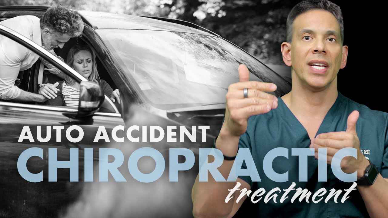

Integrative Chiropractic and Regenerative Spine Care
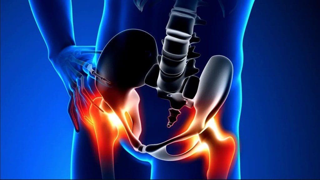
Back and neck pain can be difficult to treat because the spine is not made of bones alone. It also depends on healthy discs, muscles, ligaments, tendons, joints, nerves, and connective tissues. When several of these structures are injured, using only one type of treatment may not address the whole problem.
Integrative chiropractic and regenerative spine care takes a broader approach. Chiropractic adjustments help improve joint motion and spinal mechanics. Rehabilitation strengthens the muscles that support the spine. Spinal decompression may reduce pressure on sensitive discs and nerves. Shockwave and laser therapies may support soft-tissue recovery. Regenerative options, such as platelet-rich plasma, or PRP, may provide biological signals that support healing in selected injuries.
These treatments cannot promise to eliminate every case of pain or prevent every surgery. However, when the patient is carefully examined and the treatments are medically appropriate, a coordinated plan may reduce discomfort, improve movement, rebuild strength, and help some patients recover without surgery.
The Spine
The spine can be compared to a house in terms of its structure.
The bones and joints form the frame. The muscles and ligaments act like support beams and cables. The spinal discs are similar to cushions between the floors. Nerves are like electrical wires that carry messages throughout the body.
When the frame does not move correctly, a chiropractic adjustment may help restore joint motion. However, correcting the frame may not be enough when the supporting tissues are weak, inflamed, scarred, or injured.
A complete repair crew may also be needed:
- Chiropractic adjustments address restricted joint movement.
- Rehabilitation rebuilds strength and stability.
- Massage reduces muscle tension and guarding.
- Spinal decompression may reduce pressure on discs and nerves.
- Shockwave therapy stimulates injured soft tissues.
- Laser therapy delivers light energy to targeted tissues.
- PRP and related regenerative procedures may support tissue-healing signals.
Each treatment performs a different job. Together, they may create a better environment for recovery than a single treatment. This layered approach is described in clinical resources that combine chiropractic care, decompression, shockwave therapy, laser therapy, and rehabilitation (Oakland Spine & Physical Therapy, 2025; Sleppy Chiropractic Family Wellness Center, n.d.).
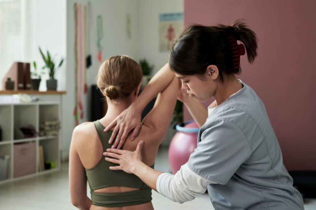
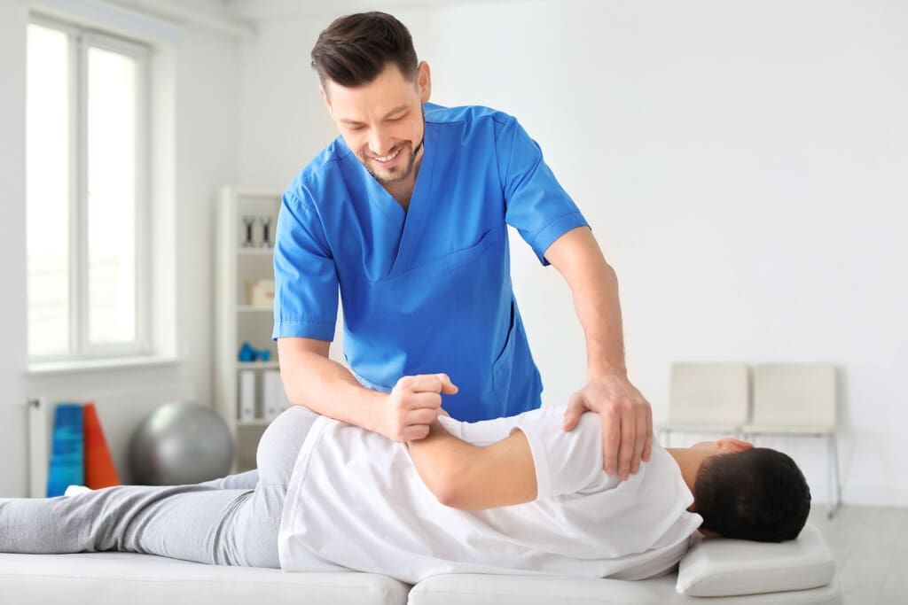
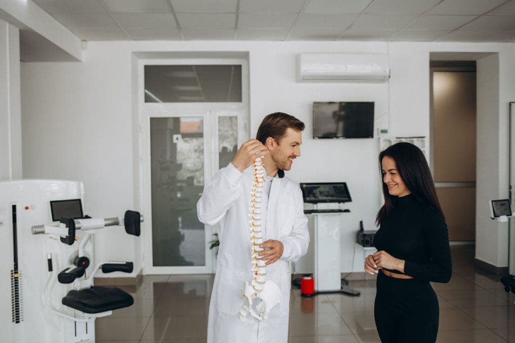
Chiropractic Care Restores Better Joint Motion
Chiropractic care commonly involves controlled adjustments to the spine or other joints. The goals may include improving restricted movement, reducing mechanical irritation, easing pain, and helping the body move more normally.
According to MedlinePlus, chiropractors may also use exercise, electrical stimulation, heat, ice, relaxation methods, and lifestyle guidance. Many people seek chiropractic care for back pain, neck pain, and headaches (MedlinePlus, n.d.).
It is more accurate to say that adjustments improve joint motion and mechanics than to say they permanently “put bones back into place.” Lasting improvement usually requires the muscles and soft tissues around the spine to become stronger and more balanced.
That is why chiropractic care is often combined with:
- Corrective exercises
- Core strengthening
- Posture training
- Mobility work
- Balance and coordination exercises
- Home activity guidance
Physical rehabilitation helps the body maintain better movement after an adjustment. Stronger muscles also reduce repeated strain on spinal joints and injured tissues (Oakland Spine & Physical Therapy, 2025).
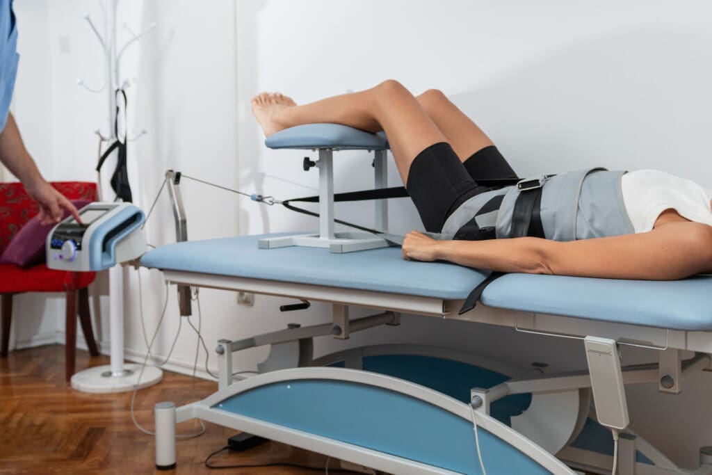
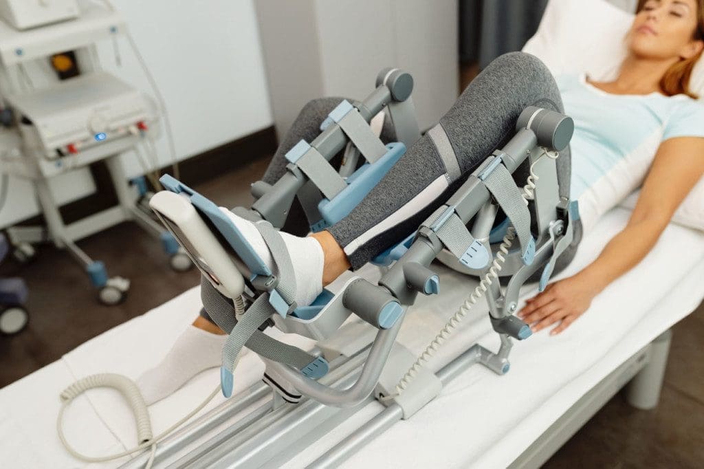
Spinal Decompression May Reduce Pressure
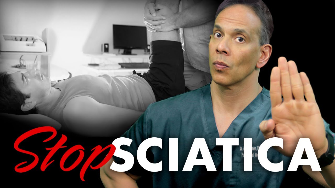
Spinal decompression uses controlled traction to gently stretch specific areas of the spine. It is commonly considered for selected patients with disc-related pain, sciatica, or nerve irritation.
The treatment is intended to temporarily increase space between spinal structures and reduce mechanical pressure. This may help some people move more comfortably while they complete rehabilitation and strengthening.
Decompression should not be presented as a treatment that automatically pulls every herniated disc back into place. Results depend on the diagnosis, severity of the injury, age of the condition, general health, and whether weakness or serious nerve damage is present.
When properly selected, decompression may complement chiropractic care:
- Decompression focuses on pressure affecting discs and nerves.
- Adjustments focus on restricted joint movement.
- Rehabilitation strengthens the muscles that protect the area.
- Massage reduces muscle tension around the spine.
This is another example of treatments performing separate but connected jobs (Sleppy Chiropractic Family Wellness Center, n.d.).
Shockwave Therapy Stimulates Soft Tissue
Extracorporeal shockwave therapy sends acoustic pressure waves into a targeted area. It is often used for chronic tendon problems, tight muscle bands, scarred tissues, and injuries that have stopped improving.
Shockwave therapy does not replace damaged tissue with new tissue overnight. Instead, the controlled mechanical energy may stimulate circulation, affect pain signals, and encourage a new healing response.
A randomized clinical trial involving people with chronic nonspecific low back pain found that one form of shockwave treatment reduced immediate pain and local sensitivity. However, the authors studied short-term effects, so shockwave should still be used as part of a wider recovery plan rather than as a guaranteed cure (Back et al., 2024).
Shockwave therapy may be especially useful when the surrounding muscles, tendons, or ligaments remain painful after spinal movement has improved. It can then be paired with gradual exercise, helping the healing tissue become stronger and better able to handle normal activity.
Laser Therapy Supports Cellular Activity
Therapeutic laser treatment, sometimes called photobiomodulation, applies specific wavelengths of light to targeted tissues. The light is absorbed by cells and may affect cellular energy production, inflammation, circulation, and pain signaling.
Laser therapy is generally used as a supportive treatment. It does not physically realign the spine, repair a major ligament tear, or replace the need for exercise. Its purpose is to support the biological environment in which healing occurs.
A practical treatment sequence may include:
- Reducing pain and irritation with laser or another supportive therapy.
- Improving restricted movement with gentle manual care.
- Reducing pressure when decompression is appropriate.
- Rebuilding strength through progressive rehabilitation.
- Teaching safer posture and movement habits.
The exact sequence should be based on examination findings rather than giving every patient the same treatment package.
PRP and Regenerative Therapies
PRP is prepared from a sample of the patient’s blood. The blood is centrifuged to obtain plasma with a higher platelet concentration. These platelets contain growth factors involved in the body’s normal healing response.
The PRP is then injected into a carefully selected joint, tendon, ligament, or other injured area. Ultrasound guidance may be used to improve accuracy.
Johns Hopkins Medicine explains that PRP may support healing in certain joint and soft-tissue injuries. However, results are not immediate or permanent for every patient, and additional treatment may be needed (Johns Hopkins Medicine, 2026).
Regenerative treatment is not magic. Research is still developing, especially for complex spinal conditions. Patients should be properly screened, and the diagnosis should be clear before an injection is considered. Imaging, medications, bleeding risks, infection risks, metabolic health, and the patient’s recovery goals must all be reviewed.
Regenerative procedures may support tissue biology, but rehabilitation remains necessary. New healing tissue must gradually learn to tolerate lifting, bending, walking, work duties, and exercise.
Massage and Rehabilitation Complete the Plan
Pain often causes the body to protect itself. Muscles tighten, movement becomes limited, and the patient begins using other areas to avoid the painful region. These compensation patterns can create additional problems in the hips, shoulders, knees, or opposite side of the back.
Massage and soft-tissue therapy may help reduce:
- Muscle guarding
- Trigger points
- Local stiffness
- Restricted movement
- Pain caused by compensation
Rehabilitation then helps the patient maintain the progress. Exercises may improve core control, hip strength, posture, balance, endurance, and spinal stability.
This is why long-term recovery is rarely based on passive treatment alone. The patient must gradually become an active part of the repair process.
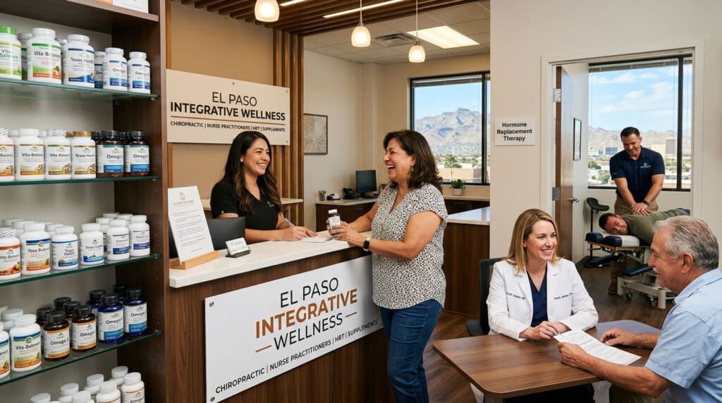
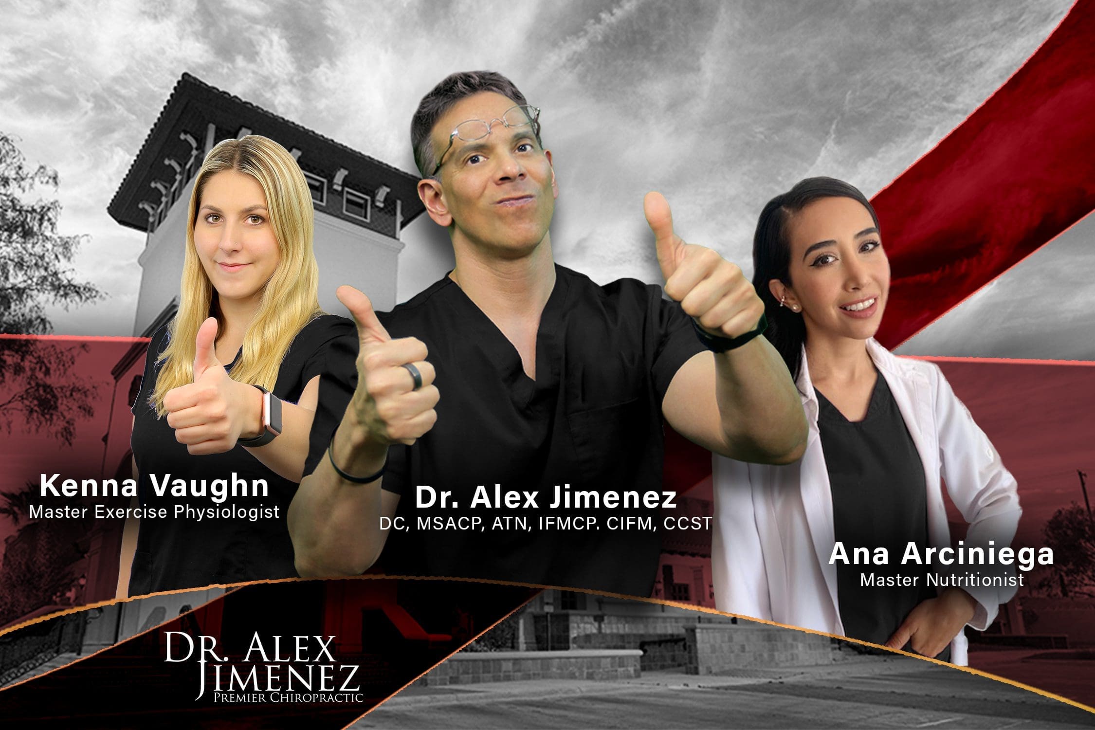
Multidisciplinary Spine Care in El Paso
At Injury Medical Clinic PA in El Paso, Texas, Dr. Alexander Jimenez, DC, APRN, FNP-BC, CCST, CFMP, IFMCP, ATN, works within a multidisciplinary model that combines chiropractic care, advanced clinical assessment, functional medicine, rehabilitation, personal injury care, and related supportive therapies.
Dr. Jimenez’s published clinical observations emphasize the need to examine both the mechanical and biological aspects of an injury. A patient may have restricted spinal movement, but that same patient may also have muscle weakness, inflammation, poor posture, nerve irritation, metabolic concerns, or damaged soft tissue. Addressing these connected problems may provide a more complete path toward recovery (Jimenez, n.d.; Personal Injury Doctor Group, 2026).
Dr. Maria Guadalupe Cardenas, MD, works with Dr. Jimenez as medical director and collaborative physician. Dr. Cardenas is a board-certified internal medicine physician who graduated from medical school in 1981 and has more than 40 years of clinical experience. Public provider information identifies her NPI as 1164426748 and lists Texas medical license J2933.
In this collaborative model:
- Dr. Jimenez leads chiropractic, neuromusculoskeletal, functional, and rehabilitative care within his professional scope.
- Dr. Cardenas provides internal medicine experience and medical direction.
- The team reviews health risks that may affect healing.
- Imaging, laboratory findings, medications, and medical conditions can be considered.
- Personal injury documentation and rehabilitation goals can be coordinated.
- Patients can be referred to surgeons or other specialists when conservative care is not appropriate.
This type of team structure allows chiropractic and rehabilitative care to occur under medical oversight, rather than each part of treatment operating separately.
When Surgery or Urgent Care May Be Necessary
Integrative care is not the right answer for every spinal problem. Some conditions require emergency treatment or a surgical consultation.
Immediate medical evaluation is important for:
- New loss of bladder or bowel control
- Numbness in the groin or saddle region
- Rapidly worsening arm or leg weakness
- Major trauma or suspected fracture
- Fever with severe spinal pain
- A history of cancer with new unexplained back pain
- Severe pain with unexplained weight loss
- Signs of infection
- Progressive spinal cord or nerve compression
A responsible integrative clinic does not promise to prevent surgery in every case. The goal is to provide the least invasive, safe, and appropriate treatment while referring patients when a higher level of care is needed.
Building Recovery From the Inside Out
The house comparison helps explain integrative spine care. Chiropractic adjustments improve the way the frame moves. Decompression may reduce pressure on sensitive structures. Shockwave and laser therapies support the soft-tissue environment. PRP may provide biological healing signals for selected injuries. Massage reduces protective tension, while rehabilitation rebuilds the strength needed to support the repairs.
The greatest benefit does not come from collecting as many treatments as possible. It comes from choosing the right treatments for the right patient at the right time.
With careful evaluation, medical oversight, realistic goals, and active rehabilitation, integrative chiropractic and regenerative spine care may help select patients reduce pain, restore mobility, improve strength, and return to daily activities without immediately resorting to surgery.

References
Back, C. G. N., Peron, R., Lopes, C. V. R., de Souza, J. V. E., and Liebano, R. E. (2024). Immediate effect of extracorporeal shockwave therapy in patients with chronic nonspecific low back pain: A randomized placebo-controlled triple-blind trial. Clinical Rehabilitation, 38(8), 1080–1090.
Health Coach Clinic. (n.d.). Poor posture and regenerative chiropractic recovery methods.
Jimenez, A. (n.d.). El Paso, TX chiropractor Dr. Alex Jimenez: Personal injury specialist.
Jimenez, A. (n.d.). Dr. Alex Jimenez’s professional profile. LinkedIn.
Johns Hopkins Medicine. (2026). Platelet-rich plasma injections.
MedlinePlus. (n.d.). Chiropractic. U.S. National Library of Medicine.
Oakland Spine & Physical Therapy. (2025, November 5). Benefits of combining chiropractic care with physical therapy.
Personal Injury Doctor Group. (2026, June 29). Regenerative therapies and chiropractic for injury recovery.
Personal Injury Doctor Group. (2026, June 30). Chiropractic and regenerative therapies for structural support.
Sciatica Pain and Treatment Clinic. (2026, June 30). Integrated posture care combining multiple therapies.
Sleppy Chiropractic Family Wellness Center. (n.d.). Beyond the adjustment: How decompression, shockwave therapy, and laser treatment work together.
Wellness Doctor Rx. (n.d.). Regenerative spine care for chronic back pain.