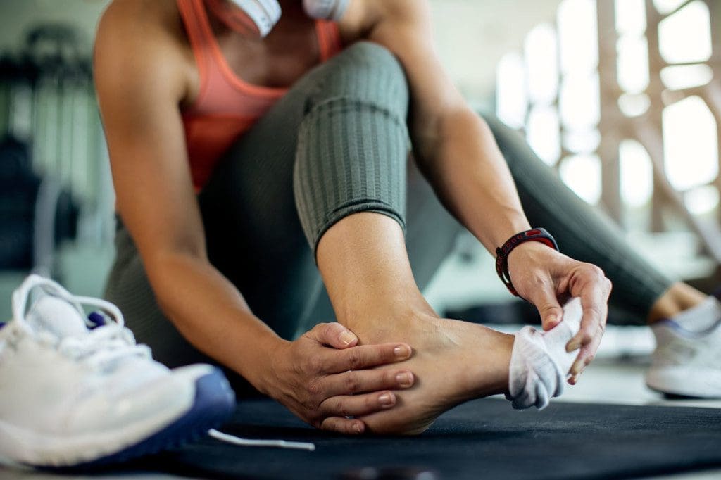

Regenerative Medicine for Joint Pain and Recovery
Abstract
Orthobiologics represents one of the most rapidly evolving frontiers in musculoskeletal medicine. This educational post explores the critical distinctions in platelet-rich plasma (PRP) formulation, specifically the role of neutrophil concentration, the clinical use of adipose-derived biologics, and subchondral bone interventions in managing degenerative joint conditions such as osteoarthritis. Drawing from peer-reviewed research and expert clinical dialogue, this post examines why cellular composition matters profoundly in regenerative injections, how microneedle patch therapy and fat grafting are expanding biologic options for patients who have exhausted conventional care, and why the “twenty percent failure rate” in subchondral treatments demands a more comprehensive, multidisciplinary approach. At Injury Medical Clinic PA in El Paso, Texas, Dr. Alex Jimenez, DC, APRN, FNP-BC, collaborates alongside Dr. Maria Guadalupe Cardenas, MD, Board Certified in Internal Medicine, to deliver exactly that kind of integrated, individualized care.

The Multidisciplinary Foundation: Dr. Jimenez and Dr. Cardenas Working Together
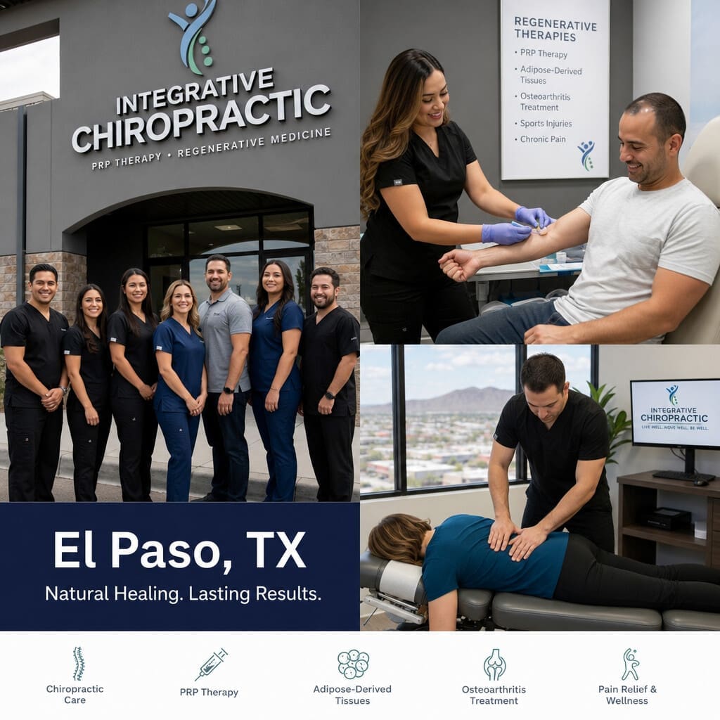
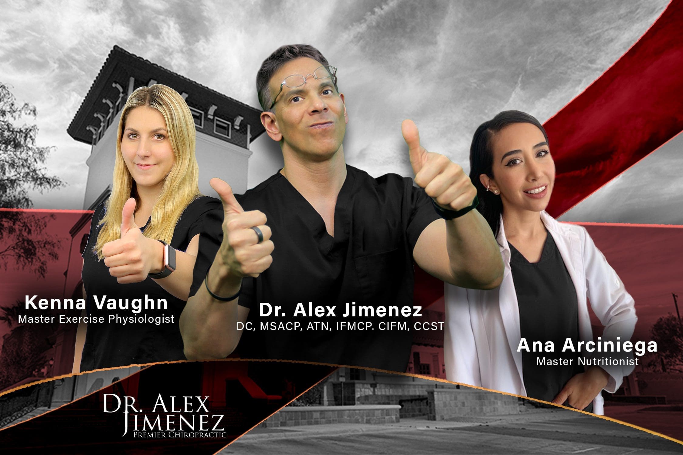
At Injury Medical Clinic PA, also known as Mission Plaza Injury Medical Clinic, located in El Paso, Texas, our clinical model is built on a powerful multidisciplinary foundation. Dr. Maria Guadalupe Cardenas, MD (NPI #1164426749, Texas MD License #J2933), Board Certified in Internal Medicine with over 40 years of experience as a practicing internist, serves as our Medical Director and Collaborative Physician. Her decades of internal medicine expertise bring essential medical oversight to every patient case we manage.
This model, where a licensed chiropractor and a board-certified internal medicine physician collaborate under one roof, is increasingly recognized as the gold standard in integrative injury care. Dr. Cardenas provides:
- Medical direction and clinical oversight for complex cases
- Diagnostic evaluation and co-management of systemic conditions affecting musculoskeletal health
- Collaborative case review for patients receiving regenerative or orthobiologic treatments
- Internal medicine consultation for patients with comorbidities such as metabolic syndrome, hypertension, or diabetes that can complicate joint degeneration
My role as a Doctor of Chiropractic and Advanced Practice Registered Nurse allows me to bridge structural, neurological, and functional medicine, while Dr. Cardenas ensures that each patient’s broader health picture is fully addressed. This is not simply co-location; it is true clinical collaboration aimed at restoring function and reducing pain at every level.
Understanding PRP Composition: Why Neutrophil Concentration Changes Everything
One of the most clinically significant and frequently misunderstood topics in regenerative medicine today is the cellular composition of PRP. Not all PRP is the same, and the difference is not merely technical; it is biologically consequential.
The Neutrophil Problem in Joint Injections
When we evaluate PRP systems, particularly those widely used in the United States, a critical observation emerges. Many commercially available centrifugation systems market their products as “leukocyte-rich” or even “leukocyte-poor” PRP, yet upon closer examination of the white blood cell differential, the neutrophil count remains the same as, or even higher than, baseline whole blood. In contrast, European protocols, particularly those practiced in Italy, where phlebotomy-based manual preparation methods are standard, tend to produce formulations that more precisely concentrate mononuclear cell populations, including monocytes and lymphocytes.
This distinction matters enormously for the following physiological reasons:
- Neutrophils are the body’s primary acute inflammatory responders. They release matrix metalloproteinases (MMPs), reactive oxygen species (ROS), and pro-inflammatory cytokines such as IL-1β and TNF-α, all of which degrade cartilage matrix and amplify synovial inflammation.
- Injecting a neutrophil-rich PRP preparation into an already inflamed joint can accelerate chondrocyte apoptosis and worsen synovitis rather than promote healing.
- Mononuclear cells, particularly monocytes and regulatory T-lymphocytes, are associated with tissue remodeling, anti-inflammatory signaling, and growth factor secretion, making them particularly desirable for intra-articular regenerative injections.
As I emphasize, understanding the cellular environment of a degenerative joint is not optional. It is the foundation of rational biologic therapy.
What Clinicians Should Ask Before Choosing a PRP System
If you are entering the field of orthobiologics, the following questions are non-negotiable:
- What is the neutrophil-to-lymphocyte ratio in the final PRP product?
- Does the system provide a white blood cell differential, not just a total WBC count?
- Are there peer-reviewed publications using this specific system, and do they report cellular composition data?
- Can preparation variables such as spin speed, spin duration, and kit design be modified to optimize the mononuclear fraction?
The take-home message is clear: know your system. A high platelet count does not guarantee a therapeutic product if neutrophil contamination undermines the anti-inflammatory environment needed for cartilage repair.
Adipose-Derived Biologics and Microneedling Patches: Second-Line Options With Real Clinical Power
The Role of Fat-Derived Stem Cells in Regenerative Care
Adipose tissue is one of the richest sources of mesenchymal stromal cells (MSCs) in the body, and its clinical application in orthobiologics is gaining substantial momentum. For patients with osteoarthritis who have not responded to PRP, hyaluronic acid, or corticosteroid injections and who are not yet ready for joint replacement, adipose-derived cell therapy offers a biologically meaningful alternative.
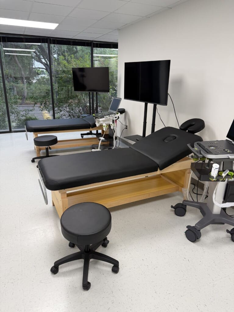
The procedure involves harvesting fat from areas such as the abdomen or flanks using a tumescent liposuction technique, performed in a dedicated special-procedure room. Critically, compelling safety data from the plastic surgery literature demonstrate that awake liposuction under local tumescent anesthesia has a significantly safer profile than procedures performed under general anesthesia. This makes in-clinic fat harvesting not only feasible but also preferable from a risk-management standpoint.
Key procedural considerations include:
- Tumescent anesthetic solution must be allowed to dwell in the tissue for a minimum of twenty minutes, ideally thirty minutes or longer, before fat harvest begins. This dwell time significantly reduces bleeding, facilitates fat separation, and improves cellular viability.
- Patients should be positioned comfortably, typically prone, in a calm environment, as the procedure, while technically invasive, is generally well tolerated under proper local anesthesia.
- Harvested fat is then processed and reinjected into the target joint, delivering a concentrated population of adipose-derived MSCs, growth factors, and anti-inflammatory mediators.
Microneedle Patch Therapy for Persistent Joint Effusions
Microneedle patch technology represents a newer frontier in biologic delivery, particularly useful in patients with persistent knee effusions or those who have undergone prior surgery and are seeking biologic augmentation of their healing environment. This is appropriately positioned as a second-line intervention rather than a first-line treatment, employed when:
- Standard orthobiologic injections have failed to produce lasting relief
- The patient has a persistent effusion despite conservative management
- The patient is post-surgical and seeks biologic enhancement of recovery
- Joint replacement is declined or deferred
While not universally effective, the clinical response in appropriately selected patients has been genuinely encouraging.
Subchondral Bone Interventions: The 80/20 Reality
What the Literature Tells Us
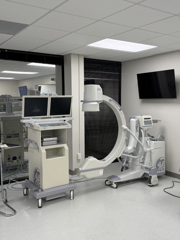
The subchondral bone is increasingly recognized as a central player in the pathophysiology of osteoarthritis. The subchondral region lies immediately beneath the articular cartilage, and dysfunction in this zone, including bone marrow lesions, vascular disruption, and increased intraosseous pressure, contributes directly to pain, cartilage degradation, and disease progression.
Multiple studies, including a notable French investigation demonstrating approximately 95% avoidance of arthroplasty at fifteen years following subchondral intervention, highlight the therapeutic potential of targeting this region. Techniques have included injection of bone marrow concentrate, calcium phosphate cement, and simple needle decompression to reduce intraosseous pressure.
However, a consistent finding across the breadth of subchondral bone literature is a 20% failure rate. Regardless of the specific biologic used, roughly one in five patients will not achieve meaningful or durable benefit. This is not a reason to abandon these techniques; it is a reason to think more comprehensively.
Modifying the Healing Environment: The Integrative Imperative
The critical clinical insight here is that injecting a needle, regardless of what is delivered, is only one part of the solution. The joint exists within a biological, mechanical, and metabolic environment. If that environment remains hostile to healing, the most sophisticated biologic preparation will underperform.
At Injury Medical Clinic PA, this is precisely where the integration of chiropractic care, functional medicine, rehabilitation, and internal medicine oversight becomes clinically decisive. Strategies to modify the subchondral and periarticular environment include:
- Osteotomy or joint offloading bracing to redistribute compressive forces across the joint surface
- Neuromuscular rehabilitation targeting quadriceps strength and lower extremity kinetic chain function, because weakened quadriceps dramatically increase compressive joint loads regardless of any biologic administered
- Weight management through functional medicine and metabolic support, reducing mechanical stress on the joint
- Chiropractic spinal and extremity adjustments to normalize biomechanical load distribution across the kinetic chain
- Anti-inflammatory nutritional protocols managed collaboratively with Dr. Cardenas to address systemic inflammatory drivers of joint degeneration
As I have observed clinically and through my professional work, the patients who achieve the most durable outcomes from orthobiologic interventions are those whose overall mechanical and metabolic environment has been optimized in parallel with the injection itself.
Integrative Chiropractic Care as the Structural Foundation of Regenerative Medicine
Chiropractic care contributes to orthobiologic outcomes in ways that are mechanistically well supported:
- Spinal manipulation and joint mobilization reduce aberrant neuromotor patterns that overload peripheral joints
- Soft tissue therapies address myofascial restrictions that perpetuate abnormal joint mechanics
- Functional rehabilitation protocols restore proprioception, muscle balance, and load-sharing capacity
- Postural and ergonomic correction reduces cumulative mechanical stress that drives cartilage breakdown
When a patient presents with knee osteoarthritis, their knee is not an isolated structure. It is the product of forces transmitted from the foot, ankle, hip, pelvis, and lumbar spine. Addressing only the knee with an injection while ignoring the rest of the kinetic chain is, physiologically speaking, incomplete medicine.
Clinical Takeaways for Patients and Practitioners
- PRP is not a single product. Cellular composition, particularly the neutrophil-to-mononuclear cell ratio, determines its therapeutic or potentially harmful effects in joint environments.
- Adipose-derived biologics offer a viable second-line option for patients with advanced osteoarthritis who are not surgical candidates.
- Subchondral interventions have an approximately 80% success rate, but maximizing that success requires comprehensive modification of the joint environment.
- Multidisciplinary care, combining chiropractic, internal medicine, functional medicine, and rehabilitation, is not a luxury; it is a clinical necessity for durable outcomes.
- At Injury Medical Clinic PA, Dr. Cardenas and I work together every day to deliver exactly this kind of comprehensive, patient-centered care.

References
- Filardo, G., Di Matteo, B., Di Martino, A., Merli, M. L., Cenacchi, A., Fornasari, P., Marcacci, M., & Kon, E. (2015). Platelet-rich plasma intra-articular knee injections show no superiority versus viscosupplementation. The American Journal of Sports Medicine, 43(7), 1575-1582.
- Kon, E., Mandelbaum, B., Buda, R., Filardo, G., Delcogliano, M., Timoncini, A., Fornasari, P. M., Giannini, S., & Marcacci, M. (2011). Platelet-rich plasma intra-articular injection versus hyaluronic acid viscosupplementation as treatments for cartilage pathology. Arthroscopy: The Journal of Arthroscopic & Related Surgery, 27(11), 1490-1501.
- Magalon, J., Bausset, O., Serratrice, N., Giraudo, L., Lutomski, D., Bellaiche, L., Veran, J., Magalon, G., & Dignat-George, F. (2014). Characterization and comparison of 5 platelet-rich plasma preparations in a single-donor model. Arthroscopy: The Journal of Arthroscopic & Related Surgery, 30(5), 629-638.
- Murray, I. R., Corselli, M., Petrigliano, F. A., Soo, C., & Peault, B. (2014). Recent insights into the identity of mesenchymal stem cells: Implications for orthopaedic applications. The Bone & Joint Journal, 96-B(3), 291-298.
- Sampson, S., Gerhardt, M., & Mandelbaum, B. (2008). Platelet rich plasma injection grafts for musculoskeletal injuries: A review. Current Reviews in Musculoskeletal Medicine, 1(3-4), 165-174.
- Spasovski, D., Spasovski, V., Baščarević, Z., Stojiljković, M., Vreća, M., Anđelković, M., & Pavlović, S. (2018). Intra-articular injection of platelet-rich plasma versus corticosteroid as treatment for knee osteoarthritis. Journal of Biological Regulators and Homeostatic Agents, 32(6), 1445-1452.
- Sundman, E. A., Cole, B. J., Karas, V., Della Valle, C., Tetreault, M. W., Mohammed, H. O., & Fortier, L. A. (2014). The anti-inflammatory and matrix restorative mechanisms of platelet-rich plasma in osteoarthritis. The American Journal of Sports Medicine, 42(1), 35-41.