
El Paso Motorcycle Brain Injury Recovery After a Helmeted Crash
Introduction: A Helmet Helps, But It Cannot Stop Every Injury
If a motorcycle rider in El Paso suffers a brain injury while wearing a helmet, it usually means the crash force was severe. The helmet may have reduced the impact and helped prevent a fatal or more severe injury. However, no helmet can fully prevent the brain from moving inside the skull during a hard crash, especially when the head, neck, and spine are subjected to sudden acceleration, deceleration, or twisting forces (Zimmerman & Frachtman, 2023; Emroch & Kilduff, n.d.).
This matters for two reasons. First, the injured rider still needs a full medical evaluation, even if the helmet looks intact. Second, if another driver caused the crash through negligence, the rider may still have the right to seek compensation for medical bills, lost income, pain and suffering, and long-term care needs (Ruhmann Law Firm, n.d.; Rodman Law Office, n.d.).

Why Brain Injuries Can Happen With a Helmet On
Motorcycle helmets are designed to reduce the impact of direct head impacts. A quality helmet has a durable outer shell and an inner liner that absorbs some of the crash energy. This can reduce skull fractures and lower the risk of severe traumatic brain injury. Still, helmets have limits. In a high-speed crash, side-impact collision, rollover, or sudden ejection, the force may exceed the limits the helmet was designed to handle (Emroch & Kilduff, n.d.).
A helmet also cannot fully prevent rotational forces. These forces happen when the head twists quickly. The brain can shift inside the skull, which may lead to concussion, diffuse axonal injury, headaches, dizziness, memory problems, and balance issues. This is why a rider can walk away from a crash wearing a helmet but still have a real brain injury (TopDog Law, 2025; CDC, 2025).
Common symptoms after a helmeted motorcycle crash may include:
- Headache or pressure in the head
- Dizziness or balance problems
- Nausea or vomiting
- Confusion or feeling “foggy”
- Memory or concentration problems
- Sensitivity to light or noise
- Neck pain, back pain, or shoulder pain
- Sleep changes, anxiety, or mood swings
The CDC explains that mild traumatic brain injury and concussion symptoms may appear right away, but some symptoms may not show up for hours or days (CDC, 2025).
Helmets Reduce Fatal Injuries, But They Do Not Make Riders Invincible
Helmet use is still one of the most important safety steps a motorcyclist can take. The National Highway Traffic Safety Administration encourages riders to wear DOT-compliant helmets and notes that motorcyclists remain highly vulnerable on the road. In 2024, motorcyclists were almost 27 times more likely than passenger car occupants to die in a crash per vehicle mile traveled (NHTSA, n.d.).
Older NHTSA data also found that motorcycle helmets reduce the likelihood of crash deaths and are effective in reducing brain injuries. This means a helmeted rider who suffers a concussion or TBI should not assume the helmet failed completely. In many cases, the helmet may have reduced a fatal injury to a survivable one (NHTSA, 2008; Rodman Law Office, n.d.).
Legal Rights After a Helmeted Motorcycle Brain Injury in El Paso
Wearing a helmet does not take away a rider’s right to file a personal injury claim. In fact, wearing a helmet may help show that the rider took reasonable steps to protect themselves. If another driver caused the crash by speeding, failing to yield, texting, making an unsafe lane change, following too closely, or turning left in front of the motorcycle, that driver’s negligence may still be the main legal issue (Ruhmann Law Firm, n.d.; Law Offices of Ruben Ortiz, n.d.).
Texas uses a modified comparative fault system. This means an injured person may recover compensation if they are not more than 50% at fault, but the amount may be reduced by their percentage of fault. Helmet use may become part of the insurance argument, especially in head injury cases, but it does not automatically decide the case (Rodman Law Office, n.d.).
Possible damages after a serious motorcycle brain injury may include:
- Emergency room care
- CT scans, MRIs, and neurological testing
- Chiropractic and rehabilitation care
- Physical therapy and occupational therapy
- Lost wages or reduced earning ability
- Long-term care needs
- Pain, suffering, and reduced quality of life
- Motorcycle repair or replacement
Ruhmann Law Firm notes that motorcycle accident claims may include current and future medical care, rehabilitation services, medical devices, lost wages, disability, property damage, and pain and suffering (Ruhmann Law Firm, n.d.).
Why an El Paso Personal Injury Lawyer Matters
Motorcycle riders often face unfair assumptions. Some insurance companies may try to blame the rider before the facts are fully reviewed. Local legal resources, including the Ruhmann Law Firm and the Law Offices of Ruben Ortiz, discuss the importance of building motorcycle injury cases around evidence, crash reports, medical records, witness statements, and the real long-term impact of the injuries (Ruhmann Law Firm, n.d.; Law Offices of Ruben Ortiz, n.d.).
A personal injury lawyer can help investigate:
- Who caused the crash
- Whether the other driver failed to yield or was distracted
- Whether road design or traffic conditions played a role
- Whether the helmet shows evidence of severe impact
- Whether medical records support the brain, neck, spine, and soft tissue injuries
- Whether future treatment costs should be included
This is especially important in TBI cases because symptoms may be invisible. A person may look “fine” but still struggle with headaches, brain fog, dizziness, mood changes, and work limitations.
The Medical Side: Brain, Neck, and Spine Must Be Evaluated Together
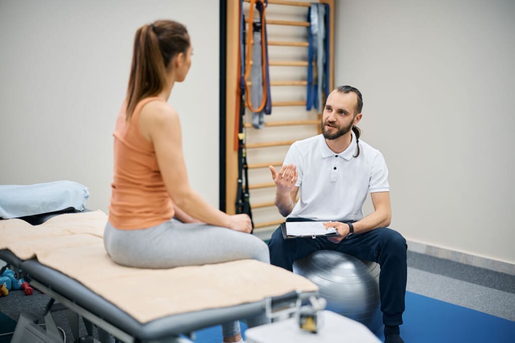
A motorcycle brain injury rarely affects only the head. The same force that injures the brain can also injure the neck, spine, shoulders, discs, ligaments, muscles, and nerves. Whiplash, cervical strain, spinal misalignment, herniated discs, and nerve irritation may appear alongside concussion symptoms.
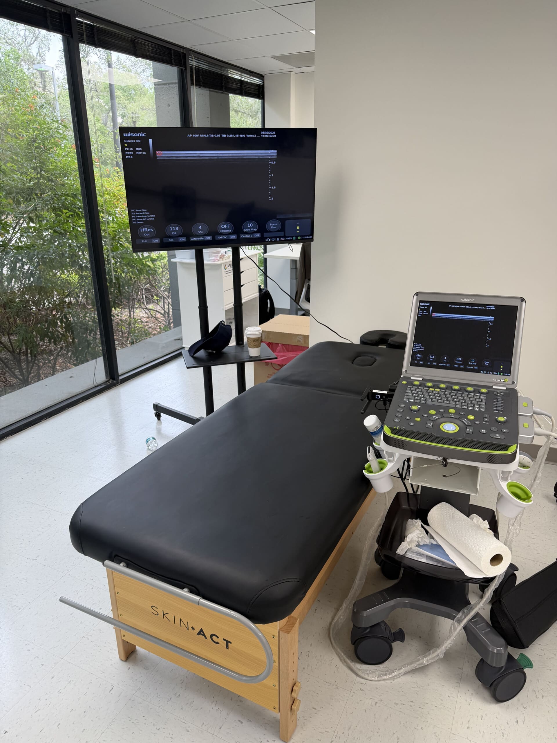
This is why a full evaluation should include both medical and musculoskeletal assessment. The first step is urgent medical care to rule out bleeding, fracture, worsening neurological symptoms, or emergency complications. After that, a coordinated recovery plan may include chiropractic care, rehabilitation, physical therapy, neurological follow-up, and, when appropriate, pain management.
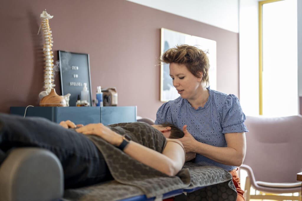
Integrative Chiropractic and Regenerative Support in El Paso
Integrative care may help after the initial diagnosis by focusing on mobility, pain control, spinal alignment, soft-tissue healing, and nervous system function. Chiropractic care does not “treat” a brain injury by itself, but it may support recovery by addressing neck trauma, spinal strain, muscle guarding, headaches linked to cervical dysfunction, and postural changes after the crash.
Dr. Alexander Jimenez, DC, APRN, FNP-BC, has written about a team-based, dual-scope approach for traumatic brain injury rehabilitation that may include spine and posture care, functional neurology concepts, medical oversight, and exercise planning (Jimenez, 2025).
In motorcycle accident recovery content, Dr. Jimenez also emphasizes that head and neck injuries are common after motorcycle crashes and that early intervention may reduce long-term complications such as chronic headaches, dizziness, and pain patterns tied to cervical trauma (Jimenez, 2025).
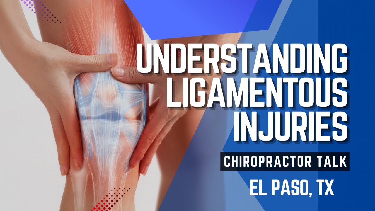
Regenerative medicine may also be considered when the rider has musculoskeletal tissue damage, such as ligament sprains, tendon injuries, joint irritation, or chronic soft-tissue pain. Regenerative medicine, also called orthobiologics, aims to stimulate the body’s ability to repair damaged muscles, joints, tendons, and other tissues (Weill Cornell Medicine, n.d.).
Examples may include:
- Platelet-rich plasma, or PRP
- Prolotherapy
- Microfragmented adipose tissue, or MFAT
- Image-guided injection planning
- Chiropractic rehabilitation
- Corrective exercise
- Nutrition support for inflammation and tissue repair
These therapies should be used only after a proper diagnosis and by qualified healthcare providers. They are not a replacement for emergency care, neurology, imaging, or legal documentation.
Local Recovery Options in the El Paso and Horizon City Area
For riders in the El Paso and Horizon City area, the best next step is a comprehensive evaluation. Clinics such as Synergy Chiropractic, Aktiv Integrative Chiropractic, and Dr. Alex Jimenez’s Injury Medical & Chiropractic Clinic are examples of local integrative or chiropractic-focused options that may help evaluate musculoskeletal injury, spine trauma, and rehabilitation needs after a crash. Synergy Chiropractic lists traumatic brain injury, car accident treatment, chiropractic adjustments, spinal decompression, soft tissue care, and shockwave therapy among its services, while Aktiv Integrative Chiropractic describes patient-centered musculoskeletal and wellness care in El Paso (Synergy Chiropractic, n.d.; Aktiv Integrative Chiropractic, n.d.).
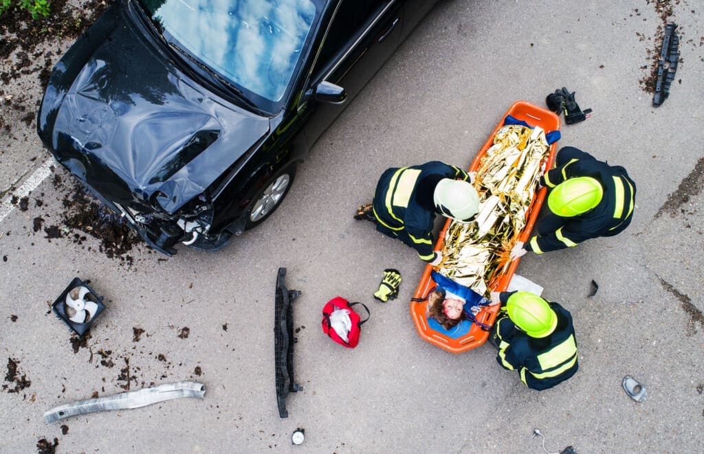
What To Do After a Helmeted Motorcycle Crash
After a helmeted crash with possible brain injury:
- Call 911 and get medical care immediately.
- Keep the helmet, even if it is damaged.
- Take photos of the motorcycle, helmet, road, vehicles, and injuries.
- Report headaches, dizziness, memory problems, neck pain, and back pain.
- Do not give a recorded statement before understanding your rights.
- Follow up with medical, neurological, chiropractic, and rehabilitation providers.
- Speak with an El Paso personal injury lawyer if another driver caused the crash.
Conclusion: A Helmeted Brain Injury Is Still Serious
A motorcycle brain injury while wearing a helmet should never be dismissed. The helmet may have prevented something worse, but the rider may still have a concussion, neck injury, spinal strain, nerve irritation, and long-term recovery needs. In El Paso, a strong recovery plan should include emergency medical evaluation, careful documentation, legal guidance when negligence is involved, and coordinated rehabilitation.
The goal is not only to survive the crash. The goal is to protect the rider’s health, legal rights, mobility, nervous system function, and long-term quality of life.

References
Aktiv Integrative Chiropractic. (n.d.). Chiropractor El Paso TX 79912.
Centers for Disease Control and Prevention. (2025). Symptoms of mild TBI and concussion.
Emroch & Kilduff. (n.d.). Can you get a head injury while wearing a helmet?
Jimenez, A. (2025). Motorcycle accident recovery with chiropractic care.
Jimenez, A. (2025). Rehabilitative sports after traumatic brain injury: Integrative care.
Law Offices of Ruben Ortiz. (n.d.). Motorcycle accident attorney in El Paso.
National Highway Traffic Safety Administration. (n.d.). Motorcycle safety: Helmets, motorists, road awareness.
National Highway Traffic Safety Administration. (2008). Traffic safety facts: Motorcycle helmet laws.
Rodman Law Office. (n.d.). Motorcycle helmet use and injury claims: What the law says.
Ruhmann Law Firm. (n.d.). Motorcycle accident lawyer in El Paso.
Synergy Chiropractic. (n.d.). Chiropractic care for traumatic brain injury recovery in El Paso.
Weill Cornell Medicine. (n.d.). Regenerative medicine.
Zimmerman & Frachtman. (2023). Can a motorcyclist suffer a head injury with a helmet on?.