
Uncover the potential advantages of hormone therapy for men’s health and how it can enhance your overall health and energy levels.
Abstract
In this educational post, I present an evidence-based, integrative approach to two common men’s health concerns: erectile dysfunction (ED) and testosterone deficiency (low T). I review the vascular-neural physiology of erections; risk factors and diagnostic pathways; and stepwise treatments, including PDE5 inhibitors, vacuum erection devices, intraurethral and intracavernosal therapies, penile prosthesis, and low-intensity shockwave therapy. I then discuss evaluating and treating testosterone deficiency, integrating endocrinology guidelines with functional medicine and rehabilitative care, including a review of testosterone replacement therapy (TRT) options, risks, and fertility-preserving alternatives. I also integrate chiropractic and functional medicine strategies, cardiometabolic risk-reduction strategies, and rehabilitative protocols into our multidisciplinary clinical model. Our medical direction is led by Dr. Maria Guadalupe Cardenas, MD (Board Certified in Internal Medicine; NPI 1164426749; Texas MD License J2933), who serves as Medical Director and Collaborative Physician at Injury Medical Clinic PA (Mission Plaza Injury Medical Clinic) in El Paso, Texas. Together, we coordinate safe, monitored care for personal injury, spine-related pain, ED, and hormonal health using modern diagnostics and research-informed protocols.
Introduction: Our Multidisciplinary Clinical Model in Men’s Health
I’m Dr. Alex Jimenez, DC, APRN, FNP-BC, CFMP, IFMCP, ATN, CCST. In our El Paso practice—Injury Medical Clinic PA (Mission Plaza Injury Medical Clinic)—we operate a multidisciplinary model common to integrative and injury care clinics. Our Medical Director and Collaborative Physician, Dr. Maria Guadalupe Cardenas, MD (Board Certified in Internal Medicine; NPI #1164426749; Texas MD License #J2933), has over 40 years of experience as an internist. Dr. Cardenas oversees medical safety, pharmacologic decisions, diagnostic appropriateness, and chronic disease management while I lead integrative chiropractic care, spine and joint biomechanics, neuro-musculoskeletal rehabilitation, and functional medicine strategies. This dual-track care model pairs MD medical direction with chiropractic and functional medicine interventions to maximize outcomes while maintaining safety and governance.
- Core pillars of our men’s health care:
-
- Medical oversight and internal medicine risk management: hypertension, diabetes, dyslipidemia, obesity, and medication interactions
- Integrative chiropractic care: optimizing spinal, pelvic, and sacral alignment; neuromechanical function; and autonomic balance
- Functional medicine: nutrition, sleep, stress, microbiome, metabolic flexibility, and cardiometabolic risk reduction
- Personal injury care: addressing pain, deconditioning, and neuro-musculoskeletal dysfunction that can undermine sexual health
- Rehabilitation: graded exercise, pelvic floor strategies, and fascia/soft tissue interventions for circulation and nerve signaling
Clinical Observations: Why Men’s Health Needs Integrated Care
In my clinical experience—reflected in the case discussions and insights I share publicly at my sciatica-focused repository and professional profile—you’ll find that men with ED and low energy often present with overlapping spine, hip, and pelvic dysfunction, metabolic syndrome, and autonomic stress dysregulation. My observations at:
- sciatica.clinic
- linkedin.com/in/dralexjimenez
repeatedly show that correcting pelvic mechanics, reducing lumbosacral nerve irritation, and improving endothelial health can meaningfully support medical therapies for men’s health. Mechanical load and pain can drive sympathetic hyperarousal and sleep fragmentation, worsening metabolic and hormonal function.
Understanding Erectile Dysfunction: Vascular-Neural Physiology
Erections are a synchronized vascular and neural event. The penis must receive adequate arterial inflow and maintain venous occlusion while neural pathways trigger smooth muscle relaxation via nitric oxide (NO) signaling.
- Key physiological steps:
-
- Sexual stimulation increases parasympathetic neural activity, releasing nitric oxide from endothelial cells and nerve terminals (Burnett, 2006; Carson et al., 2021).
- NO activates soluble guanylate cyclase, converting GTP to cyclic GMP (cGMP).
- cGMP induces smooth muscle relaxation in the corpus cavernosum, increasing arterial inflow and compressing subtunical venules, achieving rigidity (Goldstein et al., 2024).
- Phosphodiesterase type 5 (PDE5) breaks down cGMP; PDE5 inhibitors (sildenafil, tadalafil) prolong cGMP activity, sustaining erection potential (Hedlund et al., 2020).
- Why this matters: Any condition impairing endothelial function (e.g., diabetes, hypertension, dyslipidemia), autonomic control, or penile structure may blunt NO-cGMP signaling or venous trapping, reducing rigidity and durability of erections (Khera et al., 2023).
Risk Factors and Etiologies Impacting Erectile Function
ED reflects an interplay of vascular, metabolic, neurogenic, psychogenic, and iatrogenic influences.
- High-impact risk factors:
-
- Age, hypertension, diabetes, obesity, dyslipidemia, tobacco, sedentary lifestyle, and chronic alcohol use degrade endothelial health, reduce NO bioavailability, and weaken cavernosal smooth muscle response (Khera et al., 2023; Vlachopoulos & Jackson, 2014).
- Neurogenic sources (e.g., spinal cord injury, MS), pelvic trauma, and lumbosacral radiculopathy alter neural inputs essential for arousal and cavernosal relaxation.
- Medications: SSRIs, certain diuretics, and nonselective alpha-blockers can impair erection quality through vascular or central mechanisms (Khera et al., 2023).
Diagnostic Approach: Standardized Assessment and Labs
We use validated questionnaires and a structured medical evaluation to understand the severity and root causes of sexual dysfunction.
- Standardized tools:
-
- International Index of Erectile Function (IIEF) and Sexual Health Inventory for Men (SHIM) provide quantifiable measures of severity and treatment response (Rosen et al., 1997; Mulhall et al., 2007).
- Focused labs and exams:
-
- Morning total testosterone, lipid panel, A1c/fasting glucose, TSH/free T4, PSA as indicated, and a comprehensive male GU exam with prostate assessment (AUA Testosterone Guideline; Khera et al., 2023).
- Rationale: Morning testosterone captures the diurnal peak; metabolic and thyroid screening identifies reversible contributors to endothelial dysfunction and energy/libido deficits.
Evidence-Based Treatment Pathways for Erectile Dysfunction
We follow a shared decision-making model under Dr. Cardenas’s medical oversight to ensure safety, realistic expectations, and coordination with comorbid management.
- Lifestyle and cardiometabolic optimization:
-
- Smoking cessation, weight loss, glycemic control, lipid optimization, sleep restoration, and exercise improve endothelial function and NO signaling, potentiating ED therapies (Vlachopoulos & Jackson, 2014).
- Our functional medicine strategies target inflammation, insulin resistance, and oxidative stress that degrade cavernosal compliance.
- PDE5 inhibitors (sildenafil, tadalafil):
-
- Mechanism: Increase cGMP persistence, improving smooth muscle relaxation and cavernosal filling (Hedlund et al., 2020).
- Dosing: On-demand use 1 hour pre-activity; daily low-dose tadalafil may help men with ED and lower urinary tract symptoms (Carson et al., 2021). Avoid with nitrates; use caution with nonselective alpha-blockers due to the risk of hypotension.
- Clinical reasoning: First-line efficacy with favorable safety; less effective in severe diabetic vasculopathy or post-prostatectomy due to reduced NO release and structural changes.
- Vacuum erection devices (VEDs):
-
- Mechanism: Negative pressure draws blood into the penis; a constriction ring helps maintain venous trapping.
- Indications: PDE5 nonresponders or adjunct therapy; cost-effective yet variable satisfaction due to mechanical complexity and potential discomfort (Carson et al., 2021).
- Intraurethral prostaglandin (alprostadil/MUSE):
-
- Mechanism: Prostaglandin E1 induces smooth muscle relaxation; rapid onset with lower efficacy than injections; may cause urethral discomfort and hypotension (Carson et al., 2021).
- Considerations: Cost and insurance coverage vary; supervised test dosing recommended.
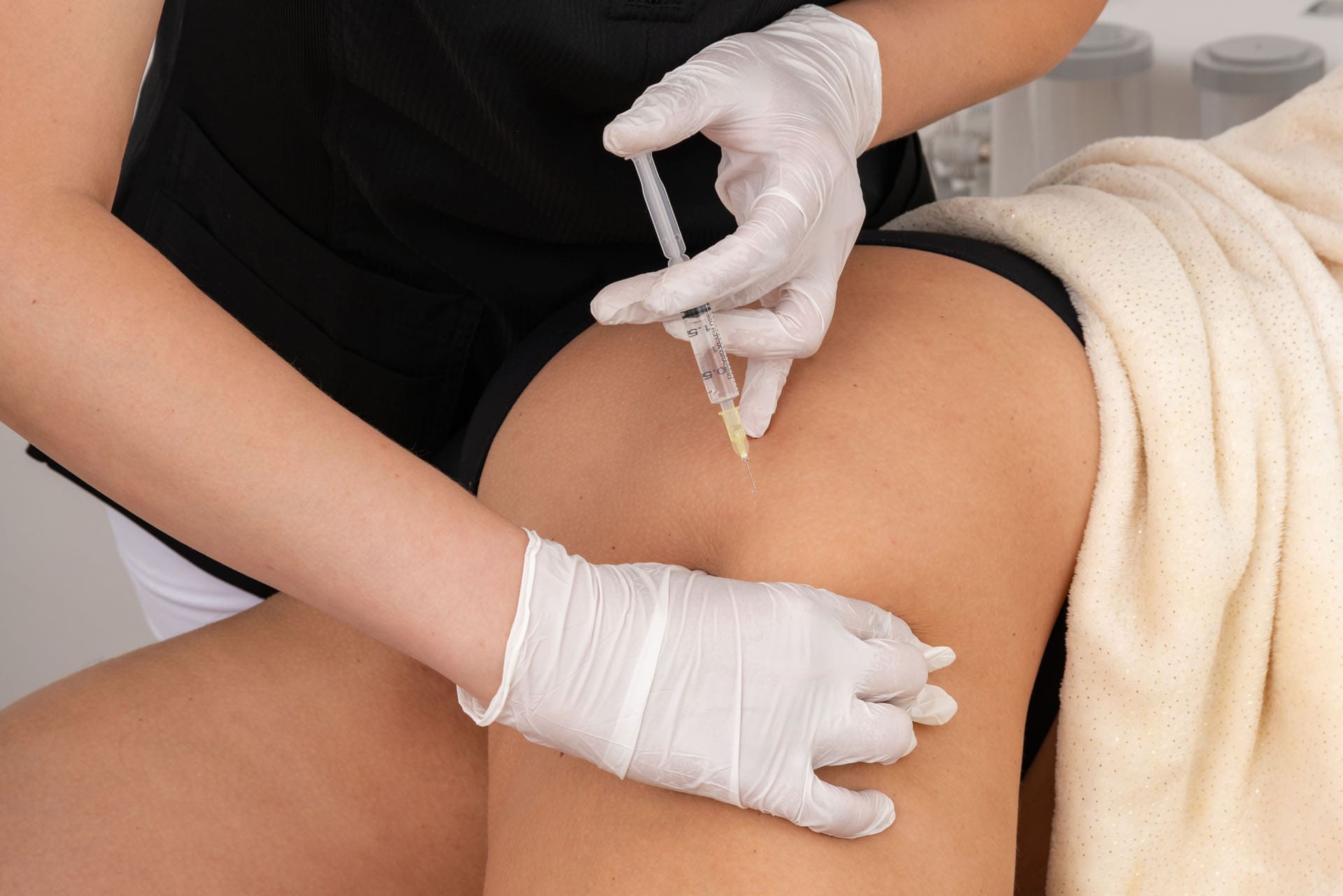
- Intracavernosal injections (alprostadil; compounded bimix/trimix):
-
- Mechanism: Direct cavernosal smooth muscle relaxation via prostaglandin and vasodilators (papaverine, phentolamine).
- Pros: High efficacy in PDE5 nonresponders; rapid onset.
- Risks: Pain, fibrosis, priapism; dose titration and injection rotation essential; not more than three times weekly (Carson et al., 2021).
- Penile prosthesis:
-
- Mechanism: Inflatable devices mechanically restore rigidity; high satisfaction (>90%) in appropriate candidates.
- Rationale: Definitive option for severe structural or refractory ED; low infection risk with modern devices; patients lose spontaneous erections but maintain orgasm and ejaculation routes (Carson et al., 2021).
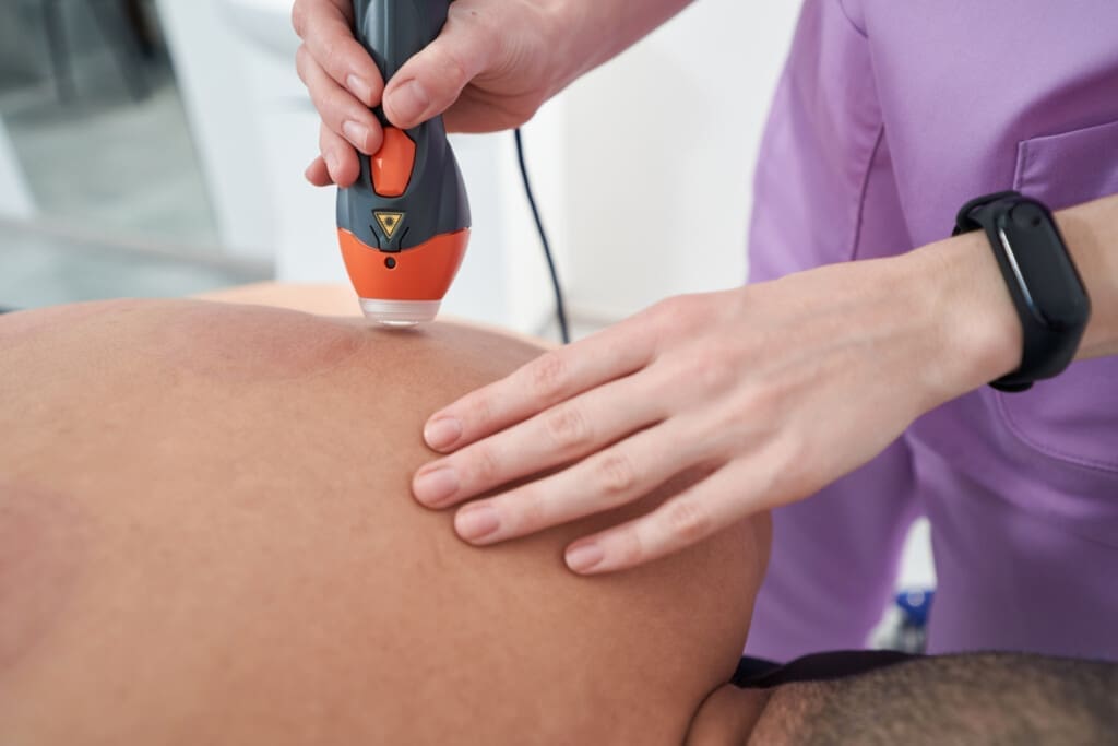
- Low-intensity shockwave therapy (LiSWT):
-
- Mechanism: Acoustic pulses induce microtrauma and neovascularization, potentially improving endothelial function and penile perfusion (Hisasue et al., 2024).
- Status: Investigational in many regions; best evidence in mild-to-moderate ED, particularly PDE5 partial responders; cost and access considerations.
- Online compounded ED options and OTC gels:
-
- Compounded combinations (e.g., sildenafil/tadalafil with add-ons) are not FDA-approved; efficacy and safety vary, requiring medical oversight to avoid interactions and counterfeit risks (FDA communications).
- OTC topical gels (e.g., evaporative thermogenic stimulation) may offer sensory-driven effects with limited and mixed user outcomes; clinical monitoring advised.
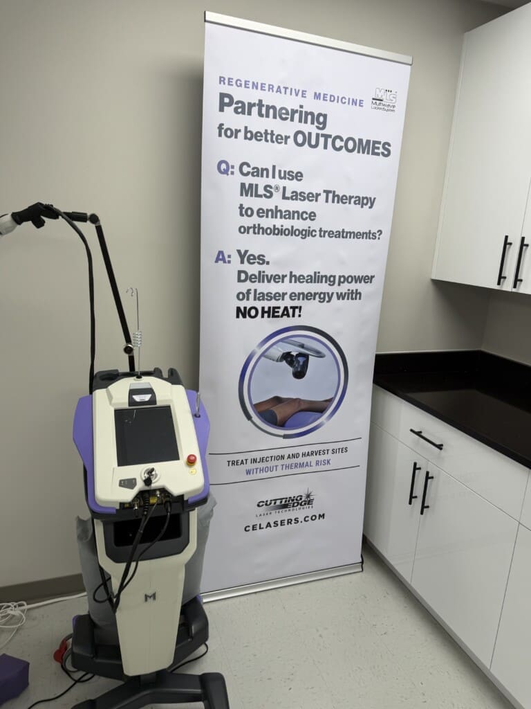
Emerging Therapies: PRP, Stem Cells, Hyperbaric Oxygen, Nutraceuticals
- PRP and stem cells:
-
- Concept: Regenerative signaling for angiogenesis and cavernosal tissue remodeling.
- Evidence: Limited rigorous randomized trials; investigational and not FDA-approved; we use caution and emphasize informed consent (Khera et al., 2023).
- Hyperbaric oxygen therapy:
-
- Mechanism: Enhanced oxygen delivery and angiogenesis, similar intent to shockwave outcomes.
- Evidence: Early data suggest perfusion benefits; protocols vary; best considered adjunctive for select cases.
- Nutraceuticals:
-
- L-arginine (NO precursor) and L-citrulline (arginine recycling) may modestly support NO pathways; quality and dosing are critical; we monitor for interactions and realistic expectations (Khera et al., 2023).
Understanding Hypogonadism and Testosterone Deficiency
As men age, a gradual decline in testosterone—often about 1–2% per year—can intersect with comorbidities such as obesity and sleep apnea. Low testosterone (hypogonadism) impacts libido, energy, mood, body composition, and cardiometabolic risk. However, ED and low T overlap variably; libido may improve with testosterone, but rigidity often remains vascularly constrained, requiring parallel ED therapies. Not all symptoms are due to low T, and overtreatment can occur.
- Defining hypogonadism:
-
- A clinical syndrome of testicular failure to produce physiological testosterone concentrations with associated signs and symptoms (Mulhall et al., 2018).
- Typical symptoms:
-
- Specific: decreased libido, erectile dysfunction (ED), fatigue, loss of muscle mass, reduced body hair.
- Nonspecific: poor concentration, memory issues, low energy, decreased endurance.
- Physiology of low testosterone:
-
- Testosterone modulates central sexual desire, nitric oxide synthase expression, and cavernosal tissue integrity. Low T can reduce NO availability and smooth muscle content while increasing fibrosis (Khera et al., 2018; AUA guideline).
- It influences skeletal muscle anabolism through androgen receptor signaling, influencing satellite cell activation and myofibrillar protein synthesis (Kadi, 2008).
- It increases hematopoiesis by stimulating erythropoietin, which requires monitoring to avoid erythrocytosis (Coviello et al., 2008).
- It impacts body composition and insulin sensitivity, influencing visceral adiposity and inflammation (Grossmann, 2011).
Diagnostic Algorithm for Testosterone Deficiency
Our approach mirrors American Urological Association (AUA) recommendations to ensure we identify true deficiency and address underlying causes (Mulhall et al., 2018).
- Measure total testosterone in the morning (typically 7–10 AM). The AUA recommends confirming deficiency with two separate morning total testosterone measurements below 300 ng/dL, accompanied by relevant clinical symptoms.
- If <300 ng/dL, repeat a morning level to confirm.
- Order LH, prolactin, hematocrit (Hct), and PSA (age-appropriate).
- Evaluate medication contributors (opioids, glucocorticoids), alcohol use, and obstructive sleep apnea (OSA). When comorbidities such as OSA and obesity increase inflammatory cytokines, hypothalamic-pituitary-gonadal axis signaling can be blunted, reducing testosterone production (Vgontzas et al., 2005).
Testosterone Treatment Options: Formulations, Physiology, and Rationale
When criteria for deficiency are met and comorbidities are optimized, we consider TRT under Dr. Cardenas’s medical direction. Formulation choice depends on patient preference, insurance coverage, and risk profile (Bhasin et al., 2018).
- Intramuscular injections: Testosterone cypionate or enanthate (e.g., 75-100 mg weekly) offers predictable dosing but can cause peaks and troughs.
- Transdermal gels (e.g., AndroGel) provide more stable daily levels but carry a risk of transference to others.
- Pellets (Testopel): Subdermal implantation every 3-4 months offers convenience but requires a minor procedure.
- Oral testosterone undecanoate: Bypasses liver metabolism but is expensive and carries black box warnings for blood pressure.
- Long-acting injections and nasal formulations are less commonly used due to specific risks, such as POME and local irritation, respectively.
TRT Monitoring and Contraindications: Safety First
Because testosterone elevates erythropoietin and can raise hematocrit, we monitor closely.
- Monitoring: We check testosterone and hematocrit at 9-12 weeks, then every 6-12 months. We also monitor PSA, blood pressure, and lipids. Our clinical goal is to pause or adjust therapy if Hct >52-54%.
- Contraindications: We avoid or defer TRT in men with untreated prostate/breast cancer, severe untreated OSA, baseline hematocrit ≥50%, severe heart failure, recent MI/stroke, or an active desire for fertility, as TRT suppresses sperm production.
Fertility-Preserving Alternative: Clomiphene Citrate
For younger men planning children or those averse to TRT, clomiphene citrate is a practical off-label option.
- Mechanism: As a selective estrogen receptor modulator, it reduces negative feedback on the pituitary, increasing LH/FSH. This stimulates the testes to produce more testosterone and maintain spermatogenesis (Taylor & Levine, 2010).
- Dosing: Typically started at 25 mg three days per week and titrated based on labs and symptoms.
A Case Journey: Translating Physiology to Practice
Consider Mr. T, a 56-year-old male with hypertension, diabetes, obesity, and OSA. He reports fatigue, low libido, weight gain, and mild ED (SHIM score of 8). His PCP measured a single afternoon testosterone of 150 ng/dL, which is suboptimal. He is noncompliant with his CPAP.
- Stepwise plan:
-
- Medical Oversight (Dr. Cardenas): Reinforce CPAP adherence for 3 months, as untreated OSA suppresses testosterone (Budweiser et al., 2013). Obtain two separate morning total testosterone levels. If ED persists, initiate a PDE-5 inhibitor. Optimize his blood pressure and lipids.
- Integrative Chiropractic and Functional Medicine (Dr. Jimenez): Apply intensive lifestyle supports: weight reduction, glycemic control, and stress modulation to restore endothelial and autonomic balance. Implement graded aerobic and resistance training to enhance endothelial function and metabolic health. Correct pelvic and lumbosacral mechanics to reduce neurogenic irritation and optimize autonomic tone.
- Decision on TRT: If confirmed deficiency persists after lifestyle optimization, discuss TRT formulations, including clomiphene, with full risk-benefit counseling.
Signs of Hormonal Imbalances In Men *THIS IS WHY*- Video
Why Integrative Chiropractic Care Fits in Men’s Health Management
- Biomechanics and vascular supply: Proper lumbo-pelvic alignment enhances neurovascular signaling to the pelvic organs. Soft tissue work reduces fascial restrictions that may limit perfusion, and spinal adjustments can alleviate sympathetic overdrive.
- Autonomic regulation: Reducing spinal joint dysfunction and nociceptive signaling can modulate autonomic balance, supporting restorative sleep and heart rate variability. Improving thoracic mobility complements OSA treatment and exercise tolerance.
- Pain reduction and activity resumption: Addressing back, hip, and pelvic pain increases activity levels, improving cardiometabolic health and endothelial function, which directly contribute to improvements in ED and testosterone.
Coordinated Care Workflow at Injury Medical Clinic PA
- Intake and triage: SHIM/IIEF scoring; cardiometabolic screening; spine/pelvic assessment; medication reconciliation.
- Collaborative plan: Medical therapy selection and monitoring by Cardenas; chiropractic and rehabilitation plan by me; functional medicine supports; personal injury protocols if applicable.
- Follow-up cadence: 6–12 weeks for ED therapy reassessment; 3-month labs for testosterone therapy; periodic SHIM/IIEF to track progress; adjust biomechanics and rehab intensity based on pain/function.
Conclusions: Modern, Evidence-Based Men’s Health Through Integration
ED and low T are not isolated issues; they sit at the crossroads of vascular health, neural control, endocrine function, biomechanics, and psychosocial factors. With internal medicine oversight by Dr. Maria Guadalupe Cardenas, MD, and integrative chiropractic, rehabilitation, and functional medicine led by me, we provide comprehensive, safe, and research-informed care. We empower patients through structured coaching in sleep, nutrition, and exercise to enhance the effectiveness of medical therapies and restore endothelial health. Our approach aligns physiology with practical strategies to help men regain confidence, performance, and well-being.
References
- American Urological Association Guideline on Erectile Dysfunction (Khera, M. et al., 2023).
- American Urological Association Guideline: Evaluation and Management of Testosterone Deficiency (Mulhall, J. P., et al., 2018).
- AUA Guideline on Testosterone Deficiency (Khera, M., et al., 2018).
- Clomiphene Citrate for Hypogonadism: Mechanisms and Outcomes (Taylor, F., & Levine, L., 2010). The Journal of Clinical Endocrinology & Metabolism.
- CPAP Therapy Effects on Testosterone and Metabolic Profiles (Budweiser, S., et al., 2013). Respiratory Research.
- Endothelial Dysfunction and ED: Clinical Correlates (Vlachopoulos, C., & Jackson, G., 2014).
- Exercise Training Improves Cardiometabolic Health (Kraus, W. E., et al., 2019). Circulation.
- IIEF Development and Validation (Rosen, R. C., et al., 1997).
- Long-acting Testosterone Undecanoate: Safety Considerations (Pastuszak, A. W., et al., 2015). The Journal of Sexual Medicine.
- Mediterranean Diet and Cardiovascular Outcomes (Estruch, R. et al., 2018). New England Journal of Medicine.
- Metabolic Dysfunction in Hypogonadal Men (Grossmann, M., 2011). Nature Reviews Endocrinology.
- NO-cGMP Pathway in Penile Physiology (Burnett, A. L., 2006).
- Obstructive Sleep Apnea and Endocrine Function (Vgontzas, A. N., et al., 2005). The Journal of Clinical Endocrinology & Metabolism.
- Princeton IV Consensus Recommendations for PDE5 Inhibitors and Cardiovascular Risk (Hedlund, P. et al., 2020).
- SHIM Validation Study (Mulhall, J. P., et al., 2007).
- Shockwave Therapy for ED: Current Evidence (Hisasue, S., et al., 2024).
- Testosterone and Skeletal Muscle: Molecular and Cellular Mechanisms (Kadi, F., 2008). Biology of Reproduction.
- Testosterone Therapy and the Risk of Adverse Events: A Review (Layton, J. B., et al., 2014). Therapeutic Advances in Drug Safety.
- Testosterone Therapy in Men With Hypogonadism: An Endocrine Society Clinical Practice Guideline (Bhasin, S. et al., 2018). The Journal of Clinical Endocrinology & Metabolism.
- Textbook of Erectile Dysfunction: Vascular and Neural Mechanisms (Carson, C. C., et al., 2021).
- The Relationship Between Testosterone and Erythrocytosis in Men (Coviello, A. D., et al., 2008). JAMA Internal Medicine.
- The Role of Testosterone in Erectile Function: Pathophysiology and Clinical Implications (Corona, G., et al., 2014). The Journal of Sexual Medicine.
- Zinc and Testicular Function: A Review (Kumar, N., & Singh, A. K., 2015). Biological Trace Element Research.
SEO tags: erectile dysfunction, low testosterone, TRT, hypogonadism, PDE5 inhibitors, tadalafil, sildenafil, shockwave therapy, penile prosthesis, intracavernosal injection, alprostadil, trimix, clomiphene, CPAP, sleep apnea, men’s health, integrative chiropractic, functional medicine, endothelial function, nitric oxide, cGMP, hematocrit monitoring, PSA, endocrine guidelines, strength training, Mediterranean diet, El Paso, Injury Medical Clinic PA, Mission Plaza Injury Medical Clinic, Dr. Maria Guadalupe Cardenas MD, Dr. Alex Jimenez DC