

Table of Contents
How to Eat Healthy on a Budget in El Paso, TX (Without Feeling Deprived)

A very common question in El Paso is, “How do I eat healthy without spending a fortune?” Groceries are pricier now, and the “healthy” aisle seems to be the most expensive. The good news is that healthy eating can be economical when you use a few smart habits: planning meals, buying seasonal or frozen produce, using beans and lentils for protein, shopping sales, and cooking at home more often.
In El Paso, you can also lean on local community resources (like city programs and community health efforts) and build routines that support both your budget and your body—especially if you’re recovering from injuries, dealing with chronic pain, or trying to reduce inflammation.
Why “Healthy” Feels Expensive (and How to Flip the Script)
Healthy eating often feels expensive for a few reasons:
-
Convenience costs money. Pre-cut fruit, ready-to-eat meals, and grab-and-go snacks are usually more expensive.
-
Food waste drains your wallet. If fresh produce goes bad in the fridge, it’s like throwing cash away.
-
Impulse shopping adds up. A few unplanned items can blow your budget fast.
The fix is not “buy fancy health foods.” The fix is building a simple system.
The Budget-Friendly Healthy Eating System (El Paso Edition)
Here’s the system many dietitians and public health programs recommend:
Plan → Shop smart → Cook simple → Waste less → Repeat.
Step 1: Plan meals before you shop
Planning is the #1 money-saver because it helps you buy only what you’ll use.
Try this weekly routine:
-
Pick 2–3 breakfasts you can repeat
-
Pick 2 lunches (often leftovers)
-
Pick 3–4 dinners
-
Add 2 simple snacks
-
Build your grocery list from your plan
Simple planning tip: Start by planning dinners only. Once that feels easy, plan lunches and snacks.
Step 2: Build a “cheap + healthy” core grocery list
A smart budget list is built around high-fiber, high-protein, long-lasting foods.
Low-cost staples that work in many meals:
-
Beans (pinto, black, lentils), chickpeas
-
Brown rice, oats, whole wheat pasta
-
Eggs
-
Frozen vegetables
-
Canned tuna or salmon (watch sodium, choose water-packed when possible)
-
Plain yogurt (can double as a probiotic-rich food for some people)
-
Peanut butter or nuts (watch portions)
-
Olive oil, salsa, spices
This is the kind of “foundation” that lets you avoid buying expensive, one-use ingredients.
The Produce Strategy: Seasonal + Frozen = Big Savings
Buy seasonal produce when possible
Seasonal produce is usually cheaper and tastes better.
Use frozen and canned produce on purpose
Frozen fruits and vegetables can be a budget hero because they last longer and reduce waste. Many reputable health organizations recommend using frozen/canned options to keep healthy eating affordable.
Quick tips for choosing frozen/canned:
-
Frozen: look for “just vegetables” (no sauce)
-
Canned veggies: choose low-sodium or rinse them
-
Canned fruit: choose water or 100% juice, not heavy syrup
Protein Without the High Price Tag
Meat can get expensive fast. One of the easiest ways to lower your grocery bill is to use plant proteins more often.
Affordable proteins that still build strong meals:
-
Beans, lentils, peas
-
Eggs
-
Canned fish
-
Chicken bought on sale (freeze portions)
-
Plain yogurt or cottage cheese (if tolerated)
Canada’s Food Guide specifically calls out beans and lentils as inexpensive sources of protein and suggests choosing plant-based proteins more often.
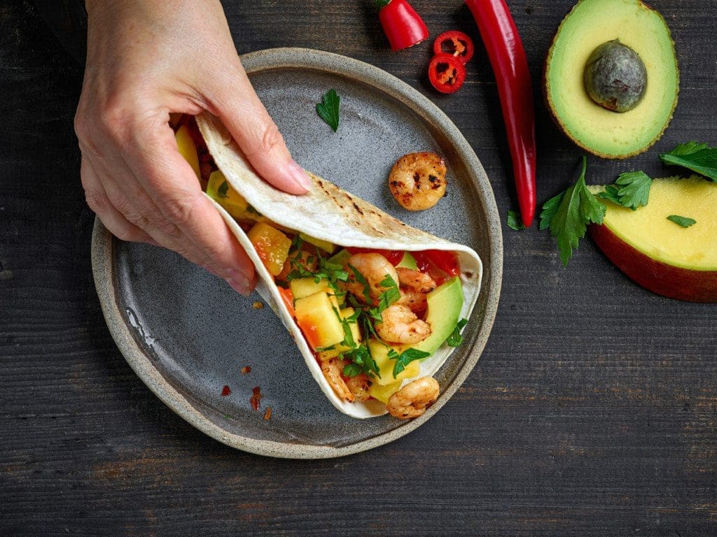
El Paso-friendly idea:
Make a big pot of charro-style beans (lighter version) and use them all week:
-
Bowl with rice + avocado
-
Taco filling
-
Soup base
-
Side dish with eggs
Shop Like a Pro (Even If You Hate Grocery Shopping)
Use “unit price” and store brands
Comparing unit prices helps you find the best deal. Store brands are often cheaper for similar nutrition.
Shop sales and stock up on staples
When shelf-stable items go on sale, buy enough for a few weeks (if you’ll actually use them).
Try “odd and imperfect” produce
Some stores sell “imperfect” produce for less, and it’s still nutritious.
Don’t shop hungry
This sounds small, but it matters—hunger increases impulse buying. Planning + a list helps you stay focused.
Batch Cooking: The Secret to Saving Money and Time
Batch cooking means you cook once and eat multiple times. It reduces:
-
Food waste
-
Takeout spending
-
Decision fatigue after a long day
Great budget-friendly batch meals:
-
Bean chili
-
Chicken and veggie soup
-
Stir-fry with frozen vegetables
-
Sheet-pan fajita bowls (use onions/peppers, beans, and a smaller amount of meat)
Simple “batch cook” checklist:
-
Cook 1 big protein (beans, chicken, eggs)
-
Cook 1 big carb (rice, potatoes, pasta)
-
Prep 1–2 vegetables (fresh or frozen)
-
Make 1 sauce (salsa, yogurt-lime, vinaigrette)
A Realistic 3-Day Budget-Friendly Meal Example
These are simple, repeatable meals that don’t require fancy ingredients.
Day 1
-
Breakfast: oatmeal + frozen berries + peanut butter
-
Lunch: bean/rice bowl + salsa + cabbage slaw
-
Dinner: egg scramble + frozen veggies + tortillas
Day 2
-
Breakfast: yogurt + oats + banana
-
Lunch: leftover scramble in a wrap
-
Dinner: lentil soup (big batch) + side salad
Day 3
-
Breakfast: eggs + toast + fruit
-
Lunch: lentil soup leftovers
-
Dinner: chicken (sale pack) + roasted seasonal vegetables + rice
Eating Out in El Paso Without Destroying Your Goals
Eating out can cost more, but you don’t have to avoid it completely. In El Paso, the City’s public health program Eat Well! El Paso focuses on improving the local food environment and includes a restaurant initiative that encourages healthier menu options (especially for families).
Budget-friendly ordering tips:
-
Choose grilled instead of fried
-
Ask for sauces/dressings on the side
-
Split an entrée or box half right away
-
Add a side salad or veggies when possible
If you want ideas for “healthier restaurant options,” directories like Tripadvisor list “healthy” categories for El Paso restaurants (always double-check menus because items change).
Local and Community Resources That Can Help
Sometimes the most economical healthy plan includes community support.
Helpful types of resources:
-
Nutrition education and cooking programs are supported by community health efforts
-
Food distribution and pantry support (especially when money is tight)
-
Federal nutrition support programs like WIC (and other tools highlighted by ODPHP)
Also, food banks and community organizations often teach “shop smart” strategies and simple meal-building methods that reduce waste and stretch staples.
How Integrative Chiropractic Care Fits In (and Why It Matters for Budgeting)
At first, chiropractic care and grocery shopping sound like two separate worlds. But in real life, they connect through one big idea:
When your body feels better, it’s easier to cook, plan, and stay consistent.
In El Paso, some clinics describe chiropractic care as part of a broader, patient-centered approach that may include exercise guidance and even dietary advice as part of a wellness plan.
Integrated clinics may also combine services like rehab, nutrition counseling, and nurse practitioner support under one roof.
Why this matters for economic healthy eating
When people are dealing with:
-
back pain
-
sciatica
-
headaches
-
stress and poor sleep
-
inflammation and gut issues
…they often spend more money on:
-
takeout (because cooking feels hard)
-
convenience foods
-
“snack grazing” due to fatigue
So part of “healthy eating on a budget” is making the plan physically doable.
Clinical Observations from Dr. Alexander Jimenez (Practical, Not Fancy)
In Dr. Alexander Jimenez’s integrative setting, the focus is often on whole-person recovery—especially when patients are dealing with pain, inflammation, injury recovery, or gut stress. His content frequently connects nutrition choices with wellness goals, including gut-support strategies (such as probiotic foods for some people) alongside integrative chiropractic care.
Here are budget-friendly nutrition habits that align with that “whole-person” approach:
-
Prioritize protein + fiber at meals (beans, lentils, eggs, oats). This supports steadier energy and fewer cravings.
-
Use anti-inflammatory basics regularly: vegetables, fruits, omega-3 sources (such as canned fish), and, when possible, fewer ultra-processed foods.
-
Support gut-friendly routines with simple options like yogurt or fermented foods (if tolerated), plus consistent meal timing.
Important note: Nutrition is individual. If you have IBS, reflux, diabetes, kidney disease, food allergies, or you’re on medications, your plan should be personalized.
The “No-Waste” Rule (This Alone Can Save a Lot)
Food waste is one of the highest hidden costs. If you waste less, you automatically spend less.
Easy no-waste habits:
-
Plan 1 weekly “use it up” meal (soup, stir-fry, omelet)
-
Freeze leftovers in single portions
-
Keep a “finish first” bin in the fridge for foods that need to be eaten soon
Quick Grocery List: One Week of Budget-Friendly Basics
Here’s a simple list you can adjust based on sales:
Proteins
-
Beans/lentils
-
Eggs
-
Chicken (sale pack) or canned fish
Carbs
-
Oats
-
Rice
-
Whole wheat tortillas/pasta
Produce
-
Seasonal fresh vegetables
-
Frozen mixed vegetables
-
Frozen berries
-
Onions/cabbage (cheap and versatile)
Flavor
-
Salsa
-
Garlic powder/chili powder
-
Olive oil (or another basic cooking oil)
Key Takeaways (Simple and Actionable)
If you want healthy eating to be economical in El Paso, focus on these:
-
Plan meals and shop with a list
-
Use beans/lentils as an affordable source of protein
-
Buy seasonal + frozen produce to reduce waste
-
Cook at home more often and batch cook
-
Use local resources and programs when needed
-
If pain and stress are blocking consistency, consider integrative care that supports movement, recovery, and lifestyle habits

References
-
Eat Healthy on a Budget by Planning Ahead (American Heart Association, 2025).
-
Cooking Healthy on a Budget (American Heart Association, 2024).
-
How to Eat Healthy on a Budget (Scripps Health, 2024).
-
Tips for Eating Healthy on a Budget (Mayo Clinic Health System, 2025).
-
Healthy Eating on a Budget (Canada’s Food Guide, n.d.).
-
Shop Smart (USDA MyPlate, n.d.).
-
Eat Healthy on a Budget (Tip Sheet) (USDA MyPlate, n.d.).
-
Tools to Help Consumers Eat Healthy on a Budget (Office of Disease Prevention and Health Promotion, 2024).
-
Eat Well! El Paso (City of El Paso Department of Public Health, n.d.).
-
Healthy Eating and Active Living (Paso del Norte Health Foundation, n.d.).
-
Shopping Smart on a Budget: Tips for Nutritious and Affordable Meals (Central Texas Food Bank, 2025).
-
10 Tips for Eating Healthy on a Budget (Mount Carmel Health, 2023).
-
Eating Healthy on a Budget (Lone Star Cares, 2024).
-
How to Stay Healthy When You’re on a Budget (Queensland Health, 2024).
-
Healthy Restaurants in El Paso (Tripadvisor, n.d.).
-
Aktiv Integrative Chiropractic (Clinic Site) (Aktiv Integrative Chiropractic, n.d.).
-
Chiropractic Services in El Paso TX (Aktiv Integrative Chiropractic, n.d.).
-
Dr. Alex Jimenez, Injury Medical & Chiropractic Clinic (Profile) (A4M, n.d.).
-
El Paso’s Guide to Probiotics and Chiropractic Healing (Jimenez, n.d.).
-
ChiroMed – Integrated Medicine (ChiroMed, n.d.).
-
Healthy Eating on a Budget (Video) (YouTube, n.d.).


:max_bytes(150000):strip_icc()/Yoga-Back-Pain-Stocksy_txp8ad835bcdJc300_Medium_1772162-95080420a6164913a60a973dc0add6d6.jpg)