
Can knowing which foods to eat help individuals recovering from food poisoning restore gut health?
Food Poisoning and Restoring Gut Health
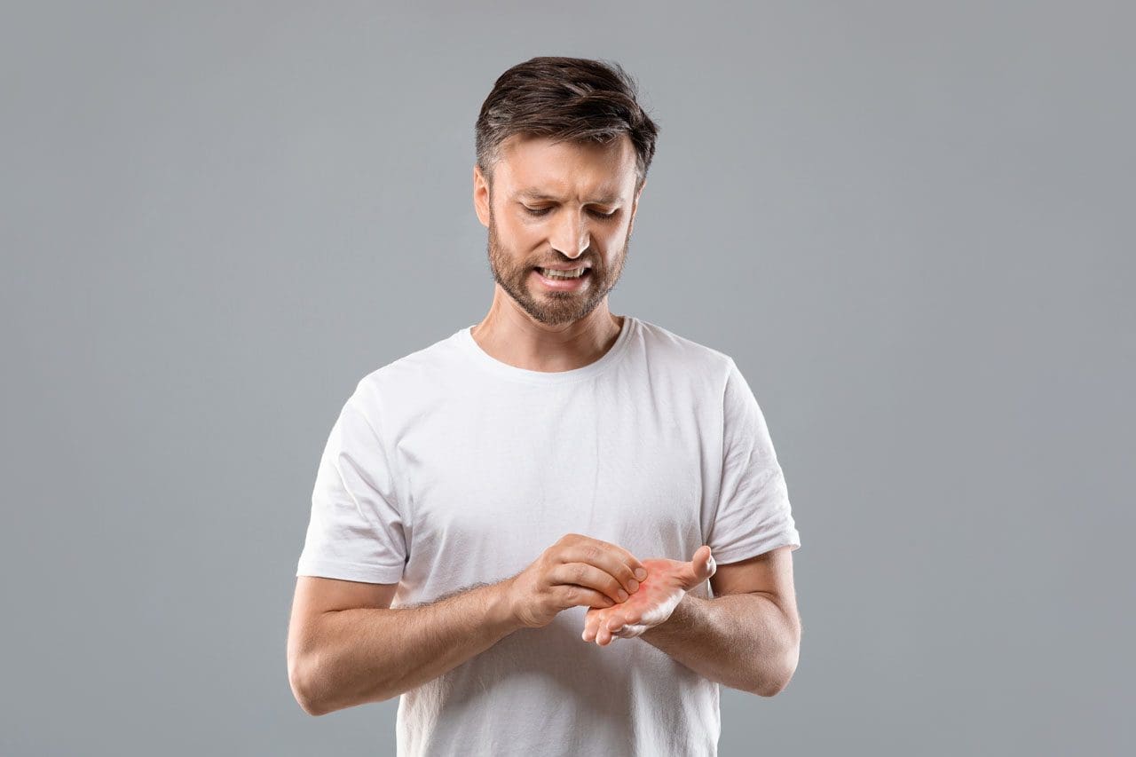
Food poisoning can be life-threatening. Fortunately, most cases are mild and short-lived and last only a few hours to a few days (Centers for Disease Control and Prevention, 2024). But even mild cases can wreak havoc on the gut, causing nausea, vomiting, and diarrhea. Researchers have found that bacterial infections, like food poisoning, can cause changes in gut bacteria. (Clara Belzer et al., 2014) Eating foods that promote gut healing after food poisoning may help the body recover and feel better faster.
Foods to Eat
After food poisoning symptoms have resolved, one may feel that returning to the usual diet is fine. However, the gut has endured quite an experience, and even though acute symptoms have subsided, individuals may still benefit from foods and drinks that are easier on the stomach. Recommended foods and beverages after food poisoning include: (National Institute of Diabetes and Digestive and Kidney Diseases. 2019)
- Gatorade
- Pedialyte
- Water
- Herbal tea
- Chicken broth
- Jello
- Applesauce
- Crackers
- Toast
- Rice
- Oatmeal
- Bananas
- Potatoes
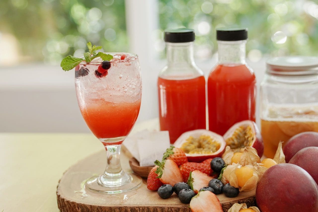
Hydration after food poisoning is crucial. Individuals should add other nutritious and hydrating foods, like chicken noodle soup, which helps because of its nutrients and fluid content. The diarrhea and vomiting that accompany the illness can leave the body severely dehydrated. Rehydrating beverages help the body replace lost electrolytes and sodium. Once the body is rehydrated and can hold down bland foods, slowly introduce foods from a regular diet. When resuming the usual diet after rehydration, eating small meals frequently, every three to four hours, is recommended instead of eating a large breakfast, lunch, and dinner meal daily. (Andi L. Shane et al., 2017) When choosing Gatorade or Pedialyte, remember that Gatorade is a sports-rehydrating drink with more sugar, which could irritate an inflamed stomach. Pedialyte is designed for rehydrating during and after illness and has less sugar, making it a better option. (Ronald J Maughan et al., 2016)
When Food Poisoning Is Active Foods To Avoid
During food poisoning, individuals typically do not feel like eating at all. However, to avoid worsening the illness, Individuals are recommended to avoid the following while actively ill (Ohio State University. 2019)
- Caffeinated drinks and alcohol can further dehydrate.
- Greasy foods and high-fiber foods are hard to digest.
- Foods and beverages high in sugar can cause the body to produce high glucose levels and weaken the immune system. (Navid Shomali et al., 2021)
Recovery Time and Resuming Regular Diet
Food poisoning doesn’t last long, and most uncomplicated cases are resolved within a few hours or days. (Centers for Disease Control and Prevention, 2024) Symptoms depend on the type of bacteria. Individuals may become ill within minutes of consuming contaminated food up to two weeks later. For example, Staphylococcus aureus bacteria generally cause symptoms almost immediately. On the other hand, listeria may take up to a couple of weeks to cause symptoms. (Centers for Disease Control and Prevention, 2024) Individuals can resume their usual diet once symptoms are gone, the body is thoroughly hydrated and can hold down bland foods. (Andi L. Shane et al., 2017)
Recommended Gut Foods Post Stomach Virus
Gut-healthy foods can help restore the gut microbiome or all the living microorganisms in the digestive system. A healthy gut microbiome is essential for immune system functioning. (Emanuele Rinninella et al., 2019) Stomach viruses can disrupt the balance of gut bacteria. (Chanel A. Mosby et al., 2022) Eating certain foods may help restore the gut balance. Prebiotics, or indigestible plant fibers, can help break down in the small intestines and allow the beneficial bacteria to grow. Prebiotic foods include: (Dorna Davani-Davari et al., 2019)
- Beans
- Onions
- Tomatoes
- Asparagus
- Peas
- Honey
- Milk
- Banana
- Wheat, barley, rye
- Garlic
- Soybean
- Seaweed
In addition, probiotics, which are live bacteria, may help increase the number of healthy bacteria in the gut. Probiotic foods include: (Harvard Medical School, 2023)
- Pickles
- Sourdough bread
- Kombucha
- Sauerkraut
- Yogurt
- Miso
- Kefir
- Kimchi
- Tempeh
Probiotics can also be taken as a supplement and come in tablets, capsules, powders, and liquids. Because they contain live bacteria, they need to be refrigerated. Healthcare providers sometimes recommend taking probiotics when recovering from a stomach infection. (National Institute of Diabetes and Digestive and Kidney Diseases, 2018) Individuals should consult their healthcare provider to see whether this option is safe and healthy.
At Injury Medical Chiropractic and Functional Medicine Clinic, we treat injuries and chronic pain syndromes by developing personalized treatment plans and specialized clinical services focused on injuries and the complete recovery process. If other treatment is needed, individuals will be referred to a clinic or physician best suited to their injury, condition, and/or ailment.
Learning About Food Substitutions
References
Centers for Disease Control and Prevention. (2024). Food poisoning symptoms. Retrieved from https://www.cdc.gov/foodsafety/symptoms.html
Belzer, C., Gerber, G. K., Roeselers, G., Delaney, M., DuBois, A., Liu, Q., Belavusava, V., Yeliseyev, V., Houseman, A., Onderdonk, A., Cavanaugh, C., & Bry, L. (2014). Dynamics of the microbiota in response to host infection. PloS one, 9(7), e95534. https://doi.org/10.1371/journal.pone.0095534
National Institute of Diabetes and Digestive and Kidney Diseases. (2019). Eating, diet, & nutrition for food poisoning. Retrieved from https://www.niddk.nih.gov/health-information/digestive-diseases/food-poisoning/eating-diet-nutrition
Shane, A. L., Mody, R. K., Crump, J. A., Tarr, P. I., Steiner, T. S., Kotloff, K., Langley, J. M., Wanke, C., Warren, C. A., Cheng, A. C., Cantey, J., & Pickering, L. K. (2017). 2017 Infectious Diseases Society of America Clinical Practice Guidelines for the Diagnosis and Management of Infectious Diarrhea. Clinical infectious diseases : an official publication of the Infectious Diseases Society of America, 65(12), e45–e80. https://doi.org/10.1093/cid/cix669
Maughan, R. J., Watson, P., Cordery, P. A., Walsh, N. P., Oliver, S. J., Dolci, A., Rodriguez-Sanchez, N., & Galloway, S. D. (2016). A randomized trial to assess the potential of different beverages to affect hydration status: development of a beverage hydration index. The American journal of clinical nutrition, 103(3), 717–723. https://doi.org/10.3945/ajcn.115.114769
Ohio State University. Kacie Vavrek, M., RD, CSSD Ohio State University. (2019). Foods to avoid when you have the flu. https://health.osu.edu/wellness/exercise-and-nutrition/foods-to-avoid-with-flu
Shomali, N., Mahmoudi, J., Mahmoodpoor, A., Zamiri, R. E., Akbari, M., Xu, H., & Shotorbani, S. S. (2021). Harmful effects of high amounts of glucose on the immune system: An updated review. Biotechnology and applied biochemistry, 68(2), 404–410. https://doi.org/10.1002/bab.1938
Rinninella, E., Raoul, P., Cintoni, M., Franceschi, F., Miggiano, G. A. D., Gasbarrini, A., & Mele, M. C. (2019). What is the Healthy Gut Microbiota Composition? A Changing Ecosystem across Age, Environment, Diet, and Diseases. Microorganisms, 7(1), 14. https://doi.org/10.3390/microorganisms7010014
Mosby, C. A., Bhar, S., Phillips, M. B., Edelmann, M. J., & Jones, M. K. (2022). Interaction with mammalian enteric viruses alters outer membrane vesicle production and content by commensal bacteria. Journal of extracellular vesicles, 11(1), e12172. https://doi.org/10.1002/jev2.12172
Davani-Davari, D., Negahdaripour, M., Karimzadeh, I., Seifan, M., Mohkam, M., Masoumi, S. J., Berenjian, A., & Ghasemi, Y. (2019). Prebiotics: Definition, Types, Sources, Mechanisms, and Clinical Applications. Foods (Basel, Switzerland), 8(3), 92. https://doi.org/10.3390/foods8030092
Harvard Medical School. (2023). How to get more probiotics. https://www.health.harvard.edu/staying-healthy/how-to-get-more-probiotics
National Institute of Diabetes and Digestive and Kidney Diseases. (2018). Treatment of viral gastroenteritis. Retrieved from https://www.niddk.nih.gov/health-information/digestive-diseases/viral-gastroenteritis/treatment