Home Blog Page 32

Advanced Oscillation Protocols For Spinal Decompression

In many individuals with spinal issues, how does spinal decompression compared with traditional care restore muscle strength?

Introduction

Many people unknowingly put pressure on their spines during daily activities, causing intervertebral disc compression and tightness in surrounding ligaments, muscles, nerve roots, and tissues. Repetitive motions and aging can also lead to intervertebral disc cracking and misalignment, resulting in pain and discomfort in the three common areas: the back, neck, and shoulders. Spinal stenosis is a spinal condition where the spinal cord is compressed and narrow and can cause symptoms of muscle weakness and pain to the upper and lower body extremities if left untreated. This article explores how non-surgical treatments like advanced oscillation and spinal decompression can restore muscle strength and alleviate the effects of spinal stenosis. By working with certified medical providers who use our patients’ information to treat individuals suffering from spinal stenosis. We inform them about non-surgical treatments to regain spinal mobility and restore muscle strength. We encourage our patients to ask essential questions while seeking education from our associated medical providers about their situation. Dr. Alex Jimenez, D.C., provides this information as an educational service. Disclaimer


Spinal Stenosis Causing Muscle Strength Issues

Do you find yourself struggling to hold onto objects while doing activities? Are you experiencing strange sensations like numbness or tingling in your arms or legs? Or you’re dealing with chronic back and neck pain that won’t go away. These issues can all be related to problems with your spine, which can cause your muscles to weaken and lead to conditions like low back pain, sciatica, and spinal stenosis.


Research shows that spinal stenosis is a common condition caused by nerve root impingement or ischemia in the spinal canal. This can lead to pain, weakness, sensory loss in your extremities, and tingling or numbness in your hands or feet. Additionally, studies have found that spinal stenosis in the lumbar spine can increase your risk of developing locomotive syndrome, which can further affect the muscle strength in your arms and legs. {Kasukawa, 2019


Strong muscles are important for daily movements, such as using your arms, legs, hands, and feet. However, spinal stenosis affects your muscle strength. In that case, it can cause various issues, including numbness or tingling in your upper and lower limbs, severe pain when walking but relief when sitting or resting, decreased grip strength, sciatic pain that mimics and reduced walking distance. While spinal stenosis can be caused by normal or traumatic factors that affect the mobility, flexibility, and stability of the upper and lower muscle quadrants in the body, several available treatments can alleviate the effects of spinal stenosis and help restore muscle strength to the body.


Discovering The Benefits Of Chiropractic Care-Video

Discovering the Benefits of Chiropractic Care | El Paso, Tx (2023)

Many people experiencing musculoskeletal pain symptoms related to spinal stenosis use over-the-counter medication, hot/cold therapy, and stretching to alleviate the referred pain. Traditional surgery is an effective option to remove the damaged disc that is aggravating the nerve root and relieve the spinal column. However, this surgery is typically only recommended when other treatments have failed and can be expensive. {Herrington, 2023} Nevertheless, numerous cost-effective non-surgical treatments are available to help reduce the pain-like symptoms caused by spinal stenosis and alleviate associated symptoms. Chiropractic care and spinal decompression are non-surgical treatments that use mechanical and manipulated techniques to realign the body and minimize nerve entrapment that causes pain-like symptoms. The video above provides more information about how non-surgical treatments can assist many individuals in maintaining mobility and flexibility by providing a personalized treatment plan to prevent the recurrence of musculoskeletal and spinal conditions.


Advanced Oscillation For Spinal Stenosis

Many people opt for non-surgical treatments such as chiropractic care, massage therapy, spinal decompression, and advanced oscillation to alleviate pain. In “The Ultimate Spinal Decompression,” written by Dr. Eric Kaplan, D.C., FIAMA, and Dr. Perry Bard, D.C., it is noted that advanced oscillation therapy can be tailored to an individual’s needs, helping to minimize pain symptoms caused by spinal stenosis. Advanced oscillation settings can help reduce inflammation and muscle spasms associated with spinal stenosis while promoting the replenishment of nutrients in the spine. In addition, advanced oscillation can help the body restructure and re-tone the targeted spinal structures, loosening them up and reducing nerve entrapment. Advanced oscillation is one of the non-surgical treatments that synergizes well with spinal decompression.


Spinal Decompression To Restore Muscle Strength

Now spinal decompression has a unique ability to reduce the effects of spinal stenosis as it is safe on the spine, cost-effective, and non-invasive. What spinal decompression therapy does to the body is like advanced oscillation. It uses gentle traction to reduce intervertebral disc pressure through negative pressure, allowing oxygen, fluids, and nutrients to the spinal disc and releasing the aggravating nerve root. {Choi, 2015} Spinal decompression can also help restore disc height from the spine, allowing the compressed disc causing spinal stenosis to be put back into its original space. {Kang, 2016} When many individuals start thinking about their health and wellness, non-surgical treatments can give them a positive experience and improve their pain.


References

Choi, J., Lee, S., & Hwangbo, G. (2015). Influences of spinal decompression therapy and general traction therapy on the pain, disability, and straight leg raising of patients with intervertebral disc herniation. Journal of Physical Therapy Science, 27(2), 481–483. doi.org/10.1589/jpts.27.481

Herrington, B. J., Fernandes, R. R., Urquhart, J. C., Rasoulinejad, P., Siddiqi, F., & Bailey, C. S. (2023). L3-L4 Hyperlordosis and Decreased Lower Lumbar Lordosis Following Short-Segment L4-L5 Lumbar Fusion Surgery is Associated With L3-L4 Revision Surgery for Adjacent Segment Stenosis. Global Spine Journal, 21925682231191414. doi.org/10.1177/21925682231191414

Kang, J.-I., Jeong, D.-K., & Choi, H. (2016). Effect of spinal decompression on the lumbar muscle activity and disk height in patients with herniated intervertebral disk. Journal of Physical Therapy Science, 28(11), 3125–3130. doi.org/10.1589/jpts.28.3125

Kaplan, E., & Bard, P. (2023). The Ultimate Spinal Decompression. JETLAUNCH.

Kasukawa, Y., Miyakoshi, N., Hongo, M., Ishikawa, Y., Kudo, D., Kijima, H., Kimura, R., Ono, Y., Takahashi, Y., & Shimada, Y. (2019). Lumbar spinal stenosis associated with progression of locomotive syndrome and lower extremity muscle weakness. Clinical Interventions in Aging, Volume 14, 1399–1405. doi.org/10.2147/cia.s201974

Munakomi, S., Foris, L. A., & Varacallo, M. (2020). Spinal Stenosis And Neurogenic Claudication. PubMed; StatPearls Publishing. www.ncbi.nlm.nih.gov/books/NBK430872/

Disclaimer

Hiking Training Tips and Preparation

Two unidentifiable female hikers walking down a desert trail in Scottsdale, Arizona at sunrise.

Hiking is a form of exercise accessible to a wide range of physical abilities, which makes it a great outdoor activity for all. The health benefits include improved blood pressure, sleep, and reduced stress and anxiety. However, being out in the elements without conditioning the body can lead to serious injuries and other health issues. Many trails are rough, uneven, and have elevation gain, so even the easiest courses require balance and strength to avoid injury. Hiking training that includes strength, cardio, and practice will help condition the body to allow the trip to be more enjoyable and safe.

Hiking Training Tips and Preparation

Hiking Training

Two of the most common hiking injuries are rolling the ankle and ankle sprains. Individuals that are out of shape or haven’t been active for some time are recommended to start with basic movements and exercises to warm up the muscles and increase heart rate.

Walk/Run Through Sand

  • This builds the muscles that protect the knees and ankles.

Increase Range of Motion

  • Using a resistance band will strengthen muscles through their full extension.
  • Standing on a tennis ball or balance disc is great as it builds the small stabilizer muscles around the ankles and knees.

Crunches

  • Building core strength will help maintain balance on uneven surfaces.

Squats and Lunges

  • Keep the back straight and take each squat and lunge slowly to strengthen the core muscles.

Push-ups

  • Sufficient upper body strength, especially the back muscles will help on long trips and when carrying a heavy pack.

Cardiovascular

  • Walking around the neighborhood, on a treadmill, or stationary bike will work to increase cardiovascular ability.
  • The objective is to get the heart rate up to build lung capacity.

Step-ups

  • Before a backpacking trip, weigh the pack – try 20 lbs. – and step up onto a park bench 16 to 18 inches high.
  • Add 5 pounds a week until the pack is as heavy as it will be on the hike.

Strength Hiking Training for Backpacking

Carrying a heavy pack activates many muscles, including those of the arms and shoulders, and back. Hiking for an extended period with a backpack requires getting used to the weight and feel of it. Nothing conditions the body for a pack better than actually experiencing it.

Shoulder and Neck

  • The trapezius muscles radiate out from the base of the neck.
  • This is where the shoulder harness of the pack sits.
  • Strong traps help prevent soreness.
  • Most of the pack’s weight should be on and around the hips, but it doesn’t always happen due to pack design and ​​body shape.

Shoulder and Arm

  • The shoulder of the arm used to put on and take off the pack does a lot of work at awkward angles.
  • The rotator cuff of the shoulder is vulnerable to these loads.

Upper Back

  • The muscles of the upper and mid back contract to stabilize the pack, especially with heavy loads.
  • Beginning hikers and backpackers tend to get a dull pain right in the center of the shoulder blades.

Lower Back

  • The lower back takes the brunt of the force from lifting and twisting the posterior chain of muscles.

Abdominal Muscles

Legs

  • Walking, squatting, and standing with the pack requires strong support from the legs.
  • Strong legs, especially thighs, make a difference.
  • An all-around beginner’s workout is a good place to begin.

Hiking Training: Preparing For A Weekend Hike

  • Go out for a walk two or three times during the week.
  • Make sure to move briskly enough to get the heart rate up, and keep it up for at least 30 minutes.
  • Wear a lightly-weighted daypack on weekday walks to prepare for the essential gear.
  • Wear the same shoes that you will be wearing on the hike.
  • A definite way to get blisters is to walk for a long time in shoes that haven’t been worn in a while or at all.

Take The Essentials

For simple day hikes, here are some essentials to have on hand:

  • Plenty of water
  • Simple first-aid kit
  • Map – paper type
  • Compass
  • Food
  • Fire-starter or matches
  • Multi-purpose knife or tool
  • Flashlight and batteries
  • Sun hat
  • Sunscreen and sunglasses

Start small and go slow until you feel comfortable in the surroundings. Start off with lightweight and short distances and gradually extend to heavier weights and longer distances. Remember to go at your own pace and don’t try to be an expert.


Hikers Strength Training


References

Chrusch, Adam, and Michelle Kavin. “Survey of Musculoskeletal Injuries, Prehike Conditioning, and On-Trail Injury Prevention Strategies Self-Reported by Long-Distance Hikers on the Appalachian Trail.” Wilderness & environmental medicine vol. 32,3 (2021): 322-331. doi:10.1016/j.wem.2021.04.004

Fleg, Jerome L. “Aerobic exercise in the elderly: a key to successful aging.” Discovery Medicine vol. 13,70 (2012): 223-8.

Gatterer, H et al. “Effect of weekly hiking on cardiovascular risk factors in the elderly.” Zeitschrift fur Gerontologie und Geriatrie vol. 48,2 (2015): 150-3. doi:10.1007/s00391-014-0622-0

Huber, Daniela, et al. “Sustainability of Hiking in Combination with Coaching in Cardiorespiratory Fitness and Quality of Life.” International journal of environmental research and public health vol. 19,7 3848. 24 Mar. 2022, doi:10.3390/ijerph19073848

Liew, Bernard, et al. “The Effect of Backpack Carriage on the Biomechanics of Walking: A Systematic Review and Preliminary Meta-Analysis.” Journal of applied biomechanics vol. 32,6 (2016): 614-629. doi:10.1123/jab.2015-0339

Li, Simon S W, et al. “Effects of a backpack and double pack loads on postural stability.” Ergonomics vol. 62,4 (2019): 537-547. doi:10.1080/00140139.2018.1552764

Li KW, Chu JC, Chen CC. Strength decrease, perceived physical exertion, and endurance time for backpacking tasks. Int J Environ Res Public Health. 2019;16(7):1296. doi:10.3390/ijerph16071296

Mitten, Denise, et al. “Hiking: A Low-Cost, Accessible Intervention to Promote Health Benefits.” American Journal of lifestyle medicine vol. 12,4 302-310. 9 Jul. 2016, doi:10.1177/1559827616658229

Radial Nerve: Peripheral Upper Extremity

Young woman getting her arm and elbow examined by male orthopedist medical appointment at clinic.

The brachial plexus is a network of nerves that begin in the cervical/neck spinal cord and travel down the cervicoaxillary canal into the armpit. Forming in the area of the shoulder joint at the branch junction of the brachial plexus, the radial nerve extends down the arm, through the elbow joint, into the forearm, across the wrist, and tips of the fingers. The nerves are susceptible to injury that can cause abnormal function leading to unusual sensations and impaired muscle function.

Radial Nerve: Peripheral Upper Extremity

Radial Nerve

One of the major nerves of the upper extremity.

  • There is one brachial plexus on each side of the body that carries the nerves to each arm.
  • The radial nerve has two major functions.
  • One is to provide sensations in the hands, forearms, arms, and fingers.
  • The other is to deliver messages to muscles about when to contract.

Motor Function

  • The radial nerve transmits signals to the muscles of the back of the arm and forearm on when to contract.
  • Individuals who have abnormal radial nerve function can experience weakness of the muscles and symptoms like wrist drop.
  • A wrist drop occurs when the back forearm muscles cannot support the wrist, causing the individual to hold the wrist in a flexed posture.
  • Abnormal radial nerve function can cause symptoms of numbness or tingling in the back of the hand.

Conditions

Associated conditions to the radial nerve include lacerations, contusions, fractures, and palsies.

Nerve Contusion

  • A contusion typically occurs through blunt force trauma that can crush and smash the nerve area.
  • This causes abnormal or no function.
  • A nerve contusion can occur from a personal, work, or sports injury or other conditions that generate intense pressure on the nerve/s.

Nerve Lacerations

  • A laceration occurs when there is a penetrating injury that cuts and/or severs the nerve.
  • This injury can occur from stab wounds or sliced by broken glass, metal, etc.

Fractures

  • Broken bones of the upper extremity can lead to extended damage to the nerves near the damaged bone.
  • The most common type of fracture associated with radial nerve malfunction is fractures to the humerus bone.
  • The nerve wraps tightly around the humerus and can be injured with a fracture.
  • Most fracture-related radial nerve injuries heal on their own and do not require surgery.
  • However, the way the injury heals can be the difference between normal function and chronic pain.

Crutch Palsy

  • Crutch palsy is pressure on the radial nerve in the armpit resulting from using crutches incorrectly.
  • To use crutches properly, the individual needs to support their body weight through the hands.
  • However, many tend to place pressure around the armpit at the top of the crutch, causing irritation to the nerve in that area.
  • Padding the top of crutches and using the proper form can prevent the condition.

Saturday Night Palsy

  • Saturday night palsy is the abnormal function of the radial nerve after sleeping in a position that causes direct pressure against the nerve.
  • This often occurs when an individual falls asleep with their arm draped over an armrest on a chair.
  • The name comes from when individuals are intoxicated and fall asleep in a location other than the bed and in awkward positions.

Treatment

Nerve injuries often cause symptoms at different locations other than where the nerve damage is, complicating diagnosis. Determining the specific location of nerve damage is the first step in developing an appropriate treatment plan. Once the location has been identified, steps can be taken to prevent worsening damage to the nerve.

  • The objective is to relieve the pressure from the irritation or compression.
  • Chiropractic treatment can relieve symptoms and restore function through:
  • Massage to relax the area and increase blood circulation.
  • Decompression to physically restore alignment.
  • Adjustments to restore body balance.
  • Exercises and stretches to maintain treatment, strengthen the muscles, and prevent injuries.
  • In cases where there is structural damage, surgery may be necessary to remove pressure or repair damage.

Avoid Surgery


References

Ansari FH, Juergens AL. Saturday Night Palsy. [Updated 2023 Apr 24]. In: StatPearls [Internet]. Treasure Island (FL): StatPearls Publishing; 2023 Jan-. Available from: www.ncbi.nlm.nih.gov/books/NBK557520/

Barton, N J. “Radial nerve lesions.” The Hand vol. 5,3 (1973): 200-8. doi:10.1016/0072-968x(73)90029-6

Daly, Michael, and Chris Langhammer. “Radial Nerve Injury in Humeral Shaft Fracture.” The Orthopedic Clinics of North America vol. 53,2 (2022): 145-154. doi:10.1016/j.ocl.2022.01.001

DeCastro A, Keefe P. Wrist Drop. [Updated 2022 Jul 18]. In: StatPearls [Internet]. Treasure Island (FL): StatPearls Publishing; 2023 Jan-. Available from: www.ncbi.nlm.nih.gov/books/NBK532993/

Eaton, C J, and G D Lister. “Radial nerve compression.” Hand Clinics vol. 8,2 (1992): 345-57.

Glover NM, Murphy PB. Anatomy, Shoulder and Upper Limb, Radial Nerve. [Updated 2022 Aug 29]. In: StatPearls [Internet]. Treasure Island (FL): StatPearls Publishing; 2023 Jan-. Available from: www.ncbi.nlm.nih.gov/books/NBK534840/

Ljungquist, Karin L et al. “Radial nerve injuries.” The Journal of hand surgery vol. 40,1 (2015): 166-72. doi:10.1016/j.jhsa.2014.05.010

Węgiel, Andrzej, et al. “Radial nerve compression: anatomical perspective and clinical consequences.” Neurosurgical review vol. 46,1 53. 13 Feb. 2023, doi:10.1007/s10143-023-01944-2

Chiropractic: Realigning the Spine for Better Health

Female physical therapist helping her patient

Chiropractors are neuromusculoskeletal system specialists who rehabilitate injuries, retrain healthy body posture, massage and relax the muscles to detoxify and rejuvenate tissue health, and realign the spine to restore the nervous system to optimal function. It is a form of complementary medicine that focuses on the relationship between the body’s neuromusculoskeletal structures, addressing injuries, conditions, and ailments that cause the systems to malfunction and restoring them into harmony.

Chiropractic: Realigning the Spine for Better Health

Chiropractic Therapeutic Neuromusculoskeletal Treatment

Chiropractors are licensed healthcare professionals who use various forms – hands, special instruments, or combination – of pressure to manipulate the body to relieve pain and discomfort symptoms from various injuries and conditions and improve overall health. Chiropractic is often used in conjunction with traditional medical care to treat health conditions that affect nerves, muscles, and bones. Chiropractic clinics often have a team of specialists that work and counsel patients on:

  • Developing healthy movement habits.
  • Diet and nutrition adjustments like an anti-inflammatory diet to reduce inflammation flare-ups.
  • Exercise and stretches to maintain chiropractic adjustments, retrain, and strengthen the body.
  • Modifications to home, school, and work behaviors to prevent worsening injuries or causing new injuries.

Conditions Treated

Chiropractic therapeutic medicine treats any kind of muscle, bone, or joint pain or dysfunction and conditions affecting the nervous and musculoskeletal systems. The most common reasons for seeing a chiropractor include:

  • Musculoskeletal pain anywhere in the body – the head and jaws, shoulders, elbows and wrists, hips, pelvis, and the knees and ankles.
  • Trigger points.
  • Compressed, injured, and/or damaged nerves.
  • Neck pain.
  • Headaches.
  • Lower back pain.
  • Herniated discs.
  • Sciatica.
  • Joint pain and dysfunction.
  • Arthritis.
  • Rehabilitation after vehicle, work, and sports accidents and collisions.

Effective Treatment

Chiropractic therapeutic adjustments are the most common alternative treatment option in the United States and are considered an effective treatment option for all ages, including both children and adults.

  • It’s important to inform a primary care physician about any pain being experienced, whether recent or chronic that lasts more than three months.
  • Individuals should receive a screening before chiropractic treatment to ensure that their injury or condition will benefit from chiropractic adjustments.
  • If chiropractic care is not appropriate for the injury or condition, the chiropractor will refer the individual to another healthcare specialist.
  • Many individuals feel almost immediate relief from symptoms after a treatment session as well as soreness for 24 hours.
  • The goal of chiropractic is to restore health over the long term, rather than to relieve symptoms short term.

Benefits

  • Increased Blood and Nerve Circulation.
  • Improved Blood Pressure.
  • Range of Motion Improved.
  • Improved Balance.
  • Increased Sleep.
  • Reduced Headaches.
  • Increased Energy.
  • Improved Digestion.
  • Improved Immune Function.
  • Enhanced Physical/Athletic Performance.
  • Allergy Symptoms Reduced.
  • Improved Asthma Management.
  • Healthier Pregnancy.

Scapular Winging


References

Hadler, N M. “Chiropractic.” Rheumatic diseases clinics of North America vol. 26,1 (2000): 97-102, ix. doi:10.1016/s0889-857x(05)70123-x

Lefebvre R, Peterson D, Haas M. Evidence-Based Practice and Chiropractic Care. (www.ncbi.nlm.nih.gov/pmc/articles/PMC3716373/) J Evid Based Complementary Altern Med. 2012;18(1):75-79. Accessed 4/25/2022.

Margach, Russell W. “Chiropractic Functional Neurology: An Introduction.” Integrative medicine (Encinitas, Calif.) vol. 16,2 (2017): 44-45.

Moore, Craig, et al. “The management of common recurrent headaches by chiropractors: a descriptive analysis of a nationally representative survey.” BMC neurology vol. 18,1 171. 17 Oct. 2018, doi:10.1186/s12883-018-1173-6

Nahin, Richard L et al. “Insurance Coverage for Complementary Health Approaches Among Adult Users: United States, 2002 and 2012.” NCHS data brief ,235 (2016): 1-8.

U.S. Department of Health and Human Services. Chiropractic: In Depth. (www.nccih.nih.gov/health/chiropractic-in-depth) Accessed 4/25/2022.

Vertical Jump Increase and Improvement

Athlete performing a long jump during a competition

For athletes, the vertical jump is a skill that can be increased and improved with proper training. To improve jumping abilities for sports like basketball, tennis, volleyball, or track and field events such as the high jump it is necessary to do both strength and power training. Research has found certain key components can help athletes become better at jumping. There are different ways to improve an individual’s vertical jump. Here we go over some of the most effective exercises including plyometrics, and exercises that build strength and power.

Vertical Jump Increase and Improvement

Vertical Jump Increase and Improvement

Jumping is an explosive movement.

  • To jump well, an individual needs a consistent powerful spring.
  • This is achieved by training the explosive/fast-twitch muscle fibers with the ability to shorten and stretch dynamically.
  • Upper body strength is important for creating upward momentum.
  1. Strength exercises involve slow, controlled movements like squats, lunges, and step-ups with weights.
  2. Power exercises involve explosive, quick movements.
  3. Plyometrics involve explosive hopping, bounding, and jumping drills that combine strength and speed.

Exercises

Plyometrics

  • Common plyometric exercises include hops, jumps, and bounding movements.
  • A popular exercise is jumping off a box and rebounding off the floor then jumping onto another, higher box.
  • Box jumps provide practice for jumping.

Single-Leg Squats

  • Single-leg squats can be done almost anywhere, without equipment.
  • They work the hips, hamstrings, quadriceps, gluteus maximus, and calves.
  • They strengthen the core and increase flexibility.

Full Squats

  • This is a barbell exercise to build strength and power.
  • It is considered one of the best total body exercises.

Weighted Step-Ups

  • The step-up is a recommended all-around exercise that can be done almost anywhere.
  • Not only will it build strength in your quadriceps, but you can also use it as part of a cardio workout.
  • It has a low risk of injury.

Overhead Walking Lunges

  • All that is needed is a weight and room to walk.
  • This exercise builds power, strength, and speed in the legs.
  • Improves core strength.

Stair Running

  • This is a high-intensity workout that builds speed, power, and cardiovascular fitness.
  • It targets the glutes, quads, and calves.

Agility Drills

  • Agility drills can include jumping to improve coordination, speed, power, and specific skills.

Sprints

  • Sprints are quick intense exercises to build muscle and increase performance.
  • Sprints use more muscle groups.

Practice

  • Build strength by performing basic weight training exercises using slow, controlled movements.
  • Build power with faster dynamic movements.
  • Improve movement speed to create power with explosive, quick exercises.
  • Work on form, by incorporating the lead-up to the jump, arm motion, and safe landing technique.
  1. Include time to practice maximum jumping and bring it all together.
  2. Always warm up before jumping or performing drills to keep the joints and body safe.
  3. Athletes jump rope to get the blood circulating and warm up their muscles.
  4. Do several slow, controlled toe raises to prepare the feet and ankles for jumping and landing.
  5. Gradually work up to a full vertical jump, by doing box and squat jumps.

Jumping

  • When finally working on the vertical jump, start with the feet hips-distance apart.
  • If measuring jump height, stand about a foot away from the measuring tape or measuring bar on the side.
  • Start with arms overhead.
  • As you drop into a squat position swing the arms behind the hips.
  • Swing back up to the starting position before going for the full jump.
  • The pre-swing helps build momentum.
  • Land with the knees bent to minimize the impact.

Jumping is a high-impact activity that can take a toll on the knees, hips, ankles, and feet. Be sure to rest the body between hard workouts so the muscles have time to recover, repair, and build up.


Improving Athletic Performance


References

Barnes, Jacque L et al. “Relationship of jumping and agility performance in female volleyball athletes.” Journal of Strength and conditioning research vol. 21,4 (2007): 1192-6. doi:10.1519/R-22416.1

Bezerra, Ewertton DE S et al. “Influence of Trunk Position during Three Lunge Exercises on Muscular Activation in Trained Women.” International journal of exercise science vol. 14,1 202-210. 1 Apr. 2021

Hedlund, Sofia, et al. “Effect of chiropractic manipulation on vertical jump height in young female athletes with talocrural joint dysfunction: a single-blind randomized clinical pilot trial.” Journal of Manipulative and physiological therapeutics vol. 37,2 (2014): 116-23. doi:10.1016/j.jmpt.2013.11.004

Hernández, Sebastian, et al. “Effects of Plyometric Training on Neuromuscular Performance in Youth Basketball Players: A Pilot Study on the Influence of Drill Randomization.” Journal of sports science & medicine vol. 17,3 372-378. 14 Aug. 2018

Karatrantou, Konstantina, et al. “Can sport-specific training affect vertical jumping ability during puberty?.” Biology of sport vol. 36,3 (2019): 217-224. doi:10.5114/biolsport.2019.85455

Markovic, Goran. “Does plyometric training improve vertical jump height? A meta-analytical review.” British Journal of sports medicine vol. 41,6 (2007): 349-55; discussion 355. doi:10.1136/bjsm.2007.035113

McLellan, Christopher P et al. “The role of rate of force development on vertical jump performance.” Journal of Strength and conditioning research vol. 25,2 (2011): 379-85. doi:10.1519/JSC.0b013e3181be305c

Rodríguez-Rosell, David, et al. “Traditional vs. Sport-Specific Vertical Jump Tests: Reliability, Validity, and Relationship With the Legs Strength and Sprint Performance in Adult and Teen Soccer and Basketball Players.” Journal of Strength and conditioning research vol. 31,1 (2017): 196-206. doi:10.1519/JSC.0000000000001476

Vanezis, Athanasios, and Adrian Lees. “A biomechanical analysis of good and poor performers of the vertical jump.” Ergonomics vol. 48,11-14 (2005): 1594-603. doi:10.1080/00140130500101262

What Causes Poor Posture: Clinical Explanation and Solutions

Call center, back pain and a black man consultant in his office, standing while holding is spine. I.

Factors that cause poor unhealthy posture can be caused by the day-to-day effects of gravity on the body, personal, work, or sports injuries, illness, genetics, or a combination of these factors is also common. This leads to neck and back pain that leads to various musculoskeletal health issues. Achieving consistent healthy posture requires technique and practice. Chiropractic treatment with massage and/or physical therapy can restore muscles to optimal mobility and function.

What Causes Poor Posture: Clinical Explanation and Solutions

Factors That Cause Unhealthy Posture

Factors that cause posture problems, like back pain, are often caused by issues with the strength and flexibility ratio between the body’s muscle groups that hold the body upright.

Injury

  • After sustaining an injury, muscles can spasm to protect the injured and the surrounding area.
  • Muscle spasms can help keep injuries stable and protect them from worsening, but they can also limit movements and cause pain symptoms.
  • Prolonged muscle spasms can lead to weakened/vulnerable muscles creating an imbalance between the muscles guarding against the injury and those still working normally.
  • This can cause the body posture to shift to compensate.

Muscle Tension/Weakness

  • Muscle weakness or tension can develop when holding a prolonged position day after day or when doing daily tasks/chores in a way that places added tension on the body.
  • When certain muscle groups are weak or tense, posture will be affected.
  • Aches and pains begin to develop from the awkward positioning and the other muscles that must work overtime.

Daily Habits

  • Compensation is when the body can still achieve its movement goal but with compromised and unhealthy alignment.
  • As the body compensates and accommodates muscle spasms, weakness, tension, and/or imbalance begin to present.
  • When this happens, the body may be forced to use alternate and less efficient patterns of muscle contraction and flexion.

Technology

  • Using technology or working with several combined devices can slowly shift the body from the correct alignment.
  • Incessant texting can cause text neck to develop, a condition in which the neck is held in too much flexion, or forward bending, for a prolonged time.
  • Discomfort, trigger points, and pain symptoms will start to develop, which leads to further posture problems.

Mental Health

  • Individuals who experience stress regularly and easily are factors that cause posture problems.
  • Stress can contribute to shallow breathing or overly-contracted muscles, causing the body to shift out of alignment.
  • Adjusting posture can help counter the stress effects.

Shoes

  • Footwear affects posture.
  • Heels extend the body’s weight forward, which can cause hip and spinal misalignment.
  • Individuals can wear down the outside or inside of their shoes faster because of things like:
  • Weight-bearing habits.
  • Imbalanced kinetic forces will be translated up the ankle, knee, hip, and lower back.
  • This can lead to pain and discomfort in any of these joints.

Genetics

  • Sometimes factors that cause unhealthy posture are hereditary.
  • For example, Scheuermann’s disease – a condition in which adolescent boys develop pronounced kyphosis in their thoracic spines.
  • It is recommended to work with the individual’s primary/specialist healthcare provider in conjunction with a chiropractic specialist team for treatment and management.

Chiropractic treatment can help individuals achieve and maintain proper posture through various massage therapies to release tightness and relax the muscles, decompression to realign the spine, adjustments to realign the body, and postural training through exercises and stretches to develop healthy postural habits.


Quick Patient Intake


References

In, Tae-Sung et al., “Spinal and Pelvic Alignment of Sitting Posture Associated with Smartphone Use in Adolescents with Low Back Pain.” International Journal of Environmental Research and Public Health vol. 18,16 8369. 7 Aug. 2021, doi:10.3390/ijerph18168369

Korakakis, Vasileios, et al. “Physiotherapist perceptions of optimal sitting and standing posture.” Musculoskeletal Science & Practice vol. 39 (2019): 24-31. doi:10.1016/j.msksp.2018.11.004

Mansfield JT, Bennett M. Scheuermann Disease. [Updated 2022 Aug 21]. In: StatPearls [Internet]. Treasure Island (FL): StatPearls Publishing; 2023 Jan-. Available from: www.ncbi.nlm.nih.gov/books/NBK499966/

Mingels, Sarah, et al. “Is There Support for the Paradigm ‘Spinal Posture as a Trigger for Episodic Headache’? A Comprehensive Review.” Current pain and headache reports vol. 23,3 17. 4 Mar. 2019, doi:10.1007/s11916-019-0756-2

Mork, Paul Jarle, and Rolf H Westgaard. “Back posture and low back muscle activity in female computer workers: a field study.” Clinical biomechanics (Bristol, Avon) vol. 24,2 (2009): 169-75. doi:10.1016/j.clinbiomech.2008.11.001

Pope, Malcolm H et al. “Spine ergonomics.” Annual review of Biomedical Engineering vol. 4 (2002): 49-68. doi:10.1146/annurev.bioeng.4.092101.122107

Shaghayegh Fard, B et al. “Evaluation of forward head posture in sitting and standing positions.” The European Spine Journal: official publication of the European Spine Society, the European Spinal Deformity Society, and the European Section of the Cervical Spine Research Society vol. 25,11 (2016): 3577-3582. doi:10.1007/s00586-015-4254-x

Tinitali, Sarah, et al. “Sitting Posture During Occupational Driving Causes Low Back Pain; Evidence-Based Position or Dogma? A Systematic Review.” Human Factors vol. 63,1 (2021): 111-123. doi:10.1177/0018720819871730

Wernli, Kevin, et al. “Movement, posture and low back pain. How do they relate? A replicated single-case design in 12 people with persistent, disabling low back pain.” European Journal of Pain (London, England) vol. 24,9 (2020): 1831-1849. doi:10.1002/ejp.1631

Easily Digestible Foods

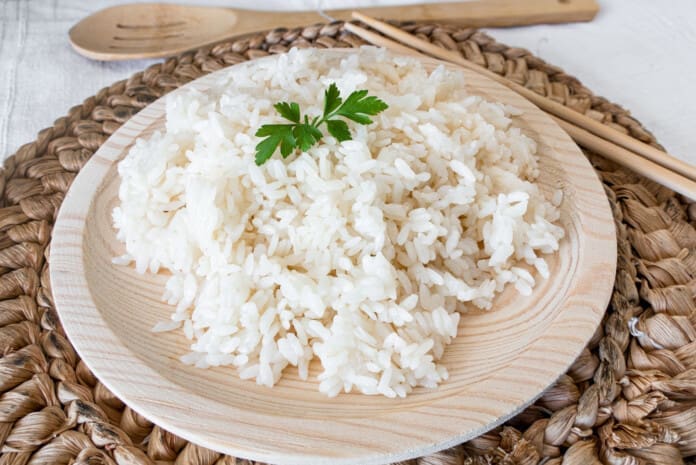
Cooked white rice garnished with mint in a ceramic bowl

With the summer heatwave blasting through, some individuals can experience digestive health problems. The relationship between the temperature outside and the temperature in the body translates to the digestive system. As the heat rises, it can make the digestive system slow down and become weakened feeling bloated, nauseated, and tired. The body’s balance may feel off because the body lowers its internal temperature to protect itself. Individuals must be careful not to overload themselves with the wrong foods. One way to avoid problems and maintain digestion working smoothly is to eat lighter, eat smaller portions for each meal, and eat easily digestible foods. Doing this will allow the body will feel cooler, and maintain alertness and energy throughout the hot day.

Easily Digestible Foods

Easily Digestible Foods

Heat-related health issues can include:

  • Loss of appetite
  • Heartburn
  • Acid buildup
  • Abdominal pain
  • Constipation
  • Gastroenteritis
  • Diarrhea
  • Irritable bowel syndrome (IBS)
  • Dehydration
  • Heat exhaustion
  • Stroke

The objective is not to skip meals but to eat regular meals, just smaller and easily digestible ones. Foods low in fiber tend to be easy to digest and can help the body feel better.

White Rice

  • White rice is low in fat and fiber, making it easy on the stomach and easy to digest.
  • It is not associated with any gastrointestinal issues and is considered a safe starch because it is an easy source of carbohydrates that provides instant energy.
  • To digest rice even more easily, eat it by itself or pair it with foods low in fat.
  • Certain foods that are high in fat, like vegetable oils, can take longer to digest and could cause discomfort.
  • A 1/2 cup of cooked white rice:
  • 210 calories
  • 4g protein
  • 0g fat
  • 49g carbohydrates
  • 1g fiber

Bananas

  • Ripe bananas are an easily digestible fruit that only contains a moderate amount of fiber.
  • They are associated with improvements in both constipation and diarrhea,
  • Individuals with a variety of digestive issues may experience relief when incorporating bananas into their diets.
  • Cooking bananas makes them even easier to digest as it makes certain nutrients easier to absorb.
  • Make sure the bananas are ripe enough.
  • Unripe bananas can be more difficult to digest.
  • 1 medium raw/ripe banana:
  • 105 calories
  • 1.3g protein
  • 0.4g fat
  • 27g carbohydrates
  • 3g fiber

Applesauce

  • Although made from apples, applesauce is low in fiber and a great source of vitamin C.
  • Cooked, canned, or processed fruits tend to be lower in fiber and easier to digest.
  • Applesauce is recommended to calm a variety of stomach-related ailments like constipation, diarrhea, and gastroparesis.
  • A 4-ounce serving of applesauce:
  • 90 calories
  • 0g protein
  • 0g fat
  • 22g carbohydrates
  • 2g fiber

White Bread

  • Plain white bread is low in fiber and easier to digest than bread made with whole-grain wheat bread.
  • It is often fortified with nutrients including folic acid, B vitamins, vitamin D3, and more.
  • Try plain toast for breakfast
  • Use low-fat fillings for an easily digestible sandwich for lunch or dinner.
  • 2 slices of plain white bread:
  • 150 calories
  • 4g protein
  • 28g carbohydrates
  • 2g fat
  • 1g fiber

Chicken and Turkey

  • Lean proteins low in fat like chicken breast and turkey are easy to digest.
  • Individuals experiencing digestive problems are recommended to consume lean protein over fattier red meats.
  • A 3-ounce serving of skinless, boneless chicken breast:
  • 128 calories
  • 26g protein
  • 2.7g fat
  • 0g carbohydrates
  • 0g fiber

Sweet Potatoes

  • Cooked potatoes of all varieties are examples of easily digestible foods.
  • Sweet potatoes are gentle on the digestive tract because they are mostly insoluble fiber, which speeds up digestion and increases regularity.
  • To make potatoes easier to digest, remove the skins and mash the inside.
  • Removing the skins decreases the fiber content, and mashing them makes digestion easier.
  • 1 medium sweet potato that is cooked and peeled:
  • 135 calories
  • 3g protein
  • 0.2g fat
  • 31g carbohydrates
  • 5g fiber

Other recommendations that could help stimulate digestion include drinking more water, getting more sleep, reducing stress levels, and exercising.


The Healing Diet


References

Howard, Sally, and Geetanjali Krishna. “How hot weather kills: the rising public health dangers of extreme heat.” BMJ (Clinical research ed.) vol. 378 o1741. 14 Jul. 2022, doi:10.1136/bmj.o1741

Kong, Fanbin, et al. “Physical changes in white and brown rice during simulated gastric digestion.” Journal of food science vol. 76,6 (2011): E450-7. doi:10.1111/j.1750-3841.2011.02271.x

Nguyen, Hoang Chinh et al. “Bioactive Compounds, Antioxidants, and Health Benefits of Sweet Potato Leaves.” Molecules (Basel, Switzerland) vol. 26,7 1820. 24 Mar. 2021, doi:10.3390/molecules26071820

Remes-Troche, José María. “Too hot” or “too cold”: effects of meal temperature on gastric function.” Digestive diseases and sciences vol. 58,9 (2013): 2439-40. doi:10.1007/s10620-013-2789-4

Salfi, Salvatore F, and Karyn Holt. “The role of probiotics in diarrheal management.” Holistic nursing practice vol. 26,3 (2012): 142-9. doi:10.1097/HNP.0b013e31824ef5a3

Singh, Balwinder, et al. “Bioactive compounds in banana and their associated health benefits – A review.” Food Chemistry vol. 206 (2016): 1-11. doi:10.1016/j.foodchem.2016.03.033

Stiffness and Pain Developing In The Shoulder

Shot of an attractive young woman suffering from a shoulder injury during her outdoor workout.

Stiffness and pain developing in the shoulder could be adhesive capsulitis, (frozen shoulder), a condition in the shoulder’s ball-and-socket joint/glenohumeral joint. It usually develops over time and limits the functional use of the arm. The pain and tightness restrict arm movement, and the duration of symptoms can persist for 12-18 months. The cause is often unknown, but it is more common in individuals over 40, individuals with diabetes, thyroid disease, and cardiac conditions have an increased risk of developing the condition, and women tend to develop the condition more than men. Chiropractic treatment can be effective at relieving pain and expediting recovery.

Stiffness and Pain Developing In The Shoulder

Stiffness and Pain

The shoulder joint allows more movement than any other joint in the body. A frozen shoulder causes the capsule surrounding the shoulder joint to contract and form scar tissue. The capsule contraction and the formation of adhesions cause the shoulder to become stiff, restrict movement, and cause pain and discomfort symptoms.

Stages

The progression is marked by three stages:

Freezing

  • Stiffness and pain begin to restrict motion.

Frozen

  • Movement and motion are severely restricted.

Thawing

  • The shoulder starts to loosen up.
  • It can take years to fully resolve symptoms.
  • In mild cases, a frozen shoulder can go away on its own but that does not mean that it is truly healed and correctly aligned.
  • Even in mild cases seeking treatment is recommended, rather than just waiting for it to go away.

Symptoms

  • Limited range of motion.
  • Stiffness and tightness.
  • Dull or aching pain throughout the shoulder.
  • Pain can radiate into the upper arm.
  • Pain can be triggered by the smallest movements.
  • The symptoms are not always due to weakness or injury, but actual joint stiffness.

Causes

Most frozen shoulders occur with no injury or discernible cause but the condition is often linked to a systemic condition or one that affects the entire body.

Age and Gender

  • Frozen shoulder most commonly affects individuals between the ages of 40 to 60, and is more common in women than in men.

Endocrine Disorders

  • Individuals with diabetes have an increased risk of developing a frozen shoulder.
  • Other endocrine abnormalities like thyroid problems can also lead to the development of this condition.

Shoulder Trauma and/or Surgery

  • Individuals who sustain a shoulder injury, or undergo surgery on the shoulder can develop a stiff and painful joint.
  • When injury or surgery is followed by prolonged immobilization/resting the arm, the risk of developing a frozen shoulder increases.

Other Systemic Conditions

Several systemic conditions such as heart disease have also been associated with an increased risk of developing the condition and can include:

  • High cholesterol
  • Adrenal disease
  • Heart and lung disease
  • Parkinson’s disease

Stiffness and pain can also be associated with damage to the joint from injuries or other shoulder problems that include:

  • Muscle or connective tissue injury
  • Rotator cuff tendinopathy
  • Calcific tendinitis
  • Dislocation
  • Fracture
  • Osteoarthritis
  • A frozen shoulder associated with any of these causes is considered secondary.

Treatment

A diagnosis is made by observing the range of motion in the shoulder, considering the two types:

Active Range

  • This is how far an individual can move a body part on their own.

Passive Range

  • This is how far another person like a therapist or doctor can move the body part.

Therapies

  • Chiropractic, massage, and physical therapy involve stretches, realignment, and exercises to relieve pain symptoms and restore mobility and function.
  • Usually, strength is not affected by a frozen shoulder but a chiropractor may want to strengthen the surrounding muscles to better support the shoulder and prevent worsening the injury or causing a new injury.
  • Anti-inflammatory medications and corticosteroid injections may help manage pain symptoms.
  • Getting a diagnosis and treatment during the freezing stage can keep the condition from progressing and expedite recovery time.

Enhancing Health: Evaluation and Treatment


References

Brun, Shane. “Idiopathic frozen shoulder.” Australian Journal of general practice vol. 48,11 (2019): 757-761. doi:10.31128/AJGP-07-19-4992

Chan, Hui Bin Yvonne, et al. “Physical therapy in the management of frozen shoulder.” Singapore medical journal vol. 58,12 (2017): 685-689. doi:10.11622/smedj.2017107

Cho, Chul-Hyun, et al. “Treatment Strategy for Frozen Shoulder.” Clinics in orthopedic surgery vol. 11,3 (2019): 249-257. doi:10.4055/cios.2019.11.3.249

Duzgun, Irem, et al. “Which method for frozen shoulder mobilization: manual posterior capsule stretching or scapular mobilization?.” Journal of Musculoskeletal & neuronal interactions vol. 19,3 (2019): 311-316.

Jain, Tarang K, and Neena K Sharma. “The effectiveness of physiotherapeutic interventions in the treatment of frozen shoulder/adhesive capsulitis: a systematic review.” Journal of back and musculoskeletal rehabilitation vol. 27,3 (2014): 247-73. doi:10.3233/BMR-130443

Kim, Min-Su, et al. “Diagnosis and treatment of calcific tendinitis of the shoulder.” Clinics in shoulder and elbow vol. 23,4 210-216. 27 Nov. 2020, doi:10.5397/cise.2020.00318

Millar, Neal L et al. “Frozen shoulder.” Nature reviews. Disease primers vol. 8,1 59. 8 Sep. 2022, doi:10.1038/s41572-022-00386-2

Massage Gun Head Attachments

Percussion massager of deep muscle tissues for athletes, relaxes. Close up handheld wireless professional therapeutic shock massage gun with massage attachments on gray background. Copy space

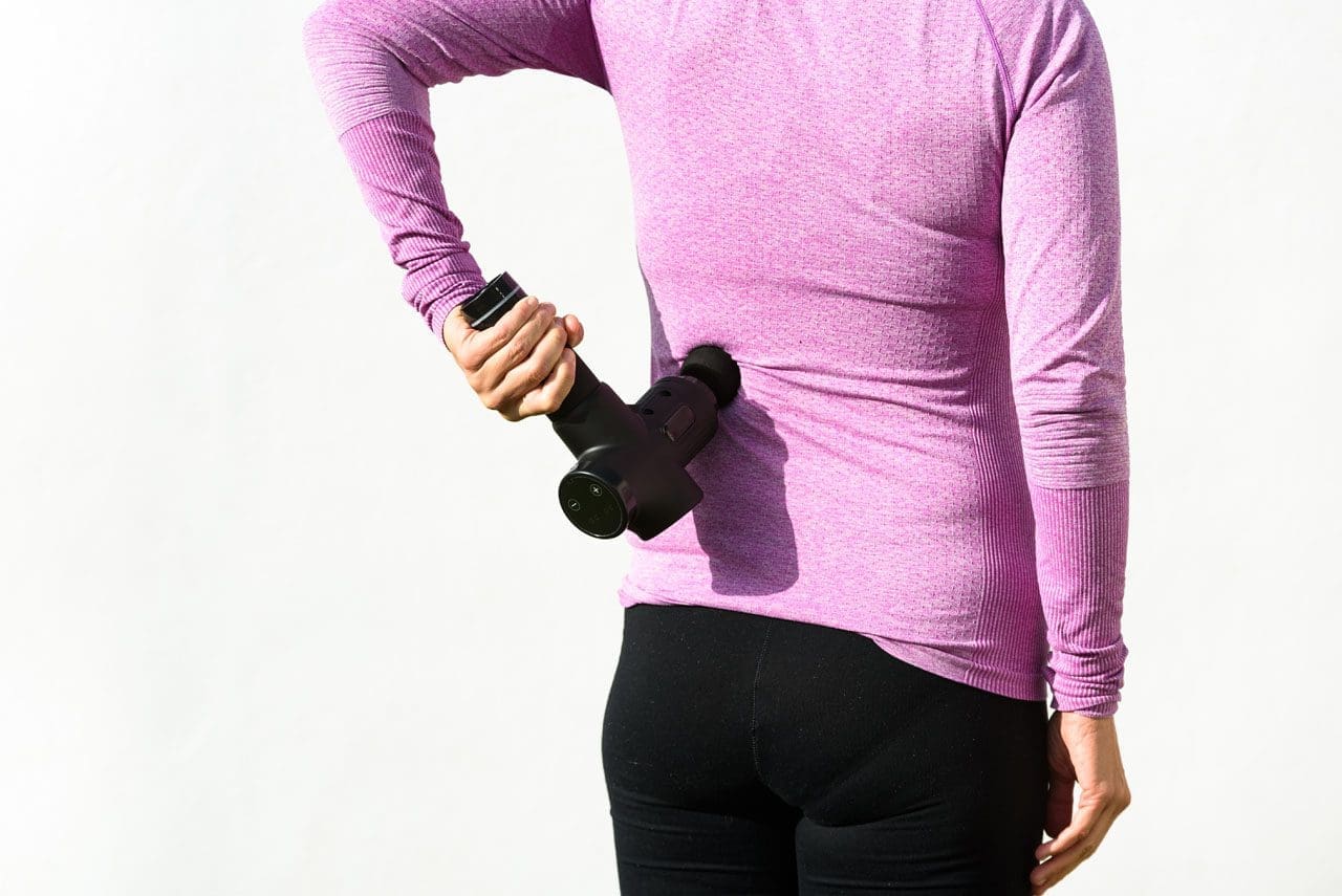
Massage guns can help relieve aching muscles and prevent soreness when used before and after physical activity, work, school, and exercise. They provide massage therapy benefits by targeting muscles with rapid burst pulses. Massage guns can be percussive or vibration-based. Percussive therapy helps increase blood flow to a targeted area, which reduces inflammation and muscle tension, and breaks up knots/trigger points that may have formed in the tissues from added stress or intense physical activity. One of the benefits is that they come with interchangeable massage gun head attachments that target different muscle groups and provide different types of massage. There are many types of interchangeable massage heads, we go over the most common to give a general idea of how they work. If experiencing joint pain, injury, acute muscle pain, or other musculoskeletal disorders, make sure to get clearance from a doctor before using a massage gun.

Massage Gun Head Attachments

Massage Gun Head Attachments

The variations of attachments/heads are designed and shaped differently to effectively apply the right amount of pressure to rejuvenate the body’s pressure points, soothe tissues, and release tight and sore muscles. The different heads are designed with a distinctive purpose based on the muscle groups targeted. This maximizes effectiveness and ensures maximum comfort and safety.

Ball Head

  • The ball attachment is for overall muscle recovery.
  • It provides a broad surface area and mimics the hands of a skilled massage therapist, delivering a soothing kneading sensation.
  • Made of durable material, the ball massage head can reach deep into the muscles.
  • Its round shape makes it more flexible to use anywhere, especially the large muscle groups like the quads and glutes.

U/Fork Shaped Head

  • A plastic, dual-pronged head also known as a fork head.
  • The attachment provides relief to areas like the shoulders, spine, neck, calves, and Achilles tendon.

Bullet Head

  • The plastic head is named as such because of its pointed shape.
  • This is recommended for tightness and discomfort in the joints, deep tissues, trigger points, and/or small muscle areas like the feet and wrist.

Flat Head

  • The multipurpose flat head is for full-body general massage.
  • It helps relieve stiffness and pain for total body muscle relaxation, including muscle groups closer to bone joints.

Shovel-Shaped Head

  • The shovel-shaped head is for the abdominal muscles and lower back.
  • The attachment provides stimulation to release stiff muscles.

Using The Right Head

Which head to use depends on the individual’s specific needs and preferences. Consider the following factors when selecting a massage gun head:

Targeted Areas

  • Identify the body areas requiring the most attention.
  • If muscle tightness or soreness is occurring in larger muscle groups, like the back or legs, the ball attachment is recommended.
  • For more precise areas like trigger points, the bullet head is recommended.
  • Heads can be used in conjunction – for example, a large surface area head is used to relax and loosen and relax a general area, then a more precise head is used to focus the massage on the actual tight spot or trigger point.

Massage Intensity

  • Massage intensity levels can be varied from a light massage to full force.
  • For a softer touch on sensitive muscles, flat head or forkhead attachments are recommended.
  • For deep muscle penetration and consistent pressure, the bullet head or shovel head attachments are recommended.

Specific Conditions

  • Consider any specific conditions or injuries previous and current.
  • For individuals recovering from an injury or with sensitive areas, it’s important to choose a massage gun head that provides the necessary relief without causing discomfort or worsening an injury.

Try Out Different Heads and Settings

  • Experiment with different massage head attachments and speeds to find the one that works best for the intended purpose.
  • Explore each to discover personal preferences.
  • Start with the lowest setting and gradually increase, based on comfort level.
  • Always consult with a qualified healthcare provider regarding any medical concerns before using a massage gun.

Choosing The Right Massage Head Attachment


References

Bergh, Anna, et al. “A Systematic Review of Complementary and Alternative Veterinary Medicine in Sport and Companion Animals: Soft Tissue Mobilization.” Animals: an open access journal from MDPI vol. 12,11 1440. 2 Jun. 2022, doi:10.3390/ani12111440

Imtiyaz, Shagufta, et al. “To Compare the Effect of Vibration Therapy and Massage in Prevention of Delayed Onset Muscle Soreness (DOMS).” Journal of Clinical and diagnostic research: JCDR vol. 8,1 (2014): 133-6. doi:10.7860/JCDR/2014/7294.3971

Konrad, Andreas, et al. “The Acute Effects of a Percussive Massage Treatment with a Hypervolt Device on Plantar Flexor Muscles’ Range of Motion and Performance.” Journal of sports science & medicine vol. 19,4 690-694. 19 Nov. 2020

Leabeater, Alana et al. “Under the Gun: The effect of percussive massage therapy on physical and perceptual recovery in active adults.” Journal of athletic training, 10.4085/1062-6050-0041.23. 26 May. 2023, doi:10.4085/1062-6050-0041.23

Lupowitz, Lewis. “Vibration Therapy – A Clinical Commentary.” International journal of sports physical therapy vol. 17,6 984-987. 1 Aug. 2022, doi:10.26603/001c.36964

Yin, Yikun, et al. “The effect of vibration training on delayed muscle soreness: A meta-analysis.” Medicine vol. 101,42 (2022): e31259. doi:10.1097/MD.0000000000031259

Facet Syndrome Protocols For Spinal Decompression

In many people with facet joint syndrome, how does spinal decompression compare to traditional spinal surgery alleviate low back pain?

Introduction

Many people around the globe experience lower back pain for various reasons, such as lifting or carrying heavy objects, sedentary jobs, or traumatic events that can cause spinal injuries. The spine has a crucial role in providing mobility and flexibility without discomfort. The facet joints and spinal discs work together to generate healthy movement and stability within each segment. However, when the muscles, ligaments, and tissues surrounding the spinal disc are overstretched or compressed due to normal or traumatic factors, it can aggravate the nerve roots and cause discomfort. As we age or carry excess weight, our spinal discs can experience wear and tear, leading to facet joint syndrome. This syndrome is often associated with lower back pain caused by injured facet joints. This article will explore how facet joint syndrome is linked to lower back pain and how non-surgical treatments can help alleviate it. We work with certified medical providers who use our patients’ valuable information to treat individuals suffering from facet joint syndrome affecting spine mobility and causes low back pain. We also inform them about non-surgical treatments to regain spinal mobility and reduce pain-like symptoms that correlate with this spinal condition. We encourage patients to ask essential questions and seek education from our associated medical providers about their situation. Dr. Jimenez, D.C., provides this information as an educational service. Disclaimer


Facet Joint Syndrome

Are you experiencing pain that radiates down to your legs, especially when standing? Do you constantly hunch over, affecting your posture during everyday activities? Have you noticed numbness or loss of sensation in your feet or buttocks? As we age or experience traumatic injuries, the facet joints on either side of our spine can become damaged, resulting in a condition called facet joint syndrome. Research indicates that environmental conditions can cause joint degeneration, leading to symptoms similar to other spinal conditions. Cartilage erosion and inflammation on the spine are common signs of facet joint syndrome, often associated with musculoskeletal disorders like low back pain.


Low Back Pain Associated With Facet Syndrome

Research studies musculoskeletal disorders like low back pain are associated with facet syndrome. When the facet joints start to degenerate from repetitive overuse motions caused by everyday activities, it can cause micro instabilities to the facet joints while compressing the surrounding nerve roots. When this happens, many individuals will experience low back pain and sciatic nerve pain conditions that cause them to be unstable while walking. Additional research studies stated that low back pain associated with facet syndrome can cause symptoms of restricted movement, reduce the quality of life and greatly impact the entire lumbar vertebral structure. Since low back pain is a common problem many individuals have, the combination with facet syndrome can trigger reactive muscle spasms, a protective mechanism in the spine to cause the individual to have difficulty moving comfortably and experience severe sudden pain. To that point, low back pain associated with facet syndrome causes the person to be dealing with constant ongoing pain, making a normal lifestyle almost difficult.


Discover The Benefits Of Chiropractic Care-Video

YouTube player

Low back pain associated with facet joint syndrome should not make life difficult. Numerous treatments pertain to relieving the pain-like symptoms and help slow the process of facet syndrome from causing more issues to the spine. Non-surgical treatments like chiropractic care can help reduce the effects of facet syndrome as they can provide benefits to restore spinal mobility. The video above explores the benefits of chiropractic care, as chiropractors will discuss the recommended course of personalized treatment with you. Non-surgical treatments are safe, gentle on the spine, and cost-effective as they help regain your body’s mobility from facet syndrome. At the same time, non-surgical treatments like chiropractic care can be combined with other non-surgical therapies that can help restart the body’s natural healing process to allow the compressed spinal disc and joint to be rehydrated.


Spinal Decompression Alleviating Facet Syndrome

According to research studies, non-surgical treatments like spinal decompression can help reduce the effects of facet syndrome as it can help improve the spine;’s mobility through gentle traction and can help stretch out the affected muscles associated with low back pain by taking pressure off the aggravating nerve root. In “The Ultimate Spinal Decompression,” Dr. Eric Kaplan, D.C., FIAMA, and Dr. Perry Bard, D.C., mentioned that when individuals are going in for spinal decompression, they might experience a “popping sensation” as the jammed facet joints are being open for treatment. This is normal for early facet arthropathy and can occur within the first few treatment sessions. At the same time, spinal decompression can gently stretch the adjacent compressed nerve root and find instant relief. After the treatment, many individuals can combine other treatments like physical therapy to reduce the painful symptoms from returning. Non-surgical treatments like spinal decompression and chiropractic care can help revitalize the spine affected by facet joint syndrome and help restore a person’s quality of life.


References

Alexander, C. E., Cascio, M. A., & Varacallo, M. (2022). Lumbosacral Facet Syndrome. PubMed; StatPearls Publishing. pubmed.ncbi.nlm.nih.gov/28722935/

Curtis, L., Shah, N., & Padalia, D. (2023). Facet Joint Disease. PubMed; StatPearls Publishing. www.ncbi.nlm.nih.gov/books/NBK541049

Du, R., Xu, G., Bai, X., & Li, Z. (2022). Facet Joint Syndrome: Pathophysiology, Diagnosis, and Treatment. Journal of Pain Research, 15, 3689–3710. doi.org/10.2147/JPR.S389602

Gose, E., Naguszewski, W., & Naguszewski, R. (1998). Vertebral axial decompression therapy for pain associated with herniated or degenerated discs or facet syndrome: An outcome study. Neurological Research, 20(3), 186–190. doi.org/10.1080/01616412.1998.11740504

Kaplan, E., & Bard, P. (2023). The Ultimate Spinal Decompression. JETLAUNCH.

Disclaimer

Sacral Plexus Rundown

Young woman doing spine checkup at vertebra clinic.

The lumbosacral plexus is located on the posterolateral wall of the lesser pelvis, next to the lumbar spine. A plexus is a network of intersecting nerves that share roots, branches, and functions. The sacral plexus is a network that emerges from the lower part of the spine. The plexus then embeds itself into the psoas major muscle and emerges in the pelvis. These nerves provide motor control to and receive sensory information from portions of the pelvis and leg. Sacral nerve discomfort symptoms, numbness, or other sensations and pain can be caused by an injury, especially if the nerve roots are compressed, tangled, rubbing, and irritated. This can cause symptoms like back pain, pain in the back and sides of the legs, sensory issues affecting the groin and buttocks, and bladder or bowel problems. Injury Medical Chiropractic and Functional Medicine Clinic can develop a personalized treatment plan to relieve symptoms, release the nerves, relax the muscles, and restore function.

Sacral Plexus Rundown

Sacral Plexus

Anatomy

  • The sacral plexus is formed by the lumbar spinal nerves, L4 and L5, and sacral nerves S1 through S4.
  • Several combinations of these spinal nerves merge together and then divide into the branches of the sacral plexus.
  • Everybody has two sacral plexi – plural of plexus – one on the right side and left side that is symmetrical in structure and function.

Structure

There are several plexi throughout the body. The sacral plexus covers a large area of the body in terms of motor and sensory nerve function.

  • Spinal nerves L4 and L5 make up the lumbosacral trunk, and the anterior rami of sacral spinal nerves S1, S2, S3, and S4 join the lumbosacral trunk to form the sacral plexus.
  • Anterior rami are the branches of the nerve that are towards the front of the spinal cord/front of the body.
  • At each spinal level, an anterior motor root and a posterior sensory root join to form a spinal nerve.
  • Each spinal nerve then divides into an anterior – ventral – and a posterior – dorsal – rami portion.
  • Each can have motor and/or sensory functions.

The sacral plexus divides into several nerve branches, which include:

  • Superior gluteal nerve – L4, L5, and S1.
  • Inferior gluteal nerve – L5, S1, and S2.
  • The sciatic nerve – is the largest nerve of the sacral plexus and among the largest nerves in the body – L4, L5, S1, S2, and S3
  • The common fibular nerve – L4 through S2, and tibial nerves – L4 through S3 are branches of the sciatic nerve.
  • Posterior femoral cutaneous nerve – S1, S2, and S3.
  • Pudendal nerve – S2, S3, and S4.
  • The nerve to the quadratus femoris muscle is formed by L4, L5, and S1.
  • The obturator internus muscle nerve – L5, S1, and S2.
  • The piriformis muscle nerve – S1 and S2.

Function

The sacral plexus has substantial functions throughout the pelvis and legs. The branches provide nerve stimulation to several muscles. The sacral plexus nerve branches also receive sensory messages from the skin, joints, and structures of the pelvis and legs.

Motor

Motor nerves of the sacral plexus receive signals from the brain that travel down the column of the spine, out to the motor nerve branches of the sacral plexus to stimulate muscle contraction and movement. Motor nerves of the sacral plexus include:

Superior Gluteal Nerve

  • This nerve provides stimulation to the gluteus minimus, gluteus medius, and tensor fascia lata, which are muscles that help move the hip away from the center of the body.

Inferior Gluteal Nerve

  • This nerve provides stimulation to the gluteus maximus, the large muscle that moves the hip laterally.

Sciatic Nerve

  • The sciatic nerve has a tibial portion and a common fibular portion, which have motor and sensory functions.
  • The tibial portion stimulates the inner part of the thigh and activates muscles in the back of the leg and the sole of the foot.
  • The common fibular portion of the sciatic nerve stimulates and moves the thigh and knee.
  • The common fibular nerve stimulates muscles in the front and sides of the legs and extends the toes to straighten them out.

Pudendal Nerve

  • The pudendal nerve also has sensory functions that stimulate the muscles of the urethral sphincter to control urination and the muscles of the anal sphincter to control defecation.
  • The nerve to the quadratus femoris stimulates the muscle to move the thigh.
  • The nerve to the obturator internus muscle stimulates the muscle to rotate the hips and stabilize the body when walking.
  • The nerve to the piriformis muscle stimulates the muscle to move the thigh away from the body.

Conditions

The sacral plexus, or areas of the plexus, can be affected by disease, traumatic injury, or cancer. Because the nerve network has many branches and portions, symptoms can be confusing. Individuals may experience sensory loss or pain in regions in the pelvis and leg, with or without muscle weakness. Conditions that affect the sacral plexus include:

Injury

  • A traumatic injury of the pelvis can stretch, tear, or harm the sacral plexus nerves.
  • Bleeding can inflame and compress the nerves, causing malfunction.

Neuropathy

  • Nerve impairment can affect the sacral plexus or parts of it.
  • Neuropathy can come from:
  • Diabetes
  • Vitamin B12 deficiency
  • Certain medications – chemotherapeutic meds
  • Toxins like lead
  • Alcohol
  • Metabolic illnesses

Infection

  • An infection of the spine or the pelvic region can spread to the sacral plexus nerves or produce an abscess, causing symptoms of nerve impairment, pain, tenderness, and sensations around the infected region.

Cancer

  • Cancer developing in the pelvis or spreading to the pelvis from somewhere else can compress or infect the sacral plexus nerves.

Treatment of the Underlying Medical Condition

Rehabilitation begins with the treatment of the underlying medical condition causing the nerve problems.

  • Cancer treatment – surgery, chemotherapy, and/or radiation.
  • Antibiotic treatment for infections.
  • Neuropathy treatment can be complicated because the cause may be unclear, and an individual can experience several causes of neuropathy simultaneously.
  • Major pelvic trauma like a vehicle collision can take months, especially if there are multiple bone fractures.

Motor and Sensory Recovery

  • Sensory problems can interfere with walking, standing, and sitting.
  • Adapting to sensory deficits is an important part of treatment, rehabilitation, and recovery.
  • Chiropractic, decompression, massage, and physical therapy can relieve symptoms, restore strength, function, and motor control.

Sciatica Secrets Revealed


References

Dujardin, Franck et al. “Extended anterolateral transiliac approach to the sacral plexus.” Orthopaedics & traumatology, surgery & research: OTSR vol. 106,5 (2020): 841-844. doi:10.1016/j.otsr.2020.04.011

Eggleton JS, Cunha B. Anatomy, Abdomen and Pelvis, Pelvic Outlet. [Updated 2022 Aug 22]. In: StatPearls [Internet]. Treasure Island (FL): StatPearls Publishing; 2023 Jan-. Available from: www.ncbi.nlm.nih.gov/books/NBK557602/

Garozzo, Debora et al. “In lumbosacral plexus injuries can we identify indicators that predict spontaneous recovery or the need for surgical treatment? Results from a clinical study on 72 patients.” Journal of brachial plexus and peripheral nerve injury vol. 9,1 1. 11 Jan. 2014, doi:10.1186/1749-7221-9-1

Gasparotti R, Shah L. Brachial and Lumbosacral Plexus and Peripheral Nerves. 2020 Feb 15. In: Hodler J, Kubik-Huch RA, von Schulthess GK, editors. Diseases of the Brain, Head and Neck, Spine 2020–2023: Diagnostic Imaging [Internet]. Cham (CH): Springer; 2020. Chapter 20. Available from: www.ncbi.nlm.nih.gov/books/NBK554335/ doi: 10.1007/978-3-030-38490-6_20

Norderval, Stig, et al. “Sacral nerve stimulation.” Tidsskrift for den Norske laegeforening : tidsskrift for praktisk medicin, ny raekke vol. 131,12 (2011): 1190-3. doi:10.4045/tidsskr.10.1417

Neufeld, Ethan A et al. “MR Imaging of the Lumbosacral Plexus: A Review of Techniques and Pathologies.” Journal of Neuroimaging: official journal of the American Society of Neuroimaging vol. 25,5 (2015): 691-703. doi:10.1111/jon.12253

Staff, Nathan P, and Anthony J Windebank. “Peripheral neuropathy due to vitamin deficiency, toxins, and medications.” Continuum (Minneapolis, Minn.) vol. 20,5 Peripheral Nervous System Disorders (2014): 1293-306. doi:10.1212/01.CON.0000455880.06675.5a

Yin, Gang, et al. “Obturator Nerve Transfer to the Branch of the Tibial Nerve Innervating the Gastrocnemius Muscle for the Treatment of Sacral Plexus Nerve Injury.” Neurosurgery vol. 78,4 (2016): 546-51. doi:10.1227/NEU.0000000000001166

Degenerative Disc Protocols Implemented For Spinal Decompression

In many individuals with degenerative disc disease, how does spinal decompression compare to spinal surgery improve spine flexibility?

Introduction

The spine is essential to the body’s musculoskeletal system, enabling individuals to perform daily movements while maintaining proper posture. The spinal cord is safeguarded by surrounding ligaments, soft tissues, muscles, and nerve roots. The spinal discs between the spinal column act as shock absorbers to reduce stress from axial overload and promote body mobility and flexibility. The spinal discs can naturally degenerate as a person ages, leading to degenerative disc disease. This condition can result in several spinal issues that can impact the spine’s flexibility. This article explores how degenerative disc disease affects the spine and the treatments available to restore its flexibility. We work with certified medical providers who use our patients’ valuable information to treat individuals suffering from degenerative disc disease affecting their spine’s flexibility. We also inform them about non-surgical treatments to regain spinal mobility and reduce pain-like symptoms. We encourage patients to ask essential questions and seek education from our associated medical providers about their condition. Dr. Jimenez, D.C., provides this information as an educational service. Disclaimer


How Does Degenerative Disc Disease Affect The Spine?


Do you experience neck or lower back pain after a long workday? After physical activity, do you find temporary relief by twisting or turning your torso? Are you experiencing radiating pain in your upper or lower extremities that worsens when standing? These symptoms are common as the body ages over time. Muscles, organs, ligaments, and joints can all be affected, including the spine and intervertebral discs. Research studies reveal that disc degeneration frequently occurs in the spine, leading to changes that can cause misalignment and spinal issues. Degenerative disc disease can disrupt the structure of spinal discs, resulting in pain-like symptoms and accelerating degenerative changes to the spine. Regardless of age, various habits and lifestyle choices can contribute to degeneration. As additional research studies have provided, this condition is characterized by a tension-resisting annulus fibrosus and compression-resisting nucleus pulposus, causing pain and discomfort.


The Symptoms Correlating With Degenerative Disc Disease

Degenerative disc disease is when the spinal disc in the spine experiences wear and tear due to natural aging. The initial indication of this disease is disc cracking caused by repetitive motion trauma. The symptoms associated with this disease are similar but may vary based on the affected spine location. Research shows that degenerative disc disease can cause micro tears in the spinal disc, leading to decreased fluids and water intake, disc space loss, disc bulging, and irritation of the adjacent nerves. This can affect surrounding muscle tissues and disc facet joints, narrowing the spinal canal. Additional studies reveal that people with degenerative disc disease may experience various symptoms that can hinder their ability to function properly. Some common symptoms include:

  • Pain in the arms, legs, and feet
  • Sensory abnormalities (loss of sensation in the hands, feet, fingers, and back)
  • Muscle tenderness and weakness
  • Instability
  • Inflammation
  • Visceral-somatic & somatic-visceral condition

If someone experiences pain-like symptoms in conjunction with degenerative disc disease can negatively impact their quality of life and potentially lead to long-term disability. Fortunately, treatments can slow the degenerative process and alleviate the pain-like symptoms.


Secrets Of Optimal Wellness- Video

Secrets of Optimal Wellness | El Paso, Tx (2023)

When individuals experience pain related to degenerative disc disease, they often seek ways to alleviate it. Some may consider spinal surgery to remove the affected disc and ease the pain caused by the irritated nerve. However, this option is typically only pursued if other treatments have failed and can be expensive. Fortunately, non-surgical treatments are cost-effective and safe, gently addressing the affected area for relief. Non-surgical treatments can be customized to the individual’s specific pain and condition, including spinal decompression, MET therapy, traction therapy, and chiropractic care. These methods work to realign the body and promote natural healing by rehydrating the spine, ultimately restoring flexibility.


Treatments To Improve Spinal Flexibility

Individuals with degenerative disc disease can benefit from non-surgical treatments tailored to their needs. These treatments involve an assessment by a pain specialist, such as a physical therapist, massage therapist, or chiropractor, who will identify the source of the pain and use various techniques to reduce pain, improve flexibility in the spine, and loosen stiff muscles that have been affected by the disease. Additionally, non-surgical treatments can help restore sensory and mobility function to the spine and address factors that may exacerbate the degenerative process.


Spinal Decompression Protocol For Degenerative Disc Disease

Research studies suggest that spinal decompression can effectively reduce the degenerative process of spinal discs through gentle traction. During a spinal decompression treatment, the individual is strapped into a traction machine. The machine gradually stretches the spine to create negative pressure on the spinal disc, which helps to rehydrate it and increase nutrient intake, thereby jumpstarting the healing process. According to Dr. Eric Kaplan, D.C., FIAMA, and Dr. Perry Bard, D.C., in their book “The Ultimate Spinal Decompression,” individuals with degenerative disc disease may require higher pressure during spinal decompression treatment due to its symptomatic problems. Spinal decompression can help restore disc height and be a viable solution for those looking to improve their health.


References

Choi, E., Gil, H. Y., Ju, J., Han, W. K., Nahm, F. S., & Lee, P.-B. (2022). Effect of Nonsurgical Spinal Decompression on Intensity of Pain and Herniated Disc Volume in Subacute Lumbar Herniated Disc. International Journal of Clinical Practice, 2022, 1–9. doi.org/10.1155/2022/6343837

Choi, Y.-S. (2009). Pathophysiology of Degenerative Disc Disease. Asian Spine Journal, 3(1), 39. doi.org/10.4184/asj.2009.3.1.39

Kaplan, E., & Bard, P. (2023). The Ulitimate Spinal Decompression. JETLAUNCH.

Liyew, W. A. (2020). Clinical Presentations of Lumbar Disc Degeneration and Lumbosacral Nerve Lesions. International Journal of Rheumatology, 2020, 1–13. doi.org/10.1155/2020/2919625

Scarcia, L., Pileggi, M., Camilli, A., Romi, A., Bartolo, A., Giubbolini, F., Valente, I., Garignano, G., D’Argento, F., Pedicelli, A., & Alexandre, A. M. (2022). Degenerative Disc Disease of the Spine: From Anatomy to Pathophysiology and Radiological Appearance, with Morphological and Functional Considerations. Journal of Personalized Medicine, 12(11), 1810. doi.org/10.1155/2020/2919625

Taher, F., Essig, D., Lebl, D. R., Hughes, A. P., Sama, A. A., Cammisa, F. P., & Girardi, F. P. (2012). Lumbar Degenerative Disc Disease: Current and Future Concepts of Diagnosis and Management. Advances in Orthopedics, 2012, 1–7. doi.org/10.1155/2012/970752

Disclaimer

Food Energy Density: EP Functional Health and Wellness Clinic

beautiful women having breakfast in the kitchen - focus on hispanic woman eating a peach -

The brain and body need macronutrients that include carbohydrates, fats, and protein in the right amounts to energize the body. About half of the calories should come from carbohydrates, 30% from fat, and 20% from protein. Food energy density is the amount of energy, represented by the number of calories, in a specific weight measurement.

Food Energy Density: EP's Functional Chiropractic Team

Food Energy Density

Energy density is determined by the proportion of macronutrients – protein, fat, carbohydrates, fiber, and water.

  • Energy-dense foods are high in calories per serving.
  • Foods with large amounts of fiber and water have a lower density.
  • Foods high in fat have an increased energy density.
  • An example of a high-energy-density food is a donut because of the high-calorie count from the sugar, fat, and small serving size.
  • An example of a low-energy-density food is spinach because it only has a few calories in a whole plate of raw spinach leaves.

Energy Dense Foods

Energy-dense foods contain a high number of calories/energy per gram. They are typically higher in fat and lower in water. Examples of energy-dense foods include:

  • Full-fat dairy
  • Butter
  • Cheese
  • Nut butter
  • Fatty cuts of meat
  • Starchy vegetables
  • Thick sauces
  • Nuts
  • Seeds

Less nutrient-dense foods include:

  • Sweets
  • Deep-fried foods
  • French fries
  • Pasta
  • Crackers
  • Chips

Foods like soups and beverages can be either high or low energy density depending on the ingredients. Broth-based soups with vegetables usually have low density while creamed soups are energy-dense. Non-fat milk is less dense than regular milk, and diet soda is less dense than regular soda.

Low Energy Dense Foods

  • Foods with low energy density include high-fiber green and colorful vegetables.
  • Foods with low energy density are often nutrient-dense, which means they have plenty of nutrients per serving size.
  • Many fruits, berries, and vegetables are low in calories, high in fiber, and packed with vitamins and minerals.
  • Foods high in water content like citrus fruits and melons are usually less energy-dense.
  • Low-calorie foods often have a low energy density, but not always.
  • It’s important to read nutrition labels to know how many calories are being provided daily.

Weight Management

  • Weight management is about watching how many calories are taken in and how many calories are burned.
  • Filling up on foods with low energy density will cause the body to feel satisfied while eating fewer high-density calories.
  • Plan all meals so they include foods with a low energy density and high in nutrients.
  • However, the opposite can happen if individuals eat mostly low-energy-dense foods, will need a larger volume of food to fill up, and as a result, will take in more calories.
  • This is not ideal for losing weight, but it could be helpful if trying to gain weight.
  • High-energy-dense foods that are nutritious include avocados, nuts, and seeds.

Adjustment Recommendations

Add More Fruits and Vegetables To The Plate

  • At least half of a plate should be covered with low-calorie fruits and vegetables.
  • Berries are sweet and delicious and provide antioxidants
  • Leave a quarter of the plate for the protein, and the remaining quarter can hold a serving of starchy foods like pasta, potatoes, or rice.
  • Eating more fruits and vegetables will partially fill the body leading to eating less high-energy-dense foods.
  • Picky eaters should try various recipes, sooner or later, they will discover something they enjoy.

Start With Salad or a Bowl of Clear Broth Soup

  • Soups and salads will fill the body before the main energy-dense course like pasta, pizza, or another high-calorie food.
  • Avoid heavy cream-based salad dressings and creamed soups.
  • Water has zero calories and drinking a few glasses can help suppress the hunger until the next meal, or a low-density snack.

From Consultation to Transformation


References

www.cdc.gov/nccdphp/dnpa/nutrition/pdf/r2p_energy_density.pdf

Fernandez, Melissa Anne, and André Marette. “Potential Health Benefits of Combining Yogurt and Fruits Based on Their Probiotic and Prebiotic Properties.” Advances in nutrition (Bethesda, Md.) vol. 8,1 155S-164S. 17 Jan. 2017, doi:10.3945/an.115.011114

Horgan, Graham W et al. “Effect of different food groups on energy intake within and between individuals.” European Journal of Nutrition vol. 61,7 (2022): 3559-3570. doi:10.1007/s00394-022-02903-1

Hubbard, Gary P et al. “A systematic review of compliance to oral nutritional supplements.” Clinical nutrition (Edinburgh, Scotland) vol. 31,3 (2012): 293-312. doi:10.1016/j.clnu.2011.11.020

Prentice, A M. “Manipulation of dietary fat and energy density and subsequent effects on substrate flux and food intake.” The American Journal of clinical nutrition vol. 67,3 Suppl (1998): 535S-541S. doi:10.1093/ajcn/67.3.535S

Slesser, M. “Energy and food.” Basic life sciences vol. 7 (1976): 171-8. doi:10.1007/978-1-4684-2883-4_15

Specter, S E et al. “Reducing ice cream energy density does not condition decreased acceptance or engender compensation following repeated exposure.” European Journal of clinical nutrition vol. 52,10 (1998): 703-10. doi:10.1038/sj.ejcn.1600627

Westerterp-Plantenga, M S. “Effects of the energy density of daily food intake on long-term energy intake.” Physiology & behavior vol. 81,5 (2004): 765-71. doi:10.1016/j.physbeh.2004.04.030

Herniated Disc Protocols Implemented For Spinal Decompression

How does non-surgical spinal decompression compare with traditional surgery to improve sensory abnornalities for individuals with herniated discs?


Introduction

The spinal column provides flexibility and stability to the body, consisting of vertebrae, spinal cord, nerve roots, and intervertebral discs. These components work with surrounding tissues, ligaments, and muscles, enabling pain-free mobility. However, activities or work that place excessive stress on the spine can cause damage, leading to misaligned discs and nerve root irritation. This can result in radiating pain in the lower extremities, which may be mistaken for other conditions such as low back pain, leg pain, or sciatica. This article will focus on the pain-like symptoms associated with herniated discs and how non-surgical spinal decompression can help people regain sensory function. We work with certified medical providers who use our patients’ valuable information to treat individuals suffering from herniated discs in their lumbar spine and inform them about non-surgical treatments to regain sensory function in their lower extremities. We encourage patients to ask essential questions and seek education from our associated medical providers about their condition. Dr. Jimenez, D.C., provides this information as an educational service. Disclaimer


The Symptoms Associated With Herniated Discs


Are you experiencing numbness or tingling down your leg that affects your walking? Does twisting or turning make your lower back uncomfortable? Or are you suffering from low back pain caused by sciatic nerve pain, making it difficult to work or do activities? Many people don’t realize they have herniated discs until repetitive movements cause wear and tear on the spine or constant compression causes the disc to crack, allowing the inner layer to protrude and press on the spinal nerve roots. Research studies reveal that herniated discs can result from various changes that cause pain-like symptoms, including limited trunk flexion, sensory abnormalities in the lower extremities, low back pain, radicular pain, sciatica, and intense distress when sitting. Herniated discs are common, and the inflammatory response from surrounding nerve roots can cause immense pain. As additional research shows, autoimmune responses released by the nucleus pulposus play a crucial role in the pathophysiology of sciatic pain and lumbar radiculopathy.


The Causes Of Herniated Disc-Video

What Causes Disc Herniation? | El Paso, Tx (2023)

Herniated discs can cause discomfort and pain depending on their severity and location. Several factors contribute to their development, including improper lifting of heavy objects, age, weight, and physical inactivity. Repetitive motions, constant stress, and autoimmune conditions also play a role. Disc herniation impacts the spinal structure and can cause sensory abnormalities in the lower extremities, muscular pain in the arms, back, foot, or leg, and affect the quality of life. However, non-surgical treatments are available to reduce disc herniation and restore the spine’s mobility, flexibility, and stability.


Treatments To Restore Sensory Function

If home remedies such as rest, hot and cold packs, and over-the-counter medications do not work, non-surgical treatments can effectively reduce the effects of disc herniation. These treatments are also safe, gentle, and cost-effective for many people who want relief without the financial burden. Chiropractic care, muscle energy techniques (MET), and spinal decompression are examples of non-surgical treatments that can help target the source of pain, reactivate the body’s natural healing process, and realign the body from herniated discs and spinal subluxation. These treatments can restore sensory function affected by herniated discs by addressing these issues.


Spinal Decompression


When dealing with herniated discs, many people turn to spinal decompression to alleviate pain and find relief in their spine. Studies have shown that spinal decompression involves using gentle traction to create negative pressure within the affected herniated disc. This can increase hydration and reduce pressure on the nerve root, easing the pain signals that affect the lower extremities. In “The Ultimate Spinal Decompression,” Dr. Eric Kaplan, D.C., FIAMA, and Dr. Perry Bard, D.C., explained that the duration of treatment for spinal decompression for herniated discs can vary depending on the severity. Mild herniation may require longer treatment, while patients with multiple herniations in different spinal locations may need additional sessions. However, the treatment should effectively reduce sensory abnormalities and relieve pain for the individual.


References

Al Qaraghli, M. I., & De Jesus, O. (2020). Lumbar Disc Herniation. PubMed; StatPearls Publishing. www.ncbi.nlm.nih.gov/books/NBK560878/

Choi, J., Lee, S., & Hwangbo, G. (2015). Influences of spinal decompression therapy and general traction therapy on the pain, disability, and straight leg raising of patients with intervertebral disc herniation. Journal of Physical Therapy Science, 27(2), 481–483. doi.org/10.1589/jpts.27.481

Cosamalón-Gan, I., Cosamalón-Gan, T., Mattos-Piaggio, G., Villar-Suárez, V., García-Cosamalón, J., & Vega-Álvarez, J. A. (2021). Inflamación en la hernia del disco intervertebral. Neurocirugía, 32(1), 21–35. doi.org/10.1016/j.neucir.2020.01.001

Kaplan, E., & Bard, P. (2023). The Ultimate Spinal Decompression. JETLAUNCH.

Ma, X. (2015). A New Pathological Classification of Lumbar Disc Protrusion and Its Clinical Significance. Orthopaedic Surgery, 7(1), 1–12. doi.org/10.1111/os.12152

Disclaimer

Ice Water Bath For Sore Muscle Recovery

Beautiful woman relaxing in the bathtub having a hydromassage therapy in the SPA

Athletes regularly take an ice-water bath after training or playing. It is known as cold water immersion/cryotherapy. It is used to relieve and reduce muscle soreness and pain after intense training or competition. From runners to professional tennis and football players, taking an ice bath is a common recovery practice. Many athletes utilize ice baths to help with faster recovery, prevent injury, and cool down the body. Here we provide some research on cold-water immersion therapy.

Ice Water Bath For Sore Muscle Recovery

Ice Water Bath

Cold Immersion After Exercise or Physical Activity

Exercise causes microtrauma/tiny tears in the muscle fibers. The microscopic damage stimulates muscle cell activity to repair the damage and strengthen the muscles/hypertrophy. However, hypertrophy is linked with delayed onset muscle soreness and pain/DOMS, between 24 and 72 hours after physical activity. An ice water bath works by:

  • Constricting the blood vessels.
  • Flushes out waste products (lactic acid), out of the muscle tissues.
  • Decreases metabolic activity.
  • Slows down physiological processes.
  • Reduces inflammation, swelling, and tissue breakdown.
  • Then, applying heat or warming up the water increases and speeds up blood circulation, improving the healing process.
  • There is no current ideal time and temperature for cold immersion, but most athletes and trainers who use the therapy recommend a water temperature between 54 to 59 degrees Fahrenheit and immersion of five to 10 minutes, and depending on the soreness, sometimes up to 20 minutes.

Pros and Cons

The effects of ice baths and cold water immersion on exercise recovery and muscle soreness.

Relieves Inflammation but Can Slow Down Muscle Growth

  • A study determined that cold water immersion can disrupt training adaptations.
  • Research suggests that icing muscles right after maximum exercise decreases inflammation, but can slow down muscle fiber growth, and delay muscle regeneration.
  • Athletes trying to increase muscle size and strength may need to adjust the therapy sessions.

Reduce Muscle Soreness

  • A review concluded there was some evidence that ice water immersion reduced delayed onset muscle soreness when compared to resting and rehabilitation or no medical treatment.
  • The most effects were seen in running athletes.
  • There was no substantial evidence to conclude whether it improved fatigue or recovery.
  • The studies did not have a standard for adverse effects or follow-up with the participants regularly.
  • There was no difference in muscle soreness between cold water immersion, active recovery, compression, or stretching.

Pain Relief

  • Cold water immersion after a physical activity offers temporary pain relief but can help with a faster recovery.
  • A study of jiu-jitsu athletes found that following a workout with cold water immersion could lead to decreased muscle aches and help reduce lactate levels.
  • Alternating cold water and warm water baths (contrast water therapy), may help athletes feel better and offer temporary pain relief.

Active Recovery Alternative

More research is needed before a firm conclusion can be reached on ice-water bath therapy. However, active recovery is a recommended alternative for athletes looking to recover faster.

  • A study suggested that ice baths were equally effective, but not more effective, as active recovery for reducing inflammation.
  • Cold water immersion is no greater than active recovery upon local and systemic inflammatory cellular stress.
  • Research determined that active recovery is still the most widely used, and currently the best way to recover after intense exercise or physical activity.
  • Low-impact workouts and stretches are still considered the most beneficial cool-down methods.

Cold Water Therapy

Ice Bath

  • Individuals can use their tub at home to perform cold water therapy.
  • Individuals may want to purchase a large bag of ice, but the cold water from the faucet will work.
  • Fill the tub with cold water, and if desired, pour in some ice.
  • Let the water and ice sit to get the cold temperature.
  • Measure the temperature if necessary before getting in.
  • Submerge the lower half of the body and adjust the temperature based on feel by adding more water, ice, or warm water if freezing.
  • It’s like icing with an ice pack, but the whole body swelling reduces and relaxes the muscles.
  • Don’t overdo it – one review found the best routine was 11 to 15 minutes of immersion at a temperature between 52 and 60 degrees Fahrenheit.

Cold Shower

  • A few minutes in a cold shower is another way to perform the therapy.
  • Individuals can get in a cold shower or start with warm water and slowly transition to cold.
  • This is the easiest and most time-efficient method of cold water therapy.

Safety

  • Consult with your doctor or a health care practitioner before practicing cold water therapy.
  • Exposure to cold water can affect blood pressure, circulation, and heart rate.
  • Cold water immersion can cause cardiac stress and can result in a heart attack.
  • Be mindful that exposure to cold temperatures can result in hypothermia.
  • Get out of the cold water if you experience numbness, tingling, discomfort, and/or pain.

Optimizing Wellness


References

Allan, R, and C Mawhinney. “Is the ice bath finally melting? Cold water immersion is no greater than active recovery upon local and systemic inflammatory cellular stress in humans.” The Journal of Physiology vol. 595,6 (2017): 1857-1858. doi:10.1113/JP273796

Altarriba-Bartes, Albert, et al. “The use of recovery strategies by Spanish first division soccer teams: a cross-sectional survey.” The Physician and sports medicine vol. 49,3 (2021): 297-307. doi:10.1080/00913847.2020.1819150

Bieuzen, François, et al. “Contrast water therapy and exercise-induced muscle damage: a systematic review and meta-analysis.” PloS one vol. 8,4 e62356. 23 Apr. 2013, doi:10.1371/journal.pone.0062356

Fonseca, Líllian Beatriz et al. “Use of Cold-Water Immersion to Reduce Muscle Damage and Delayed-Onset Muscle Soreness and Preserve Muscle Power in Jiu-Jitsu Athletes.” Journal of athletic training vol. 51,7 (2016): 540-9. doi:10.4085/1062-6050-51.9.01

Forcina, Laura, et al. “Mechanisms Regulating Muscle Regeneration: Insights into the Interrelated and Time-Dependent Phases of Tissue Healing.” Cells vol. 9,5 1297. 22 May. 2020, doi:10.3390/cells9051297

Shadgan, Babak, et al. “Contrast Baths, Intramuscular Hemodynamics, and Oxygenation as Monitored by Near-Infrared Spectroscopy.” Journal of athletic training vol. 53,8 (2018): 782-787. doi:10.4085/1062-6050-127-17

Sutkowy, Paweł, et al. “Postexercise impact of ice-cold water bath on the oxidant-antioxidant balance in healthy men.” BioMed research international vol. 2015 (2015): 706141. doi:10.1155/2015/706141