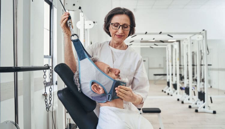

For individuals dealing with posture problems causing neck, back, and shoulder pain, can pectoralis minor stretches designed to work these areas be a part of physical therapy or as regular exercises at home?
Pectoralis Minor Muscle Stretches
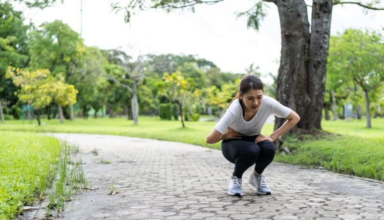
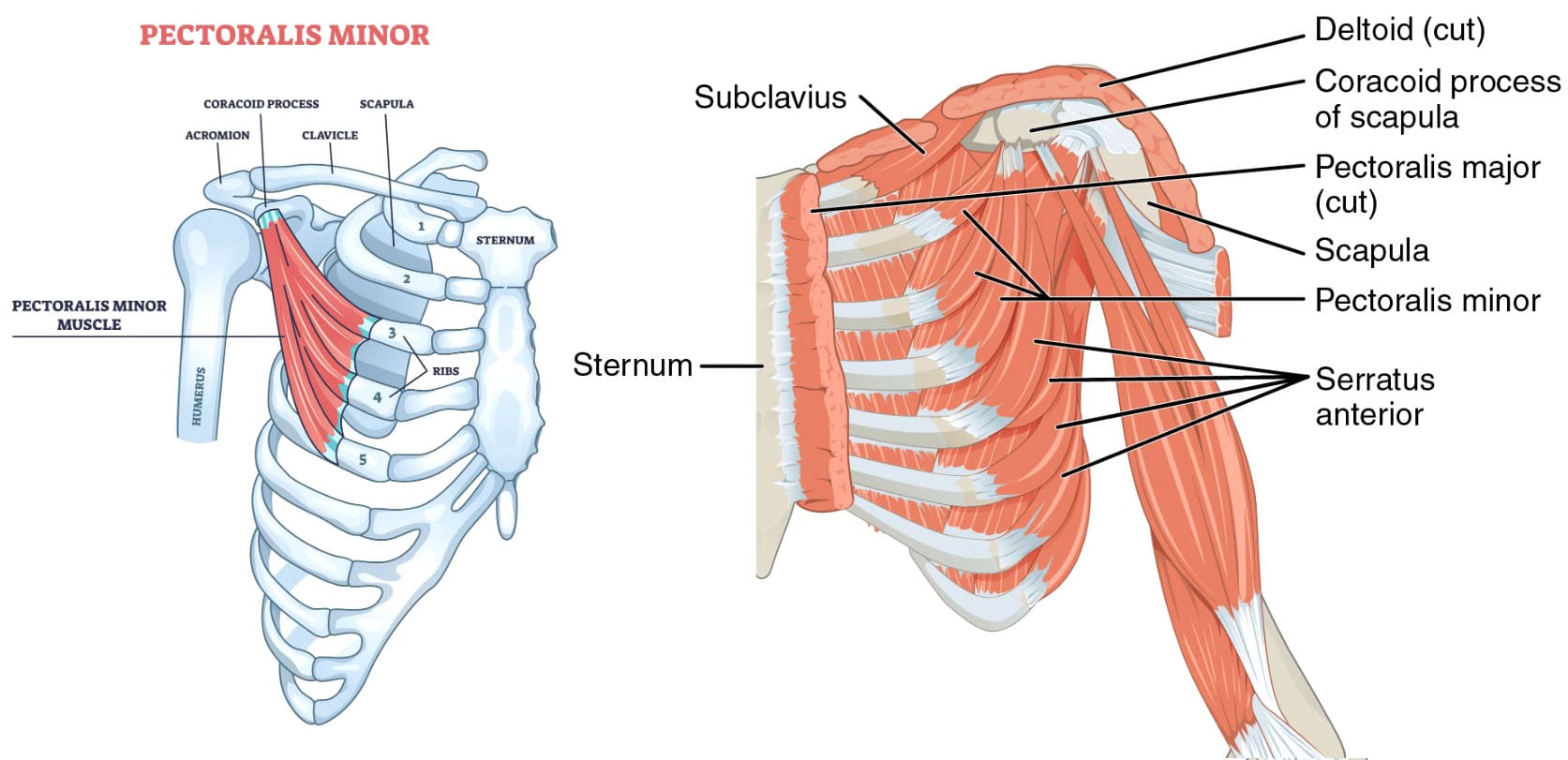
The pectoralis minor is a small, triangular muscle situated deep to the pectoralis major in the anterior chest wall. It originates from the margins of the third to fifth ribs adjacent to the costochondral junction and connects to the coracoid process of the scapula. The pectoralis minor helps with posture, mobility, and shoulder stability and aids breathing. Muscle tightness can cause pain in the chest, shoulder, and neck and a restricted range of motion. Strain and injuries can occur during activities involving overhead movements or forceful pushing. Pectoralis minor stretches are designed to work these muscles that span the ribs and connect to the shoulder to help improve posture and relieve pain and chest weakness. They can help reduce muscle tightness and other conditions like thoracic outlet syndrome. (Kaur U. et al., 2023) (Wagner E. R. et al., 2023) Talk with a healthcare provider Before starting any exercise or stretching program.
Corner Pectoralis Stretch
A corner pec stretch is similar to a wall push-up, except the emphasis is on staying in a position that lengthens the chest muscles. It’s important to move the whole body as a unit and not bend.
- Stand facing a corner with a relaxed, upright posture.
- Place your feet so they are parallel, and bend your knees slightly.
- Stay as relaxed as possible during the movement to protect your joints.
- Keep your gaze forward.
- Place your forearms and palms over the walls where two walls connect at a right angle.
- With your elbows bent to 90 degrees, move forward into the corner of the wall until you feel a comfortable stretch in the pectorals.
- Keep the hips straight.
- Hold the position for up to 30 seconds.
- Return to starting position.
- If you need a deeper stretch, move the arm position up or down. (University of North Carolina School of Medicine, 2020)
Doorway Stretch
The doorway stretch is similar to the corner stretch. It works the pectoralis major and the minor muscles and helps with mobility. To perform: (Maryland Pain & Wellness Center, 2025)
- Stand in a doorway with your feet placed together.
- Place the palms and forearms on either side of the doorway.
- Your elbows should be even with your shoulders and bend at a 90-degree angle.
- Keep your back straight.
- Take a step forward, leaning into the doorway.
- You should feel the stretch in the muscle.
- Repeat the stretch with the other foot.
Exercise and ergonomic changes to your chair or desk height can help improve posture and relieve muscle tightness. (Kaur U. et al., 2023)
T Stretch
The T stretch stretches the front of the chest and is done on the floor, typically with a foam roller placed directly under the spine. To perform: (OrthoCarolina, N.D.)
- Lie down on your back with the foam roller aligned to the spine.
- Make sure your head and tailbone are supported.
- Open your arms straight out like a T.
- Hold the position while stretching.
Y Stretch
The Y stretch is similar to the T stretch; both reduce chest muscle tightness and discomfort. To perform: (OrthoCarolina, N.D.)
- Use the same foam roll position, lying on your back with the head and tailbone supported and aligned.
- Stretch the arms out above your head, placing them into the shape of a Y.
- Allow the chest muscles that connect to the arms to relax.
Studies have examined how quickly a prone scapular retraction can help stretch the back and shoulders. Results suggest the exercises must be performed longer before the pectoralis minor is lengthened to improve symptoms. (Dye J., Allyn M., & Frank C. 2024) However, further research is needed.
Health Conditions
Pectoralis minor stretches may be part of a personalized therapy program to improve mobility, posture, and/or breathing and sleep quality with health conditions that include:
- Thoracic outlet syndrome
- Kyphosis (Chankavee N. et al., 2023)
- Obstructive sleep apnea (Liao Y. X. et al., 2025)
- Chronic obstructive pulmonary disease (COPD) (Thongchote K., Chinwaro U., & Lapmanee S. 2024)
Stretching and strengthening exercises can help improve their flexibility and function. Exercises can improve strength and function by standing or lying down, depending on the stretch.
Injury Medical Chiropractic and Functional Medicine Clinic
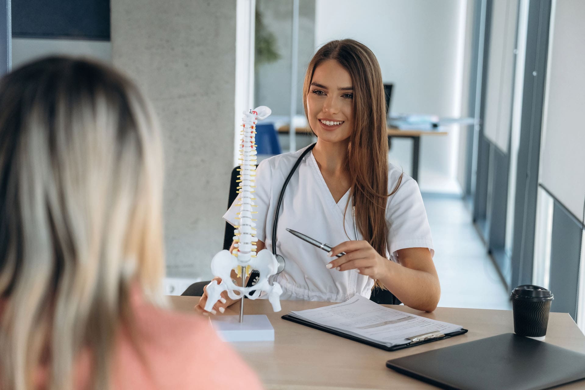
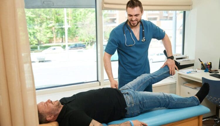
The pectoralis minor muscles are often overlooked in clinical examinations but can contribute to musculoskeletal pain and dysfunction. A healthcare provider can teach about stretches, how they can help, and whether they are safe for the individual’s injury and/or condition. Injury Medical Chiropractic and Functional Medicine Clinic works with primary healthcare providers and specialists to build optimal health and wellness solutions. Regarding musculoskeletal pain, specialists like chiropractors, acupuncturists, and massage therapists can help mitigate the pain through spinal adjustments that help the body realign itself. The clinic can also work with other medical professionals to integrate a treatment plan to resolve musculoskeletal problems.
Doorway Stretching Routine
References
Kaur, U., Shrestha, D., Hussain, M. A., Dalal, P., Kalita, M., Sharma, V., & Sharma, S. (2023). Prompt Impact of Muscle Energy Technique on Pectoralis Muscle Tightness in Computer Users: A Quasi-Experimental Study. Journal of Lifestyle Medicine, 13(2), 123–128. https://doi.org/10.15280/jlm.2023.13.2.123
Wagner, E. R., Gottschalk, M. B., Ahmed, A. S., Graf, A. R., & Karzon, A. L. (2023). Novel Diagnostic and Treatment Techniques for Neurogenic Thoracic Outlet Syndrome. Techniques in hand & upper extremity surgery, 27(2), 100–114. https://doi.org/10.1097/BTH.0000000000000419
University of North Carolina School of Medicine. (2020). Upper Body Stretching. https://www.med.unc.edu/htcenter/wp-content/uploads/sites/711/2020/04/Upper-Body-Stretching.pdf
Maryland Pain & Wellness Center. (2025). Stretches to Help with Strained Chest Muscles. Maryland Pain & Wellness Center Restoring Hope, Rebuilding Lives. https://www.marylandpainandwellnesscenter.com/blog/stretches-to-help-with-strained-chest muscles#:~:text=With%20your%20knees%20bent%20and,assist%20in%20deepening%20the%20stretch.
OrthoCarolina. (N.D.). Stretching Guide to Ease Tight Muscles. https://www.orthocarolina.com/storage/wysiwyg/stretching_guide_1.pdf
Dye, J., Allyn, M., & Frank, C. (2024). Is there an immediate effect on pectoralis minor length after performing a prone scapular retraction exercise using typical sets and repetitions in pain-free participants? Journal of bodywork and movement therapies, 40, 1014–1019. https://doi.org/10.1016/j.jbmt.2024.07.026
Chankavee, N., Amatachaya, S., Hunsawong, T., Thaweewannakij, T., & Mato, L. (2023). Effects of modified long stick exercise on hyperkyphosis, muscle imbalance, and balance control in elderly community-dwelling women with hyperkyphosis. Journal of back and musculoskeletal rehabilitation, 36(5), 1151–1162. https://doi.org/10.3233/BMR-220350
Liao, Y. X., Saiken, A., Chang, X., Guo, Y. F., Tan, Z., Deng, F., Meng, Q. L., Zhen, H., Li, Y. M., & Fang, B. M. (2025). Associations of fat, bone, and muscle indices with disease severity in patients with obstructive sleep apnea-hypopnea syndrome. Sleep & breathing = Schlaf & Atmung, 29(1), 82. https://doi.org/10.1007/s11325-024-03241-8
Thongchote, K., Chinwaro, U., & Lapmanee, S. (2024). Effects of scapulothoracic exercises on chest mobility, respiratory muscle strength, and pulmonary function in male COPD patients with forward shoulder posture: A randomized controlled trial. F1000Research, 11, 1284. https://doi.org/10.12688/f1000research.126832.2