Home Blog Page 2

Maximizing Fitness Progress with Effective Assessment Testing

Fitness coach talking to fit female client when explaining new training plan

For individuals looking to improve their fitness health, can a fitness assessment test identify potential areas and help evaluate overall health and physical status?

Maximizing Fitness Progress with Effective Assessment Testing

Fitness Assessment

A fitness test, also known as a fitness assessment, helps evaluate an individual’s overall and physical health. It comprises a series of exercises to design an appropriate exercise program for general health and fitness. (National Strength and Conditioning Association. 2017) Fitness assessment testing benefits include:

  • Identifying areas that need improvement.
  • Assisting professionals in understanding what types of exercise are safest and most effective.
  • Helping measure fitness progress over time.
  • Allowing for an individualized plan that can help prevent injuries and maintain the body’s overall health.

An assessment can comprise a wide range of tests, including:

  • Body composition tests.
  • Cardiovascular stress tests.
  • Endurance tests.
  • Range of motion tests.

They are meant to ensure the individual won’t be at risk of injury and provide the trainer with the insights needed to establish clear and effective fitness goals. Individuals who wonder whether fitness testing would benefit them should consult their healthcare provider.

General Health

Before starting a fitness program, it is important to inform the trainer of individual medical history and get the necessary approval from a primary healthcare provider. (Harvard Health Publishing. Harvard Medical School. 2012) Fitness specialists usually use one or more screening tools to help determine individual baseline health.
This may include obtaining vital sign measurements like height and weight, resting heart rate/RHR, and resting blood pressure/RBP. Many trainers will also use a physical activity readiness questionnaire/PAR-Q comprising questions about general health. (National Academy of Sports Medicine. 2020) Among the questions, individuals may​ be asked about the medications being taken, any problems with dizziness or pain, or medical conditions that may impair their ability to exercise.

Body Composition

Body composition describes total body weight components, including muscles, bones, and fat. The most common methods for estimating body composition include:

Bioelectrical Impedance Analysis – BIA

  • During BIA, electrical signals are sent from electrodes through the soles of the feet to the abdomen to estimate body composition. (Doylestown Health. 2024)

Body Mass Index – BMI

Skinfold Measurements

  • These measurements use calipers to estimate the amount of body fat in a fold of skin.

Cardiovascular Endurance

Cardiovascular endurance testing, also known as stress testing, measures how efficiently the heart and lungs work to supply oxygen and energy to the body during physical activity. (UC Davis Health, 2024) The three most common tests used include:

12-minute Run Tests

  • Twelve-minute run tests are performed on a treadmill, and an individual’s pre-exercise heart and respiration rates are compared with post-exercise heart and respiration rates.

Exercise Stress

  • Exercise stress testing is performed on a treadmill or stationary bike.
  • It involves using a heart monitor and blood pressure cuff to measure vital signs during exercise.

VO2 Max Testing

  • Performed on a treadmill or stationary bike.
  • V02 max testing uses a breathing device to measure the maximum rate of oxygen consumption during physical activity (UC Davis Health, 2024)
  • Some trainers will incorporate exercises like sit-ups or push-ups to measure response to specific exercises.
  • These baseline results can be used later to see if health and fitness levels have improved.

Strength and Endurance

Muscle endurance testing measures the length of time a muscle group can contract and release before it fatigues. Strength testing measures the maximal amount of force a muscle group can exert. (American Council on Exercise, Jiminez C., 2018) The exercises used include:

  • The push-up test.
  • Core strength and stability test.

Sometimes, a trainer will use a metronome to measure how long the individual can keep up with the rhythm. The results are then compared to individuals of the same age group and sex to establish a baseline level. Strength and endurance tests are valuable as they help the trainer spot which muscle groups are stronger, vulnerable, and need focused attention. (Heyward, V. H., Gibson, A. L. 2014).
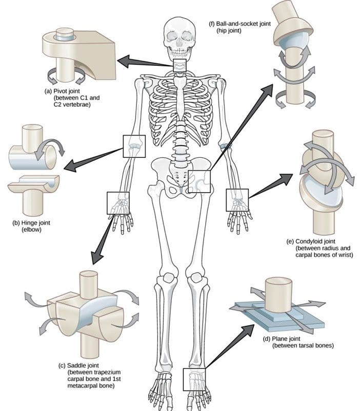
Flexibility

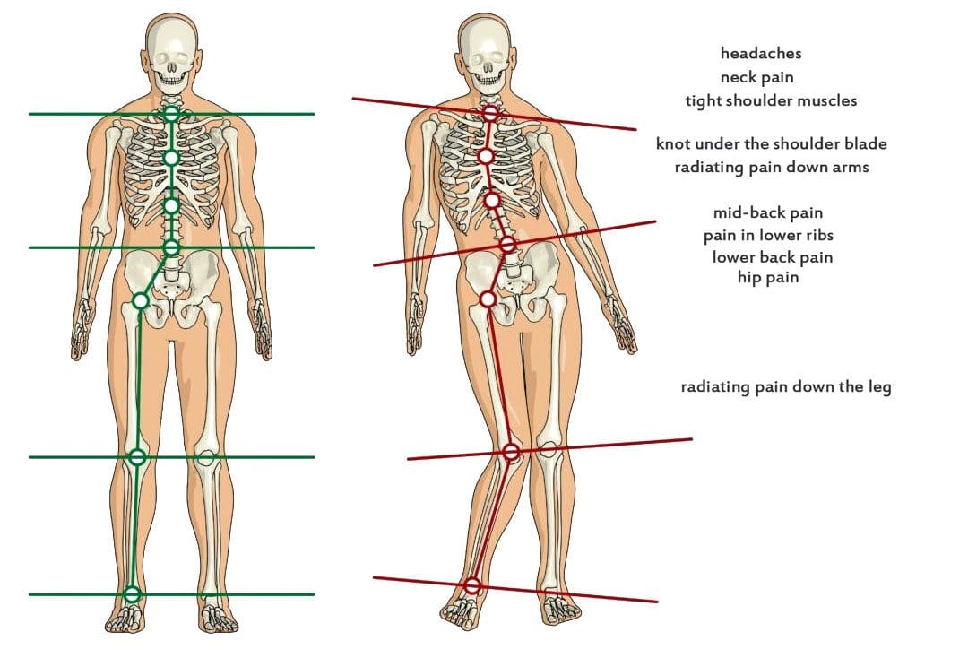
  • Measuring the flexibility of joints is vital in determining whether individuals have postural imbalances, foot instability, or limitations in range of motion. (Pate R, Oria M, Pillsbury L, 2012)

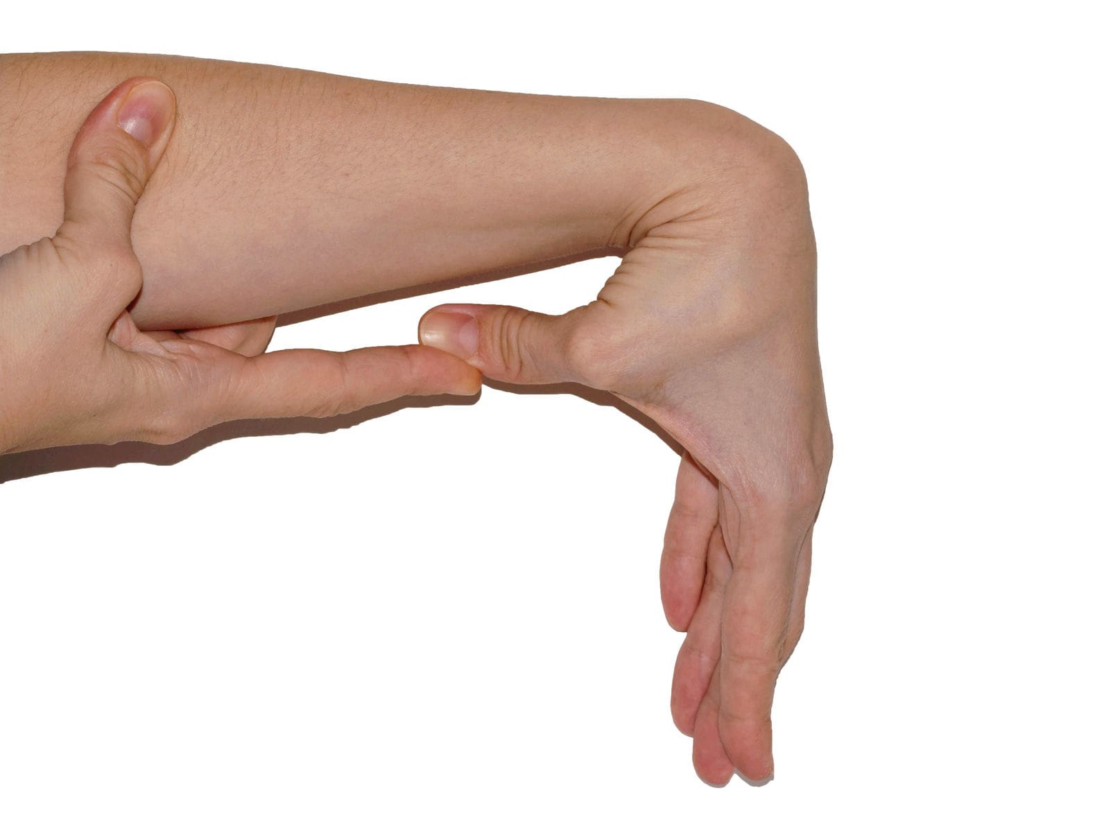
Shoulder Flexibility

  • Shoulder flexibility testing evaluates the flexibility and mobility of the shoulder joint.
  • It is performed by using one hand to reach behind the neck, between the shoulders, and the other hand to reach behind the back, toward the shoulders, to measure how far apart the hands are. (Baumgartner TA, PhD, Jackson AS, PhD et al., 2015)

Sit-And-Reach

  • This test measures tightness in the lower back and hamstring muscles. (American Council of Exercise, Metcalf A. 2014)
  • The sit-and-reach test is performed on the floor with the legs fully extended.
  • Flexibility is measured by how many inches the hands are from the feet when reaching forward.

Trunk Lift

  • Trunk lift testing is used to measure tightness in the lower back.
  • It is performed while lying face-down on the floor with arms at your side.
  • The individual will be asked to lift their upper body with just the back muscles.
  • Flexibility is measured by how many inches the individual can lift themselves off the ground. (Baumgartner TA, PhD, Jackson AS, PhD et al., 2015)

Fitness assessment testing has various benefits. It can help trainers design a personalized workout program, help individuals identify fitness areas that need improvement, measure progress, and add intensity and endurance to their routine, which can help prevent injuries and help maintain overall health. We focus on what works for you and strive to better the body through researched methods and total wellness programs. These natural programs use the body’s ability to achieve improvement goals. Ask a healthcare professional or fitness professional for guidance if you need advice.


PUSH Fitness


References

National Strength and Conditioning Association. (2017). Purposes of assessment. www.nsca.com/education/articles/kinetic-select/purposes-of-assessment/

Harvard Health Publishing. Harvard Medical School. (2012). Do you need to see a doctor before starting your exercise program? HealthBeat. www.health.harvard.edu/healthbeat/do-you-need-to-see-a-doctor-before-starting-your-exercise-program

National Academy of Sports Medicine. (2020). PAR-Q-+ The Physical Activity Readiness Questionnaire for Everyone. www.nasm.org/docs/pdf/parqplus-2020.pdf?sfvrsn=401bf1af_24

Doylestown Health. (2024). Bio-Electrical Impedance Analysis (BIA)-Body Mass Analysis. www.doylestownhealth.org/service-lines/nutrition#maintabbed-content-tab-2BDAD9F8-F379-403C-8C9C-75D7BFA6E596-1-1

National Heart, Lung, and Blood Institute. U.S. Department of Health and Human Services. (N.D.). Calculate your body mass index. Retrieved from www.nhlbi.nih.gov/health/educational/lose_wt/BMI/bmicalc.htm

UC Davis Health. (2024). VO2max and Aerobic Fitness. health.ucdavis.edu/sports-medicine/resources/vo2description

American Council on Exercise. Jiminez C. (2018). Understanding 1-RM and Predicted 1-RM Assessments. ACE Fitness. www.acefitness.org/fitness-certifications/ace-answers/exam-preparation-blog/2894/understanding-1-rm-and-predicted-1-rm-assessments/

Heyward, V. H., Gibson, A. L. (2014). Advanced Fitness Assessment and Exercise Prescription. United Kingdom: Human Kinetics. www.google.com/books/edition/Advanced_Fitness_Assessment_and_Exercise/PkdoAwAAQBAJhl=en&gbpv=1&dq=Strength+and+endurance+tests+muscle+groups+are+stronger+and+weaker&pg=PA173&printsec=frontcover#v=onepage&q=Strength%20and%20endurance%20tests%20muscle%20groups%20are%20stronger%20and%20weaker&f=false

Pate R, Oria M, Pillsbury L, (Eds). (2012). Health-related fitness measures for youth: Flexibility. In R. Pate, M. Oria, & L. Pillsbury (Eds.), Fitness Measures and Health Outcomes in Youth. doi.org/10.17226/13483

Baumgartner, T. A., Jackson, A. S., Mahar, M. T., Rowe, D. A. (2015). Measurement for Evaluation in Kinesiology. United States: Jones & Bartlett Learning. www.google.com/books/edition/Measurement_for_Evaluation_in_Kinesiolog/_oCHCgAAQBAJ?hl=en&gbpv=1&dq=Measurement+for+Evaluation+in+Kinesiology+(9th+Edition).&printsec=frontcover#v=onepage&q&f=false

American Council of Exercise. Metcalf A. (2014). How to improve flexibility and maintain it. ACE Fitness. www.acefitness.org/resources/everyone/blog/3761/how-to-improve-flexibility-and-maintain-it/

Overview of Ehlers-Danlos Syndrome: Symptoms and Diagnosis

Can individuals with Ehlers-Danlos syndrome find relief through various non-surgical treatments to reduce joint instability?

Introduction

The joints and ligaments surrounding the musculoskeletal system allow the upper and lower extremities to stabilize the body and be mobile. The various muscles and soft connective tissues that surround the joints help protect them from injuries. When environmental factors or disorders start to affect the body, many people develop issues that cause overlapping risk profiles, which then affect the stability of the joints. One of the disorders that affect the joints and connective tissue is EDS or Ehlers-Danlos syndrome. This connective tissue disorder can cause the joints in the body to be hypermobile. It can cause joint instability in the upper and lower extremities, thus leaving the individual to be in constant pain. Today’s article focuses on Ehlers-Danlos syndrome and its symptoms and how there are non-surgical ways to manage this connective tissue disorder. We discuss with certified medical providers who consolidate our patients’ information to assess how Ehlers-Danlos syndrome can correlate with other musculoskeletal disorders. We also inform and guide patients on how various non-surgical treatments can help reduce pain-like symptoms and manage Ehlers-Danlos syndrome. We also encourage our patients to ask their associated medical providers many intricate and important questions about incorporating various non-surgical therapies as part of their daily routine to manage the effects of Ehlers-Danlos syndrome. Dr. Jimenez, D.C., includes this information as an academic service. Disclaimer.


What Is Ehlers-Danlos Syndrome?


Do you often feel extremely tired throughout the day, even after a full night of sleep? Do you bruise easily and wonder where these bruises are coming from? Or have you noticed that you have an increased range in your joints? Many of these issues are often correlated with a disorder known as Ehlers-Danlos syndrome or EDS that affects their joints and connective tissue. EDS affects the connective tissues in the body. The connective tissues in the body help provide strength and elasticity to the skin, joints, as well as blood vessel walls, so when a person is dealing with EDS, it can cause a significant disruption to the musculoskeletal system. EDS is largely diagnosed clinically, and many doctors have identified that the gene coding of the collagen and proteins that interact in the body can help determine what type of EDS affects the individual. (Miklovic & Sieg, 2024)


The Symptoms

When understanding EDS, it is essential to know the complexities of this connective tissue disorder. EDS is classified into numerous types with distinct features and challenges that vary depending on the severity. One of the most common types of EDS is hypermobile Ehlers-Danlos syndrome. This type of EDS is characterized by general joint hypermobility, joint instability, and pain. Some of the symptoms that are associated with hypermobile EDS include subluxation, dislocations, and soft tissue injuries that are common and may occur spontaneously or with minimal trauma. (Hakim, 1993) This can often cause acute pain to the joints in the upper and lower extremities. With its broad range of symptoms and the personal nature of the condition itself, many often don’t realize that joint hypermobility is common in the general population and may present no complications that indicate that it is a connective tissue disorder. (Gensemer et al., 2021) Additionally, hypermobile EDS can lead to spinal deformity due to the hyperextensibility of the skin, joints, and various tissue fragility. The pathophysiology of spinal deformity associated with hypermobile EDS is primarily due to muscle hypotonia and ligament laxity. (Uehara et al., 2023) This causes many people to reduce their quality of life and daily living activities significantly. However, there are ways to manage EDS and its correlating symptoms to reduce joint instability.


Movement Medicine: Chiropractic Care-Video

Movement Medicine: Chiropractic Care | El Paso, Tx (2024)

Ways To Manage EDS

When it comes to looking for ways to manage EDS to reduce pain and joint instability, non-surgical treatments can help address the physical and emotional aspects of the condition. Non-surgical treatments for individuals with EDS commonly focus on optimizing the body’s physical function while improving muscular strength and joint stabilization. (Buryk-Iggers et al., 2022) Many individuals with EDS will try to incorporate pain management techniques and physical therapy and use braces and assistive devices to reduce the effects of EDS and improve their quality of life.


Non-surgical Treatments For EDS

Various non-surgical treatments like MET (muscle energy technique), electrotherapy, light physical therapy, chiropractic care, and massages can help strengthen while toning the surrounding muscles around the joints, provide sufficient pain relief, and limit long-term dependence on medications. (Broida et al., 2021) Additionally, individuals dealing with EDS aim to strengthen the affected muscles, stabilize the joints, and improve proprioception. Non-surgical treatments allow the individual to have a customized treatment plan for the severity of EDS symptoms and help reduce the pain associated with the condition. Many individuals, when going through their treatment plan consecutively to manage their EDS and reduce the pain-like symptoms, will notice improvement in symptomatic discomfort. (Khokhar et al., 2023) This means that non-surgical treatments allow individuals to be more mindful of their bodies and reduce the pain-like effects of EDS, thus allowing many individuals with EDS to lead fuller, more comfortable lives without feeling pain and discomfort.


References

Broida, S. E., Sweeney, A. P., Gottschalk, M. B., & Wagner, E. R. (2021). Management of shoulder instability in hypermobility-type Ehlers-Danlos syndrome. JSES Rev Rep Tech, 1(3), 155-164. doi.org/10.1016/j.xrrt.2021.03.002

Buryk-Iggers, S., Mittal, N., Santa Mina, D., Adams, S. C., Englesakis, M., Rachinsky, M., Lopez-Hernandez, L., Hussey, L., McGillis, L., McLean, L., Laflamme, C., Rozenberg, D., & Clarke, H. (2022). Exercise and Rehabilitation in People With Ehlers-Danlos Syndrome: A Systematic Review. Arch Rehabil Res Clin Transl, 4(2), 100189. doi.org/10.1016/j.arrct.2022.100189

Gensemer, C., Burks, R., Kautz, S., Judge, D. P., Lavallee, M., & Norris, R. A. (2021). Hypermobile Ehlers-Danlos syndromes: Complex phenotypes, challenging diagnoses, and poorly understood causes. Dev Dyn, 250(3), 318-344. doi.org/10.1002/dvdy.220

Hakim, A. (1993). Hypermobile Ehlers-Danlos Syndrome. In M. P. Adam, J. Feldman, G. M. Mirzaa, R. A. Pagon, S. E. Wallace, L. J. H. Bean, K. W. Gripp, & A. Amemiya (Eds.), GeneReviews((R)). www.ncbi.nlm.nih.gov/pubmed/20301456

Khokhar, D., Powers, B., Yamani, M., & Edwards, M. A. (2023). The Benefits of Osteopathic Manipulative Treatment on a Patient With Ehlers-Danlos Syndrome. Cureus, 15(5), e38698. doi.org/10.7759/cureus.38698

Miklovic, T., & Sieg, V. C. (2024). Ehlers-Danlos Syndrome. In StatPearls. www.ncbi.nlm.nih.gov/pubmed/31747221

Uehara, M., Takahashi, J., & Kosho, T. (2023). Spinal Deformity in Ehlers-Danlos Syndrome: Focus on Musculocontractural Type. Genes (Basel), 14(6). doi.org/10.3390/genes14061173

Disclaimer

Exploring the Functions and Movements of Hinge Joints

Female caucasian doctor talking with her patient

 Can understanding the body’s hinge joints and how they operate help with mobility and flexibility problems and manage conditions for individuals with difficulty fully bending or extending their fingers, toes, elbows, ankles, or knees?

Exploring the Functions and Movements of Hinge Joints

Hinge Joints

A joint forms where one bone connects to another, allowing motion. Different types of joints differ in structure and movement depending on their location. These include hinge, ball and socket, planar, pivot, saddle, and ellipsoid joints. (Boundless. General Biology, N.D.) Hinge joints are synovial joints that move through one plane of motion: flexion and extension. Hinge joints are found in the fingers, elbows, knees, ankles, and toes and control movement for various functions. Injuries, osteoarthritis, and autoimmune conditions can affect hinge joints. Rest, medication, ice, and physical therapy can help alleviate pain, improve strength and range of motion, and help manage conditions.

Anatomy

A joint is formed by the joining of two or more bones. The human body has three main classifications of joints, categorized by the degree to which they can move. These include: (Boundless. General Biology, N.D.)

Synarthroses

  • These are fixed, immovable joints.
  • Formed by two or more bones.

Amphiarthroses

  • Also known as cartilaginous joints.
  • A fibrocartilage disc separates the bones that form the joints.
  • These movable joints allow for a slight degree of movement.

Diarthroses

  • Also known as synovial joints.
  • These are the most common freely mobile joints that allow movement in multiple directions.
  • The bones that form the joints are lined with articular cartilage and enclosed in a joint capsule filled with synovial fluid that allows for smooth motion.

Synovial joints are classified into different types depending on differences in structure and the number of motion planes they allow. A hinge joint is a synovial joint that allows movement in one plane of motion, similar to a door hinge that moves forward and backward. Within the joint, the end of one bone is typically convex/pointed outward, with the other concave/rounded inward to allow the ends to fit smoothly. Because hinge joints only move through one plane of movement, they tend to be more stable than other synovial joints. (Boundless. General Biology, N.D.) Hinge joints include:

  • The finger and toe joints – allow the fingers and toes to bend and extend.
  • The elbow joint – allows the elbow to bend and extend.
  • The knee joint – allows the knee to bend and extend.
  • The talocrural joint of the ankle – allows the ankle to move up/dorsiflexion and down/plantarflexion.

Hinge joints allow the limbs, fingers, and toes to extend away and bend toward the body. This movement is essential for activities of daily living, such as showering, getting dressed, eating, walking, standing up, and sitting down.

Conditions

Osteoarthritis and inflammatory forms of arthritis can affect any joint (Arthritis Foundation. N.D.) Autoimmune inflammatory forms of arthritis, including rheumatoid and psoriatic arthritis, can cause the body to attack its own joints. These commonly affect the knees and fingers, resulting in swelling, stiffness, and pain. (Kamata, M., Tada, Y. 2020) Gout is an inflammatory form of arthritis that develops from elevated levels of uric acid in the blood and most commonly affects the hinge joint of the big toe. Other conditions that affect hinge joints include:

  • Injuries to the cartilage within the joints or ligaments that stabilize the outside of the joints.
  • Ligament sprains or tears can result from jammed fingers or toes, rolled ankles, twisting injuries, and direct impact on the knee.
  • These injuries can also affect the meniscus, the tough cartilage within the knee joint that helps cushion and absorb shock.

Rehabilitation

Conditions that affect hinge joints often cause inflammation and swelling, resulting in pain and limited mobility.

  • After an injury or during an inflammatory condition flare-up, limiting active movement and resting the affected joint can reduce increased stress and pain.
  • Applying ice can decrease inflammation and swelling.
  • Pain-relieving medications like NSAIDs can also help reduce pain. (Arthritis Foundation. N.D.)
  • Once the pain and swelling start to subside, physical and/or occupational therapy can help rehabilitate the affected areas.
  • A therapist will provide stretches and exercises to help improve the joint range of motion and strengthen the supporting muscles.
  • For individuals experiencing hinge joint pain from an autoimmune condition, biologic medications to decrease the body’s autoimmune activity are administered through infusions delivered every several weeks or months. (Kamata, M., Tada, Y. 2020)
  • Cortisone injections may also be used to decrease inflammation.

At Injury Medical Chiropractic and Functional Medicine Clinic, we passionately focus on treating patients’ injuries and chronic pain syndromes and improving ability through flexibility, mobility, and agility programs tailored to the individual. Our providers use an integrated approach to create personalized care plans that include Functional Medicine, Acupuncture, Electro-Acupuncture, and Sports Medicine protocols. Our goal is to relieve pain naturally by restoring health and function to the body. If the individual needs other treatment, they will be referred to a clinic or physician best suited for them. Dr. Jimenez has teamed up with the top surgeons, clinical specialists, medical researchers, and premier rehabilitation providers to provide the most effective clinical treatments.


Chiropractic Solutions


References

Boundless. General Biology. (N.D.). 38.12: Joints and Skeletal Movement – Types of Synovial Joints. In. LibreTexts Biology. bio.libretexts.org/Bookshelves/Introductory_and_General_Biology/Book%3A_General_Biology_%28Boundless%29/38%3A_The_Musculoskeletal_System/38.12%3A_Joints_and_Skeletal_Movement_-_Types_of_Synovial_Joints

Arthritis Foundation. (N.D.). Osteoarthritis. Arthritis Foundation. www.arthritis.org/diseases/osteoarthritis

Kamata, M., & Tada, Y. (2020). Efficacy and Safety of Biologics for Psoriasis and Psoriatic Arthritis and Their Impact on Comorbidities: A Literature Review. International journal of molecular sciences, 21(5), 1690. doi.org/10.3390/ijms21051690

Non-Surgical Treatments for Sciatica Pain Management

For individuals dealing with sciatica, can non-surgical treatments like chiropractic care and acupuncture reduce pain and restore function?

Introduction

The human body is a complex machine that allows the host to be mobile and stable when resting. With various muscle groups in the upper and lower body portions, the surrounding muscles, tendons, nerves, and ligaments serve a purpose for the body as they all have specific jobs in keeping the host functional. However, many individuals have developed various habits that cause strenuous activities that cause repetitive motions to their muscles and nerves and affect their musculoskeletal system. One of the nerves that many individuals have been dealing with pain is the sciatic nerve, which causes many issues in the lower body extremities and, when not treated right away, leads to pain and disability. Luckily, many individuals have sought non-surgical treatments to reduce sciatica and restore body function to the individual. Today’s article focuses on understanding sciatica and how non-surgical therapies like chiropractic care and acupuncture can help reduce the sciatic pain-like effects that are causing overlapping risk profiles in the lower body extremities. We discuss with certified medical providers who consolidate with our patients’ information to assess how sciatica is often correlated with environmental factors that cause dysfunction in the body. We also inform and guide patients on how various non-surgical treatments can help reduce sciatica and its correlating symptoms. We also encourage our patients to ask their associated medical providers many intricate and important questions about incorporating various non-surgical therapies as part of their daily routine to reduce the chances and effects of sciatica from returning. Dr. Jimenez, D.C., includes this information as an academic service. Disclaimer.


Understanding Sciatica

Do you often feel radiating pain that travels down one or both legs when sitting down for a long period? How often have you experienced tingling sensations that cause you to shake your leg to reduce the effect? Or have you noticed that stretching your legs causes temporary relief? While these overlapping pain symptoms can affect the lower extremities, many individuals may think it is low back pain, but in actuality, it is sciatica. Sciatica is a common musculoskeletal condition that affects many people worldwide by causing pain to the sciatic nerve and radiating down to the legs. The sciatic nerve is pivotal in providing direct and indirect motor function to the leg muscles. (Davis et al., 2024) When the sciatic nerve is compressed, many people state that the pain can vary in intensity, accompanied by symptoms like tingling, numbness, and muscle weakness that can affect a person’s ability to walk and function. 


However, some of the root causes that lead to the development of sciatica can play into the factor that causes the pain in the lower extremities. Several inherent and environmental factors are often associated with sciatica, causing lumbar nerve root compression on the sciatic nerve. Factors like poor health status, physical stress, and occupational work are correlated with the development of sciatica and can impact a person’s routine. (Gimenez-Campos et al., 2022) Additionally, some of the root causes of sciatica can include musculoskeletal conditions like herniated discs, bone spurs, or spinal stenosis, which can correlate with these inherent and environmental factors that can reduce many individuals’ motility and life quality. (Zhou et al., 2021) This causes many individuals to seek out treatments to relieve sciatica pain and its correlating symptoms. While the pain caused by sciatica can vary, many individuals often seek non-surgical treatments to alleviate their discomfort and pain from sciatica. This allows them to incorporate effective solutions to managing sciatica. 


Beyond Adjustments: Chiropractic & Integrative Healthcare- Video

Beyond Adjustments: Chiropractic and Integrative Healthcare | El Paso, Tx (2024)

Chiropractic Care For Sciatica

When it comes to seeking non-surgical treatments to reduce sciatica, non-surgical treatments can reduce the pain-like effects while helping restore body function and mobility. At the same time, non-surgical treatments are customized to the individual’s pain and can be incorporated into a person’s routine. Some non-surgical treatments like chiropractic care are excellent in reducing sciatica and its associated pain symptoms. Chiropractic care is a form of non-surgical therapy that focuses on restoring the body’s spinal movement while improving body function. Chiropractic care utilizes mechanical and manual techniques for sciatica to realign the spine and help the body heal naturally without surgery or medication. Chiropractic care can help decrease intradiscal pressure, increase intervertebral disc space height, and improve the range of motion in the lower extremities. (Gudavalli et al., 2016) When dealing with sciatica, chiropractic care can alleviate the unnecessary pressure on the sciatic nerve and help reduce the risk of reoccurrence through consecutive treatments. 


The Effects of Chiropractic Care For Sciatica

Some of the effects of chiropractic care for reducing sciatica can provide insight to the person as chiropractors work with associated medical providers to devise a personalized plan to relieve the pain-like symptoms. Many people who utilize chiropractic care to reduce the effects of sciatica can incorporate physical therapy to strengthen the weak muscles that surround the lower back, stretch to improve flexibility and be more mindful of what factors are causing sciatic pain in their lower extremities. Chiropractic care may guide many people on proper poster ergonomics, and various exercises to reduce the chances of sciatica returning while offering positive effects to the lower body.


Acupuncture For Sciatica

Another form of non-surgical treatment that can help reduce the pain-like effects of sciatica is acupuncture. As a key component in traditional Chinese medicine, acupuncture therapy involves professionals placing thin, solid needles at specific points on the body. When it comes to reducing sciatica, acupuncture therapy can exert analgesic effects on the body’s acupoints, regulate the microglia, and modulate certain receptors along the pain pathway to the nervous system. (Zhang et al., 2023) Acupuncture therapy focuses on restoring the body’s natural energy flow or Qi to promote healing.


The Effects of Acupuncture For Sciatica

 Regarding the effects of acupuncture therapy on reducing sciatica, acupuncture therapy can help reduce the pain signals that sciatica produces by changing the brain signal and rerouting the corresponding motor or sensory disturbance of the affected area. (Yu et al., 2022) Additionally, acupuncture therapy can help provide pain relief by releasing endorphins, the body’s natural pain reliever, to the specific acupoint that correlates with the sciatic nerve, reducing inflammation around the sciatic nerve, thus alleviating pressure and pain and helping improve nerve function. Both chiropractic care and acupuncture offer valuable non-surgical treatment options that can provide aid in the healing process and reduce pain caused by sciatica. When many people are dealing with sciatica and looking for numerous solutions to reduce the pain-like effects, these two non-surgical treatments can help many people address the underlying causes of sciatica, enhance the body’s natural healing process, and help provide significant relief from the pain.


References

Davis, D., Maini, K., Taqi, M., & Vasudevan, A. (2024). Sciatica. In StatPearls. www.ncbi.nlm.nih.gov/pubmed/29939685

Gimenez-Campos, M. S., Pimenta-Fermisson-Ramos, P., Diaz-Cambronero, J. I., Carbonell-Sanchis, R., Lopez-Briz, E., & Ruiz-Garcia, V. (2022). A systematic review and meta-analysis of the effectiveness and adverse events of gabapentin and pregabalin for sciatica pain. Aten Primaria, 54(1), 102144. doi.org/10.1016/j.aprim.2021.102144

Gudavalli, M. R., Olding, K., Joachim, G., & Cox, J. M. (2016). Chiropractic Distraction Spinal Manipulation on Postsurgical Continued Low Back and Radicular Pain Patients: A Retrospective Case Series. J Chiropr Med, 15(2), 121-128. doi.org/10.1016/j.jcm.2016.04.004

Yu, F. T., Liu, C. Z., Ni, G. X., Cai, G. W., Liu, Z. S., Zhou, X. Q., Ma, C. Y., Meng, X. L., Tu, J. F., Li, H. W., Yang, J. W., Yan, S. Y., Fu, H. Y., Xu, W. T., Li, J., Xiang, H. C., Sun, T. H., Zhang, B., Li, M. H., . . . Wang, L. Q. (2022). Acupuncture for chronic sciatica: protocol for a multicenter randomised controlled trial. BMJ Open, 12(5), e054566. doi.org/10.1136/bmjopen-2021-054566

Zhang, Z., Hu, T., Huang, P., Yang, M., Huang, Z., Xia, Y., Zhang, X., Zhang, X., & Ni, G. (2023). The efficacy and safety of acupuncture therapy for sciatica: A systematic review and meta-analysis of randomized controlled trails. Front Neurosci, 17, 1097830. doi.org/10.3389/fnins.2023.1097830

Zhou, J., Mi, J., Peng, Y., Han, H., & Liu, Z. (2021). Causal Associations of Obesity With the Intervertebral Degeneration, Low Back Pain, and Sciatica: A Two-Sample Mendelian Randomization Study. Front Endocrinol (Lausanne), 12, 740200. doi.org/10.3389/fendo.2021.740200

Disclaimer

Healing Time: Factors and Importance in Sports Injuries

Physiotherapist having therapy session with mature client with injured knee

What are the healing times of common sports injuries for athletes and individuals who engage in recreational sports activities?

Healing Time: Factors and Importance in Sports Injuries
A young, happy sportswoman is getting tens-electrotherapy treatments at a medical clinic.

Healing Times for Sports Injuries

Healing time from sports injuries depends on various factors, such as the location and extent of the injury and the health of the skin, joints, tendons, muscles, and bones. It is also important to take the time to recuperate or not rush back into physical sports activities before the bones or tissues have fully healed. To prevent re-injury, ensure the doctor clears health before returning to sports or strenuous physical activity.

According to CDC research, an average of 8.6 million sports and recreation-related injuries occur annually. (Sheu, Y., Chen, L. H., and Hedegaard, H. 2016) However, most sports injuries are superficial or caused by low-grade strains or sprains; at least 20% of injuries result from bone fractures or more serious injuries. Bone fractures take longer than sprains or strains, and complete tendon or muscle ruptures can take months before one can fully return to activities. Individuals in decent physical shape with no underlying illness or impairment, here is what they can expect when recovering from the following sports injuries:

Bone Fractures

In sports, the highest rate of bone fractures occurs with football and contact sports. Most are centered around the lower extremities but can involve the neck and shoulder blades, arms, and ribs.

Simple Fractures

  • Depends on the individual’s age, health, type, and location.
  • Generally, takes at least six weeks to heal.

Compound Fractures

  • In this case, a bone is broken in several places.
  • It may require surgery to stabilize the bone.
  • Healing time can take up to eight months.

Fractured Clavicle/Collarbone

  • It may require the immobilization of the shoulder and upper arm.
  • It can take five to ten weeks to heal fully.
  • Fractured fingers or toes can heal in three to five weeks.

Fractured Ribs

  • Part of the treatment plan includes breathing exercises.
  • Painkillers may be needed short term.
  • Usually, it takes around six weeks to heal.

Neck Fractures

  • It may involve any one of the seven neck vertebrae.
  • A neck brace or a halo device that is screwed into the skull for stability may be used.
  • It can take up to six weeks to heal.

Sprains and Strains

According to the CDC report, sprains and strains account for 41.4% of all sports injuries. (Sheu, Y., Chen, L. H., and Hedegaard, H. 2016)

  • A sprain is the stretching or tearing of ligaments or the tough bands of fibrous tissue that connect two bones at a joint.
  • A strain is the overstretching or tearing of muscles or tendons.

Sprained Ankles

  • It can heal in five days if there are no complications.
  • Severe sprains involving torn or ruptured tendons can take three to six weeks to heal.

Calf Strains

  • Classified as grade 1 – a mild strain can heal in two weeks.
  • A grade 3 – severe strain may require three months or more to heal completely.
  • The use of calf suppression sleeves can expedite the recovery of strains and sprains in the lower leg.

Acute Neck Strain

  • A tackle, impact, fall, quick shifting, or whipping motion can cause a whiplash injury.
  • Healing time can take a couple of weeks to six weeks.

Other Injuries

ACL Tears

  • Involving the anterior cruciate ligament.
  • Usually, it requires months of recuperation and rehabilitation, depending on several factors, including the type of sports activity.
  • Full recovery from surgery takes six to 12 months.
  • Without surgery, there is no specific timeline for rehabilitation.

Achilles Tendon Ruptures

  • It is a serious injury.
  • These occur when the tendon is either partially or completely torn.
  • Individuals will more than likely require surgery.
  • Recovery time is four to six months.

Cuts and Lacerations

  • Depends on the depth and location of the injury.
  • It can take anywhere from a week to a month to heal.
  • If there are no accompanying injuries, stitches can be removed within two to three weeks.
  • If a deep cut requires stitches, more time is necessary.

Mild Contusions/Bruises

  • Are caused by a trauma to the skin, causing blood vessels to break.
  • In most cases, a contusion will take five to seven days to heal.

Shoulder Separations

  • When treated properly, it usually takes around two weeks of rest and recovery before the patient returns to activity.

Multidisciplinary Treatment

After the initial inflammation and swelling have subsided, a doctor will recommend a treatment plan that usually involves physical therapy, self-performed physical rehabilitation, or supervision by a physical therapist or team. Fortunately, athletes and individuals who regularly exercise tend to have a faster healing time because they are in top physical shape, and their cardiovascular system provides a stronger blood supply that speeds up the healing process. At El Paso’s Chiropractic Rehabilitation Clinic & Integrated Medicine Center, we passionately focus on treating patients’ injuries and chronic pain syndromes. We focus on improving ability through flexibility, mobility, and agility programs tailored to the individual. We use in-person and virtual health coaching and comprehensive care plans to ensure every patient’s personalized care and wellness outcomes.

Our providers use an integrated approach to create personalized care plans that include Functional Medicine, Acupuncture, Electro-Acupuncture, and Sports Medicine principles. Our goal is to relieve pain naturally by restoring health and function to the body.

If the chiropractor feels the individual needs other treatment, they will be referred to a clinic or physician best suited for them. Dr. Jimenez has teamed up with the top surgeons, clinical specialists, medical researchers, and premier rehabilitation providers to provide the top clinical treatments for our community. Providing highly noninvasive protocols is our priority, and our personalized patient-based clinical insight is what we provide.


Lumbar Spine Injuries in Sports: Chiropractic Healing


References

Sheu, Y., Chen, L. H., & Hedegaard, H. (2016). Sports- and Recreation-related Injury Episodes in the United States, 2011-2014. National health statistics reports, (99), 1–12.

The Role of Pudendal Nerve in Chronic Pelvic Pain

Athletic couple riding road bicycles outside. Healthy lifestyle and fun concept. Fit couple riding in the nature while wearing sports clothing, protective helmets and sunglasses.

For individuals experiencing pelvic pain, it could be a disorder of the pudendal nerve known as pudendal neuropathy or neuralgia that leads to chronic pain. The condition can be caused by pudendal nerve entrapment, where the nerve becomes compressed or damaged. Can knowing the symptoms help healthcare providers correctly diagnose the condition and develop an effective treatment plan?

The Role of Pudendal Nerve in Chronic Pelvic Pain

Pudendal Neuropathy

The pudendal nerve is the main nerve that serves the perineum, which is the area between the anus and the genitalia – the scrotum in men and the vulva in women. The pudendal nerve runs through the gluteus muscles/buttocks and into the perineum. It carries sensory information from the external genitalia and the skin around the anus and perineum and transmits motor/movement signals to various pelvic muscles. (Origoni, M. et al., 2014) Pudendal neuralgia, also referred to as pudendal neuropathy, is a disorder of the pudendal nerve that can lead to chronic pelvic pain.

Causes

Chronic pelvic pain from pudendal neuropathy can be caused by any of the following (Kaur J. et al., 2024)

  • Excessive sitting on hard surfaces, chairs, bicycle seats, etc. Bicyclists tend to develop pudendal nerve entrapment.
  • Trauma to the buttocks or pelvis.
  • Childbirth.
  • Diabetic neuropathy.
  • Bony formations that push against the pudendal nerve.
  • Thickening of ligaments around the pudendal nerve.

Symptoms

Pudendal nerve pain can be described as stabbing, cramping, burning, numbness, or pins and needles and can present (Kaur J. et al., 2024)

  • In the perineum.
  • In the anal region.
  • In men, pain in the scrotum or penis.
  • In women, pain in the labia or vulva.
  • During intercourse.
  • When urinating.
  • During a bowel movement.
  • When sitting and goes away after standing up.

Because the symptoms are often hard to distinguish, pudendal neuropathy can often be hard to differentiate from other types of chronic pelvic pain.

Cyclist’s Syndrome

Prolonged sitting on a bicycle seat can cause pelvic nerve compression, which can lead to chronic pelvic pain. The frequency of pudendal neuropathy (chronic pelvic pain caused by entrapment or compression of the pudendal nerve) is often referred to as Cyclist’s Syndrome. Sitting on certain bicycle seats for long periods places significant pressure on the pudendal nerve. The pressure can cause swelling around the nerve, which causes pain and, over time, can lead to nerve trauma. Nerve compression and swelling can cause pain described as burning, stinging, or pins and needles. (Durante, J. A., and Macintyre, I. G. 2010) For individuals with pudendal neuropathy caused by bicycling, symptoms can appear after prolonged biking and sometimes months or years later.

Cyclist’s Syndrome Prevention

A review of studies provided the following recommendations for preventing Cyclist’s Syndrome (Chiaramonte, R., Pavone, P., Vecchio, M. 2021)

Rest

  • Take breaks at least 20–30 seconds after each 20 minutes of riding.
  • While riding, change positions frequently.
  • Stand up to pedal periodically.
  • Take time off between riding sessions and races to rest and relax the pelvic nerves. 3–10 day breaks can help in recovery. (Durante, J. A., and Macintyre, I. G. 2010)
  • If pelvic pain symptoms are barely starting to develop, rest and see a healthcare provider or specialist for an examination.

Seat

  • Use a soft, wide seat with a short nose.
  • Have the seat level or tilted slightly forward.
  • Seats with cutout holes place more pressure on the perineum.
  • If numbness or pain is present, try a seat without holes.

Bike Fitting

  • Adjust the seat height so the knee is slightly bent at the bottom of the pedal stroke.
  • The body’s weight should rest on the sitting bones/ischial tuberosities.
  • Keeping the handlebar height below the seat can reduce pressure.
  • The Triathlon bike’s extreme-forward position should be avoided.
  • A more upright posture is better.
  • Mountain bikes have been associated with an increased risk of erectile dysfunction than road bikes.

Shorts

  • Wear padded bike shorts.

Treatments

A healthcare provider may use a combination of treatments.

  • The neuropathy can be treated with rest if the cause is excessive sitting or cycling.
  • Pelvic floor physical therapy can help relax and lengthen the muscles.
  • Physical rehabilitation programs, including stretches and targeted exercises, can release nerve entrapment.
  • Chiropractic adjustments can realign the spine and pelvis.
  • The active release technique/ART involves applying pressure to muscles in the area while stretching and tensing. (Chiaramonte, R., Pavone, P., Vecchio, M. 2021)
  • Nerve blocks may help relieve pain caused by nerve entrapment. (Kaur J. et al., 2024)
  • Certain muscle relaxers, antidepressants, and anticonvulsants may be prescribed, sometimes in combination.
  • Nerve decompression surgery may be recommended if all conservative therapies have been exhausted. (Durante, J. A., and Macintyre, I. G. 2010)

Injury Medical Chiropractic and Functional Medicine Clinic care plans and clinical services are specialized and focused on injuries and the complete recovery process. Our areas of practice include Wellness and nutrition, Chronic Pain, Personal Injury, Auto Accident Care, Work Injuries, Back Injury, Low Back Pain, Neck Pain, Migraine Headaches, Sports Injuries, severe sciatica, Scoliosis, Complex Herniated Discs, Fibromyalgia, Chronic Pain, Complex Injuries, Stress Management, and Functional Medicine Treatments. If the individual requires other treatment, they will be referred to a clinic or physician best suited for their condition, as Dr. Jimenez has teamed with the top surgeons, clinical specialists, medical researchers, therapists, trainers, and premiere rehabilitation providers.


Pregnancy and Sciatica


References

Origoni, M., Leone Roberti Maggiore, U., Salvatore, S., & Candiani, M. (2014). Neurobiological mechanisms of pelvic pain. BioMed research international, 2014, 903848. doi.org/10.1155/2014/903848

Kaur, J., Leslie, S. W., & Singh, P. (2024). Pudendal Nerve Entrapment Syndrome. In StatPearls. www.ncbi.nlm.nih.gov/pubmed/31334992

Durante, J. A., & Macintyre, I. G. (2010). Pudendal nerve entrapment in an Ironman athlete: a case report. The Journal of the Canadian Chiropractic Association, 54(4), 276–281.

Chiaramonte, R., Pavone, P., & Vecchio, M. (2021). Diagnosis, Rehabilitation and Preventive Strategies for Pudendal Neuropathy in Cyclists, A Systematic Review. Journal of functional morphology and kinesiology, 6(2), 42. doi.org/10.3390/jfmk6020042

How Laser Spine Surgery Can Help Relieve Chronic Pain

Handsome Caucasian male doctor and an African senior man interacting with each other in clinic

For individuals who have exhausted all other treatment options for low back pain and nerve root compression, can laser spine surgery help alleviate nerve compression and provide long-lasting pain relief?

How Laser Spine Surgery Can Help Relieve Chronic Pain

Laser Spine Surgery

Laser spine surgery is a minimally invasive surgical procedure that uses a laser to cut through and remove spinal structures that are compressing nerves and causing intense pain. The minimally invasive procedure often results in less pain, tissue damage, and faster recovery than more extensive surgeries.

How It Works

Minimally invasive procedures result in less scarring and damage to surrounding structures, often reducing pain symptoms and a shorter recovery time. (Stern, J. 2009) Small incisions are made to access spinal column structures. With open-back surgery, a large incision is made down the back to access the spine. The surgery differs from other surgeries in that a laser beam, rather than other surgical instruments, is used to cut structures in the spine. However, the initial incision through the skin is made with a surgical scalpel. Laser is an acronym for Light Amplification Stimulated by Emission of Radiation. A laser can generate intense heat to cut through soft tissues, especially those with a high water content, like spinal column discs. (Stern, J. 2009) For many spine surgeries, the laser cannot be used to cut through bone as it generates instant sparks that can damage surrounding structures. Rather, laser spine surgery is primarily used to perform a discectomy, which is a surgical technique that removes a portion of a bulging or herniated disc that is pushing against the surrounding nerve roots, causing nerve compression and sciatic pain. (Stern, J. 2009)

Surgical Risks

Laser spine surgery may help resolve the cause of nerve root compression, but there is an increased risk of damage to nearby structures. Associated risks include: (Brouwer, P. A. et al., 2015)

  • Infection
  • Bleeding
  • Blood clots
  • Remaining symptoms
  • Returning symptoms
  • Further nerve damage
  • Damage to the membrane around the spinal cord.
  • Need for additional surgery

A laser beam is not precise like other surgical tools and requires practiced mastery and control to avoid damage to the spinal cord and nerve roots. (Stern, J. 2009) Because lasers cannot cut through bone, other surgical instruments are often used around corners and at different angles because they are more efficient and allow greater accuracy. (Atlantic Brain and Spine, 2022)

Purpose

Laser spine surgery is performed to remove structures that are causing nerve root compression. Nerve root compression is associated with the following conditions (Cleveland Clinic. 2018)

  • Bulging discs
  • Herniated discs
  • Sciatica
  • Spinal stenosis
  • Spinal cord tumors

Nerve roots that are injured or damaged and constantly send chronic pain signals can be ablated with laser surgery, known as nerve ablation. The laser burns and destroys the nerve fibers. (Stern, J. 2009) Because laser spine surgery is limited in treating certain spinal disorders, most minimally invasive spine procedures do not use a laser. (Atlantic Brain and Spine. 2022)

Preparation

The surgical team will provide more detailed instructions on what to do in the days and hours before surgery. To promote optimal healing and a smooth recovery, it is recommended that the patient stay active, eat a healthy diet, and stop smoking prior to the operation. Individuals may need to stop taking certain medications to prevent excess bleeding or interaction with anesthesia during the operation. Inform the healthcare provider about all prescriptions, over-the-counter drugs, and supplements being taken.

Laser spine surgery is an outpatient procedure at a hospital or outpatient surgical center. The patient will likely go home on the same day of the operation. (Cleveland Clinic. 2018) Patients cannot drive to or from the hospital before or after their surgery, so arrange for family or friends to provide transportation. Minimizing stress and prioritizing healthy mental and emotional well-being is important to lowering inflammation and aiding recovery. The healthier the patient goes into surgery, the easier the recovery and rehabilitation will be.

Expectations

The surgery will be decided by the patient and healthcare provider and scheduled at a hospital or outpatient surgical center. Arrange for a friend or family member to drive to the surgery and home.

Before Surgery

  • The patient will be taken to a pre-operative room and asked to change into a gown.
  • The patient will undergo a brief physical examination and answer questions about medical history.
  • The patient lies on a hospital bed, and a nurse inserts an IV to deliver medication and fluids.
  • The surgical team will use the hospital bed to transport the patient in and out of the operating room.
  • The surgical team will assist the patient in getting onto the operating table, and the patient will be administered anesthesia.
  • The patient may receive general anesthesia, which will cause the patient to sleep for the surgery, or regional anesthesia, injected into the spine to numb the affected area. (Cleveland Clinic. 2018)
  • The surgical team will sterilize the skin where the incision will be made.
  • An antiseptic solution will be used to kill bacteria and prevent the risk of infection.
  • Once sanitized, the body will be covered with sterilized linens to keep the surgical site clean.

During Surgery

  • For a discectomy, the surgeon will make a small incision less than one inch in length with a scalpel along the spine to access the nerve roots.
  • A surgical tool called an endoscope is a camera inserted into the incision to view the spine. (Brouwer, P. A. et al., 2015)
  • Once the problematic disc portion causing the compression is located, the laser is inserted to cut through it.
  • The cut disc portion is removed, and the incision site is sutured.

After Surgery

  • After surgery, the patient is brought to a recovery room, where vital signs are monitored as the effects of the anesthesia wear off.
  • Once stabilized, the patient can usually go home one or two hours after the operation.
  • The surgeon will determine when the individual is clear to resume driving.

Recovery

Following a discectomy, the individual can return to work within a few days to a few weeks, depending on the severity, but it can take up to three months to return to normal activities. Length of recovery can range from two to four weeks or less to resume a sedentary job or eight to 12 weeks for a more physically demanding job that requires heavy lifting. (University of Wisconsin School of Medicine and Public Health, 2021) During the first two weeks, the patient will be given restrictions to facilitate the spine’s healing until it becomes more stable. Restrictions can  include: (University of Wisconsin School of Medicine and Public Health, 2021)

  • No bending, twisting, or lifting.
  • No strenuous physical activity, including exercise, housework, yard work, and sex.
  • No alcohol in the initial stage of recovery or while taking narcotic pain medications.
  • No driving or operating a motor vehicle until discussed with the surgeon.

The healthcare provider may recommend physical therapy to relax, strengthen, and maintain musculoskeletal health. Physical therapy may be two to three times weekly for four to six weeks.

Process

Optimal recovery recommendations include:

  • Getting enough sleep, at least seven to eight hours.
  • Maintaining a positive attitude and learning how to cope and manage stress.
  • Maintaining body hydration.
  • Following the exercise program as prescribed by the physical therapist.
  • Practicing healthy posture with sitting, standing, walking, and sleeping.
  • Staying active and limiting the amount of time spent sitting. Try to get up and walk every one to two hours during the day to stay active and prevent blood clots. Gradually increase the amount of time or distance as recovery progresses.
  • Do not push to do too much too soon. Overexertion can increase pain and delay recovery.
  • Learning correct lifting techniques to utilize the core and leg muscles to prevent increased pressure on the spine.

Discuss treatment options for managing symptoms with a healthcare provider or specialist to determine if laser spine surgery is appropriate. Injury Medical Chiropractic and Functional Medicine Clinic care plans and clinical services are specialized and focused on injuries and the complete recovery process. Dr. Jimenez has teamed with the top surgeons, clinical specialists, medical researchers, therapists, trainers, and premiere rehabilitation providers. We focus on restoring normal body functions after trauma and soft tissue injuries using Specialized Chiropractic Protocols, Wellness Programs, Functional and integrative Nutrition, Agility and mobility Fitness Training, and Rehabilitation Systems for all ages. Our areas of practice include Wellness & Nutrition, Chronic Pain, Personal Injury, Auto Accident Care, Work Injuries, Back Injury, Low Back Pain, Neck Pain, Migraine Headaches, Sports Injuries, Severe Sciatica, Scoliosis, Complex Herniated Discs, Fibromyalgia, Chronic Pain, Complex Injuries, Stress Management, Functional Medicine Treatments, and in-scope care protocols.


The Non-Surgical Approach


References

Stern, J. SpineLine. (2009). Lasers in Spine Surgery: A Review. Current Concepts, 17-23. www.spine.org/Portals/0/assets/downloads/KnowYourBack/LaserSurgery.pdf

Brouwer, P. A., Brand, R., van den Akker-van Marle, M. E., Jacobs, W. C., Schenk, B., van den Berg-Huijsmans, A. A., Koes, B. W., van Buchem, M. A., Arts, M. P., & Peul, W. C. (2015). Percutaneous laser disc decompression versus conventional microdiscectomy in sciatica: a randomized controlled trial. The spine journal : official journal of the North American Spine Society, 15(5), 857–865. doi.org/10.1016/j.spinee.2015.01.020

Atlantic Brain and Spine. (2022). The Truth About Laser Spine Surgery [2022 Update]. Atlantic Brain and Spine Blog. www.brainspinesurgery.com/blog/the-truth-about-laser-spine-surgery-2022-update?rq=Laser%20Spine%20Surgery

Cleveland Clinic. (2018). Can Laser Spine Surgery Fix Your Back Pain? health.clevelandclinic.org/can-laser-spine-surgery-fix-your-back-pain/

University of Wisconsin School of Medicine and Public Health. (2021). Home Care Instructions after Lumbar Laminectomy, Decompression or Discectomy Surgery. patient.uwhealth.org/healthfacts/4466

Unveiling the Mystery of Back Mice: Causes and Solutions

Individuals may discover a lump, bump, or nodule under the skin around their lower back, hips, and sacrum that can cause pain by compressing nerves and damaging the fascia. Can knowing the conditions linked to them and their symptoms help healthcare providers determine a correct diagnosis and develop an effective treatment plan for them?

Unveiling the Mystery of Back Mice: Causes and Solutions

Painful Bumps, Nodules Around Low Back, Hips, and Sacrum

Painful masses in and around the hips, the sacrum, and the lower back are lumps of fat or lipomas, fibrous tissue, or other types of nodules that move when pressed on. Some healthcare providers and chiropractors, in particular, use the non-medical term back mice (In 1937, the term was used to describe lumps associated with episacroiliac lipoma) to describe the bumps. Some healthcare professionals argue against calling the masses mice because it is not specific and could lead to misdiagnoses or incorrect treatment.

  • Most show up in the lower back and hip area.
  • In some cases, they protrude or herniate through the lumbodorsal fascia or the network of connective tissue that covers the deep muscles of the lower and middle back.
  • Other lumps can develop in the tissue under the skin.

Today, many conditions are associated with back mice lumps, including:

  • Iliac crest pain syndrome
  • Multifidus triangle syndrome
  • Lumbar fascial fat herniation
  • Lumbosacral (sacrum) fat herniation
  • Episacral lipoma

Related Conditions

Iliac Crest Pain Syndrome

  • Also known as iliolumbar syndrome, iliac crest pain syndrome develops when a tear in the ligament occurs.
  • The ligament band connects the fourth and fifth lumbar vertebrae with the ilium on the same side. (Dąbrowski, K. Ciszek, B. 2023)
  • Causes include:
  • Tearing the ligament from repeated bending and twisting.
  • Trauma or fracture of the ilium bone caused by a fall or vehicle collision accident.

Multifidus Triangle Syndrome

  • Multifidus triangle syndrome develops when the multifidus muscles along the spine weaken and diminish function or ability.
  • These muscles can atrophy, and intramuscular fatty tissue can replace the muscle.
  • Atrophied muscles decrease the spine’s stability and can cause lower back pain. (Seyedhoseinpoor, T. et al., 2022)

Lumbar Facial Fat Herniation

  • The lumbodorsal fascia is a thin fibrous membrane covering the back’s deep muscles.
  • Lumbar fascial fat herniation is a painful mass of fat that protrudes or herniates through the membrane, gets trapped and inflamed, and causes pain.
  • The causes of this type of herniation are currently unknown.

Lumbosacral (Sacrum) Fat Herniation

  • Lumbosacral describes where the lumbar spine meets the sacrum.
  • Lumbosacral fat herniation is a painful mass like lumbar facial herniation in a different location around the sacrum.
  • The causes of this type of herniation are currently unknown.

Episacral Lipoma

Episacral lipoma is a small painful nodule under the skin that primarily develops over the top outer edges of the pelvic bone. These lumps occur when a portion of the dorsal fat pad protrudes through a tear in the thoracodorsal fascia, the connective tissue that helps hold the back muscles in place. (Erdem, H. R. et al., 2013) A healthcare provider may refer an individual to an orthopedist or orthopedic surgeon for this lipoma. An individual may also find pain relief from a massage therapist familiar with the condition. (Erdem, H. R. et al., 2013)

Symptoms

Back lumps can often be seen under the skin. They are typically tender to the touch and can make sitting in a chair or lying on the back difficult, as they often appear on the hip bones and sacroiliac region. (Bicket, M. C. et al., 2016) The nodules may:

  • Be firm or tight.
  • Have an elastic feel.
  • Move under the skin when pressed.
  • Cause intense, severe pain.
  • The pain results from pressure on the lump, which compresses the nerves.
  • Damage to the underlying fascia can also cause pain symptoms.

Diagnosis

Some individuals do not realize they have nodules or lumps until pressure is applied. Chiropractors and massage therapists often find them during treatments but do not diagnose the abnormal fatty growth. The chiropractor or massage therapist will refer the patient to a qualified dermatologist or medical professional who can perform imaging studies and a biopsy. Determining what the lumps are can be challenging because they are non-specific. Healthcare providers sometimes diagnose the nodules by injecting them with a local anesthetic. (Bicket, M. C. et al., 2016)

Differential Diagnosis

The fatty deposits can be any number of things, and the same applies to the sources of nerve pain. A healthcare provider may further diagnose by ruling out other causes, which can include:

Sebaceous Cysts

  • A benign, fluid-filled capsule between the layers of skin.

Subcutaneous Abscess

  • A collection of pus beneath the skin.
  • Usually painful.
  • It can become inflamed.

Sciatica

  • Radiating nerve pain down one or both legs that is caused by a herniated disc, bone spur, or spasming muscles in the lower back.

Liposarcoma

  • Malignant tumors can sometimes appear as fatty growths in the muscles.
  • Liposarcoma is typically diagnosed by biopsy, where some tissue is removed from the nodule and examined for cancer cells. (Johns Hopkins Medicine. 2024)
  • An MRI or CT scan may also be performed to determine the exact location of the nodule.
  • Painful lipomas are also associated with fibromyalgia.

Treatment

Back nodules are usually benign, so there’s no reason to remove them unless they’re causing pain or mobility problems (American Academy of Orthopedic Surgeons: OrthoInfo. 2023). However, they should be examined to make sure they are not cancerous. Treatment usually involves injected anesthetics, such as lidocaine or corticosteroids, as well as over-the-counter pain relievers like NSAIDs.

Surgery

If pain is severe, surgical removal may be recommended. This involves cutting out the mass and repairing the fascia for lasting relief. However, removal may not be recommended if there are many nodules, as some individuals can have hundreds. Liposuction may be effective if the lumps are smaller, more extensive, and comprise more fluid. (American Family Physician. 2002) Complications of surgical removal can include:

  • Scarring
  • Bruising
  • Uneven skin texture
  • Infection

Complementary and Alternative Treatment

Complimentary and Alternative Medicine treatments like acupuncture, dry needling, and spinal manipulation can help. Many chiropractors believe back nodules can be successfully treated with complementary and alternative therapies. A common approach uses acupuncture and spinal manipulation in combination. A case study reported that anesthetic injections followed by dry needling, like acupuncture, improved pain relief. (Bicket, M. C. et al., 2016)

Injury Medical Chiropractic and Functional Medicine Clinic specializes in progressive therapies and functional rehabilitation procedures focused on restoring normal body functions after trauma and soft tissue injuries and the complete recovery process. Our areas of practice include Wellness & Nutrition, Chronic Pain, Personal Injury, Auto Accident Care, Work Injuries, Back Injury, Low Back Pain, Neck Pain, Migraine Headaches, Sports Injuries, Severe Sciatica, Scoliosis, Complex Herniated Discs, Fibromyalgia, Chronic Pain, Complex Injuries, Stress Management, Functional Medicine Treatments, and in-scope care protocols. If the individual requires other treatment, they will be referred to a clinic or physician best suited for their condition, as Dr. Jimenez has teamed with the top surgeons, clinical specialists, medical researchers, therapists, trainers, and premiere rehabilitation providers.


Beyond the Surface


References

Dąbrowski, K., & Ciszek, B. (2023). Anatomy and morphology of iliolumbar ligament. Surgical and radiologic anatomy : SRA, 45(2), 169–173. doi.org/10.1007/s00276-022-03070-y

Seyedhoseinpoor, T., Taghipour, M., Dadgoo, M., Sanjari, M. A., Takamjani, I. E., Kazemnejad, A., Khoshamooz, Y., & Hides, J. (2022). Alteration of lumbar muscle morphology and composition in relation to low back pain: a systematic review and meta-analysis. The spine journal : official journal of the North American Spine Society, 22(4), 660–676. doi.org/10.1016/j.spinee.2021.10.018

Erdem, H. R., Nacır, B., Özeri, Z., & Karagöz, A. (2013). Episakral lipoma: Bel ağrısının tedavi edilebilir bir nedeni [Episacral lipoma: a treatable cause of low back pain]. Agri : Agri (Algoloji) Dernegi’nin Yayin organidir = The journal of the Turkish Society of Algology, 25(2), 83–86. doi.org/10.5505/agri.2013.63626

Bicket, M. C., Simmons, C., & Zheng, Y. (2016). The Best-Laid Plans of “Back Mice” and Men: A Case Report and Literature Review of Episacroiliac Lipoma. Pain physician, 19(3), 181–188.

Johns Hopkins Medicine. (2024). Liposarcoma. www.hopkinsmedicine.org/health/conditions-and-diseases/sarcoma/liposarcoma

American Academy of Orthopedic Surgeons: OrthoInfo. (2023). Lipoma. orthoinfo.aaos.org/en/diseases–conditions/lipoma

American Family Physician. (2002). Lipoma excision. American Family Physician, 65(5), 901-905. www.aafp.org/pubs/afp/issues/2002/0301/p901.html

A Comprehensive Guide to Spinal Nerve Roots and Their Functions

Middle aged male doctor pointing on human spine model with a pen and explaining patient his problem in medical clinic. Spinal healthcare and back pain treatment concept. Selective focus, copy space.

When sciatica or other radiating nerve pain presents, can learning to distinguish between nerve pain and different types of pain help individuals recognize when spinal nerve roots are irritated or compressed or more serious problems that require medical attention?

A Comprehensive Guide to Spinal Nerve Roots and Their Functions

Spinal Nerve Roots and Dermatomes

Spinal conditions such as herniated discs and stenosis can lead to radiating pain that travels down one arm or leg. Other symptoms include weakness, numbness, and/or shooting or burning electrical sensations. The medical term for pinched nerve symptoms is radiculopathy (National Institutes of Health: National Institute of Neurological Disorders and Stroke. 2020). Dermatomes could contribute to irritation in the spinal cord, where the nerve roots cause symptoms in the back and limbs.

Anatomy

The spinal cord has 31 segments.

  • Each segment has nerve roots on the right and left that supply motor and sensory functions to the limbs.
  • The anterior and posterior communicating branches combine to form the spinal nerves that exit the vertebral canal.
  • The 31 spine segments result in 31 spinal nerves.
  • Each one transmits sensory nerve input from a specific skin region on that side and area of the body.
  • These regions are called dermatomes.
  • Except for the first cervical spinal nerve, dermatomes exist for each spinal nerve.
  • The spinal nerves and their associated dermatomes form a network all over the body.

Dermatomes Purpose

Dermatomes are the body/skin areas with sensory input assigned to individual spinal nerves. Each nerve root has an associated dermatome, and various branches supply each dermatome off that single nerve root. Dermatomes are pathways through which sensational information in the skin transmits signals to and from the central nervous system. Sensations that are physically felt, like pressure and temperature, get transmitted to the central nervous system. When a spinal nerve root becomes compressed or irritated, usually because it comes into contact with another structure, it results in radiculopathy. (National Institutes of Health: National Institute of Neurological Disorders and Stroke. 2020).

Radiculopathy

Radiculopathy describes symptoms caused by a pinched nerve along the spine. Symptoms and sensations depend on where the nerve is pinched and the extent of the compression.

Cervical

  • This is a syndrome of pain and/or sensorimotor deficiencies when nerve roots in the neck are compressed.
  • It often presents with pain that goes down one arm.
  • Individuals may also experience electrical sensations like pins and needles, shocks, and burning sensations, as well as motor symptoms like weakness and numbness.

Lumbar

  • This radiculopathy results from compression, inflammation, or injury to a spinal nerve in the lower back.
  • Sensations of pain, numbness, tingling, electrical or burning sensations, and motor symptoms like weakness traveling down one leg are common.

Diagnosis

Part of a radiculopathy physical examination is testing the dermatomes for sensation. The practitioner will use specific manual tests to determine the spinal level from which the symptoms originate. Manual exams are often accompanied by diagnostic imaging tests like MRI, which can show abnormalities in the spinal nerve root. A complete physical examination will determine if the spinal nerve root is the source of the symptoms.

Treating Underlying Causes

Many back disorders can be treated with conservative therapies to provide effective pain relief. For a herniated disk, for example, individuals may be recommended to rest and take a nonsteroidal anti-inflammatory medication. Acupuncture, physical therapy, chiropractic, non-surgical traction, or decompression therapies may also be prescribed. For severe pain, individuals may be offered an epidural steroid injection that can provide pain relief by reducing inflammation. (American Academy of Orthopaedic Surgeons: OrthoInfo. 2022) For spinal stenosis, a provider may first focus on physical therapy to improve overall fitness, strengthen the abdominals and back muscles, and preserve motion in the spine. Pain-relieving medications, including NSAIDs and corticosteroid injections, can reduce inflammation and relieve pain. (American College of Rheumatology. 2023) Physical therapists provide various therapies to decrease symptoms, including manual and mechanical decompression and traction. Surgery may be recommended for cases of radiculopathy that don’t respond to conservative treatments.

Injury Medical Chiropractic and Functional Medicine Clinic care plans and clinical services are specialized and focused on injuries and the complete recovery process. Our areas of practice include Wellness & Nutrition, Chronic Pain, Personal Injury, Auto Accident Care, Work Injuries, Back Injury, Low Back Pain, Neck Pain, Migraine Headaches, Sports Injuries, Severe Sciatica, Scoliosis, Complex Herniated Discs, Fibromyalgia, Chronic Pain, Complex Injuries, Stress Management, Functional Medicine Treatments, and in-scope care protocols. We focus on restoring normal body functions after trauma and soft tissue injuries using Specialized Chiropractic Protocols, Wellness Programs, Functional and integrative Nutrition, Agility, and mobility Fitness Training, and Rehabilitation Systems for all ages. If the individual requires other treatment, they will be referred to a clinic or physician best suited for their condition. Dr. Jimenez has teamed with the top surgeons, clinical specialists, medical researchers, therapists, trainers, and premiere rehabilitation providers to bring El Paso, the top clinical treatments, to our community.


Reclaim Your Mobility: Chiropractic Care For Sciatica Recovery


References

National Institutes of Health: National Institute of Neurological Disorders and Stroke. (2020). Low back pain fact sheet. Retrieved from www.ninds.nih.gov/sites/default/files/migrate-documents/low_back_pain_20-ns-5161_march_2020_508c.pdf

American Academy of Orthopaedic Surgeons: OrthoInfo. (2022). Herniated disk in the lower back. orthoinfo.aaos.org/en/diseases–conditions/herniated-disk-in-the-lower-back/

American College of Rheumatology. (2023). Spinal stenosis. rheumatology.org/patients/spinal-stenosis

Treating Cervicogenic Migraine Headaches with Physical Therapy

Upset female seated at desk before neurologist touching side of neck with hand during medical consultation

For individuals who suffer from migraine headaches, can incorporating physical therapy help decrease pain, improve mobility, and manage future attacks?

Treating Cervicogenic Migraine Headaches with Physical Therapy

Migraine Physical Therapy

Cervicogenic migraine headaches can cause pain, limited motion, or confusing symptoms like dizziness or nausea. They may originate from the neck or cervical spine and be called cervicogenic headaches. A chiropractic physical therapy team can assess the spine and offer treatments that help improve mobility and decrease pain. Individuals may benefit from working with a migraine physical therapy team to perform treatments for specific conditions, quickly and safely relieving pain and returning to their previous level of activity.

Cervical Spine Anatomy

The neck is comprised of seven stacked cervical vertebrae. The cervical vertebrae protect the spinal cord and allow the neck to move through:

  • Flexion
  • Extension
  • Rotation
  • Side bending

The upper cervical vertebrae help support the skull. There are joints on either side of the cervical level. One connects to the back of the skull and allows motion. This suboccipital area is home to several muscles that support and move the head, with nerves that travel from the neck through the suboccipital area into the head. The nerves and muscles in this area may be a source of neck pain and/or headaches.

Symptoms

Sudden motions can trigger symptoms of cervicogenic migraine, or they may come on during sustained neck postures. (Page P. 2011) The symptoms are often dull and non-throbbing and may last several hours to days. Symptoms of cervicogenic migraine headache may include:

  • Pain on both sides of the back of the head.
  • Pain in the back of the head that radiates to one shoulder.
  • Pain on one side of the upper neck that radiates to the temple, forehead, or eye.
  • Pain in one side of the face or cheek.
  • Reduced range of motion in the neck.
  • Sensitivity to light or sound
  • Nausea
  • Dizziness or vertigo

Diagnosis

Tools a physician may use may include:

  • X-ray
  • MRI
  • CT scan
  • Physical examination includes neck range of motion and palpation of the neck and skull.
  • Diagnostic nerve blocks and injections.
  • Neck imaging studies may also show:
  • Lesion
  • Bulging or herniated disc
  • Disc degeneration
  • Arthritic changes

Cervicogenic headache diagnosis is usually made with one-sided, non-throbbing headache pain and a loss of neck range of motion. (Headache Classification Committee of the International Headache Society. 2013) A healthcare provider may refer the individual to physical therapy to treat cervicogenic headaches once diagnosed. (Rana M. V. 2013)

Physical Therapy

When first visiting a physical therapist, they will go through medical history and conditions, and questions will be asked about the onset of pain, symptom behavior, medications, and diagnostic studies. The therapist will also ask about previous treatments and review medical and surgical history. Components of the evaluation may include:

  • Palpation of the neck and skull
  • Measures of neck range of motion
  • Strength measurements
  • Postural assessment

Once the evaluation is completed, the therapist will work with the individual to develop a personalized treatment program and rehabilitation goals. Various treatments are available.

Exercise

Exercises to improve neck motion and decrease pressure on cervical nerves may be prescribed and may include. (Park, S. K. et al., 2017)

  • Cervical rotation
  • Cervical flexion
  • Cervical side bending
  • Cervical retraction

The therapist will train the individual to move slowly and steadily and avoid sudden or jerky movements.

Postural Correction

If forward head posture is present, the upper cervical spine and the suboccipital area could compress the nerves that travel up the back of the skull. Correcting posture may be an effective strategy for treatment and can include:

  • Performing targeted postural exercises.
  • Utilizing a supportive neck pillow for sleep.
  • Using a lumbar support when sitting.
  • Kinesiology taping may help increase tactile awareness of back and neck position and improve overall postural awareness.

Heat/Ice

  • Heat or ice may be applied to the neck and skull to help decrease pain and inflammation.
  • Heat can help relax tight muscles and improve circulation and may be used before performing neck stretches.

Massage

  • If tight muscles are limiting neck motion and causing head pain, a massage can help improve mobility.
  • A special technique called suboccipital release loosens the muscles that attach the skull to the neck for improved motion and decreased nerve irritation.

Manual and Mechanical Traction

  • Part of the migraine physical therapy plan may involve mechanical or manual traction to decompress the neck’s discs and joints, improve motion in the neck, and decrease pain.
  • Joint mobilizations may be used to improve neck motion and manage pain. (Paquin, J. P. 2021)

Electrical Stimulation

  • Electrical stimulation, like electro-acupuncture or transcutaneous neuromuscular electrical stimulation, may be used on the neck muscles to decrease pain and improve headache symptoms.

Therapy Duration

Most migraine physical therapy sessions for cervicogenic headaches last about four to six weeks. Individuals may experience relief within a few days of starting therapy, or symptoms may come and go in different phases for weeks. Some experience continued migraine headache pain for months after starting treatment and use techniques they learned to help control symptoms.

Injury Medical Chiropractic and Functional Medicine Clinic specializes in progressive therapies and functional rehabilitation procedures focused on restoring normal body functions after trauma and soft tissue injuries. We use Specialized Chiropractic Protocols, Wellness Programs, Functional and integrative Nutrition, Agility and mobility Fitness Training, and Rehabilitation Systems for all ages. Our natural programs use the body’s ability to achieve specific measured goals. We have teamed up with the city’s premier doctors, therapists, and trainers to provide high-quality treatments that empower our patients to maintain the healthiest way of living and live a functional life with more energy, a positive attitude, better sleep, and less pain.


Chiropractic Care For Migraines


References

Page P. (2011). Cervicogenic headaches: an evidence-led approach to clinical management. International journal of sports physical therapy, 6(3), 254–266.

Headache Classification Committee of the International Headache Society (IHS) (2013). The International Classification of Headache Disorders, 3rd edition (beta version). Cephalalgia : an international journal of headache, 33(9), 629–808. doi.org/10.1177/0333102413485658

Rana M. V. (2013). Managing and treating headache of cervicogenic origin. The Medical clinics of North America, 97(2), 267–280. doi.org/10.1016/j.mcna.2012.11.003

Park, S. K., Yang, D. J., Kim, J. H., Kang, D. H., Park, S. H., & Yoon, J. H. (2017). Effects of cervical stretching and cranio-cervical flexion exercises on cervical muscle characteristics and posture of patients with cervicogenic headache. Journal of physical therapy science, 29(10), 1836–1840. doi.org/10.1589/jpts.29.1836

Paquin, J. P., Tousignant-Laflamme, Y., & Dumas, J. P. (2021). Effects of SNAG mobilization combined with a self-SNAG home-exercise for the treatment of cervicogenic headache: a pilot study. The Journal of manual & manipulative therapy, 29(4), 244–254. doi.org/10.1080/10669817.2020.1864960

Understanding the Nutritional Differences: Fresh Fruit vs Dried Fruit

Dried fruits - best start of day with yogurt - bananas apricots, papaya, crenberries, bio quality.

Can knowing the serving size help lower sugar and calories for individuals who enjoy eating dried fruits?

Understanding the Nutritional Differences: Fresh Fruit vs Dried Fruit

Dried Fruits

Dried fruits, like cranberries, dates, raisins, and prunes, are great because they last a long time and are healthy sources of fiber, minerals, and vitamins. However, dried fruits contain more sugar and calories per serving because they lose volume when dehydrated, allowing more to be consumed. This is why the serving size matters to ensure one does not overeat.

Serving Size

Fruits are dried in dehydrators or left in the sun to dehydrate naturally. They are ready once most of the water has disappeared. The loss of water decreases their physical size, which allows individuals to eat more, increasing sugar and calorie intake. For example, around 30 grapes fit in a single measuring cup, but 250 raisins can fill one cup once dehydrated. Nutritional information for fresh and dried fruit.

Sugar

  • Ten grapes have 34 calories and about 7.5 grams of sugar. (FoodData Central. U.S. Department of Agriculture. 2018)
  • Thirty raisins have 47 calories and under 10 grams of sugar.
  • Grapes’ natural sugar content varies, so different types can be subject to nutritional value assessments.
  • Some fruits, like cranberries, can be very tart, so sugar or fruit juices are added during drying.

Ways to Use

Fresh fruit may be higher in certain vitamins, but mineral and fiber content are retained during drying. Dried fruits are versatile and can be made part of a healthy, balanced diet that can include:

Trail Mix

  • Mix dried fruits, nuts, and seeds.
  • Monitor portion size.

Oatmeal

  • Lightly sweeten oatmeal with a small serving of dried fruits for a hearty and healthy breakfast.

Salads

  • Toss dark, leafy greens, fresh apple slices, dried cranberries or raisins, and cheeses.

Main Course

  • Use dried fruit as an ingredient in savory entrees.

Protein Bar Substitutes

  • Raisins, dried blueberries, apple chips, and dried apricots are convenient and last longer than fresh fruit, making them perfect when protein bars are unavailable.

At Injury Medical Chiropractic and Functional Medicine Clinic, our areas of practice include Wellness & Nutrition, Chronic Pain, Personal Injury, Auto Accident Care, Work Injuries, Back Injury, Low Back Pain, Neck Pain, Migraine Headaches, Sports Injuries, Severe Sciatica, Scoliosis, Complex Herniated Discs, Fibromyalgia, Chronic Pain, Complex Injuries, Stress Management, Functional Medicine Treatments, and in-scope care protocols. We focus on what works for you to achieve improvement goals and create an improved body through research methods and total wellness programs.


Functional Medicine’s Influence Beyond Joints


References

FoodData Central. U.S. Department of Agriculture. (2017). Raisins. Retrieved from fdc.nal.usda.gov/fdc-app.html#/food-details/530717/nutrients

FoodData Central. U.S. Department of Agriculture. (2018). Grapes, American type (slip skin), raw. Retrieved from fdc.nal.usda.gov/fdc-app.html#/food-details/174682/nutrients

FoodData Central. U.S. Department of Agriculture. (2018). Grapes, red or green (European type, such as Thompson seedles), raw. Retrieved from fdc.nal.usda.gov/fdc-app.html#/food-details/174683/nutrients

Choosing the Right Size and Firmness of an Exercise Stability Ball

Time to get the ball rolling. Portrait of a handsome man standing with an exercise ball

For individuals wanting to improve core stability, can using the right size exercise or stability ball help improve workouts and achieve goals?

Choosing the Right Size and Firmness of an Exercise Stability Ball

Exercise Stability Ball

An exercise ball, stability ball, or Swiss ball is a piece of fitness equipment used in gyms, Pilates and yoga studios, and HIIT classes. (American Council on Exercise. 2014) It is inflated with air to supplement bodyweight workouts or improve posture and balance. It can also be used as a chair. They add a core stability challenge to almost any exercise (American Council on Exercise, N.D.) Getting the appropriate exercise ball size and firmness for your body and purpose will ensure an optimal workout.

Size

  • The exercise ball size should be proportional to individual height.
  • Individuals should be able to sit on the ball with their legs at a 90-degree angle or slightly more, but not less.
  • The thighs should be parallel to the ground or angled slightly down.
  • With the feet flat on the floor and the spine straight, not leaning forward, backward, or sideways, the knees should be even with or slightly lower than the hips.

Here is the American Council on Exercise guide when choosing. (American Council on Exercise. 2001)

Height – Ball Size

  • Under 4’6”/137 cm – 30 cm/12 inches
  • 4’6” – 5’0”/137-152 cm – 45 cm/18 inches
  • 5’1”-5’7”/155-170 cm –  55 cm/22 inches
  • 5’8”-6’2”/173-188 cm – 65 cm/26 inches
  • Over 6’2”/188 cm – 75 cm/30 inches

Getting the right exercise ball for weight is also important. Individuals who are heavy for their height may need a larger ball to keep the knees and legs at the correct angle. It is recommended to check the weight rating of the ball, its durability, and its high burst resistance before buying.

Inflation

Individuals want a little give on the ball’s surface for exercise. When sitting on the exercise stability ball, body weight should create a little seat and provide more stability. More importantly, it allows sitting evenly on the ball, which is essential for exercising with proper spinal alignment. (Rafael F. Escamilla et al., 2016) Inflation is a matter of preference, but the more inflated the ball is, the more difficult it will be to balance the body, whether sitting or in other positions. It is recommended not to over-inflate the ball at the risk of bursting. The ball may require reinflation occasionally, so many are sold with a small pump for this purpose.

Exercises and Stretches

Exercise balls are highly versatile, inexpensive, and easy-to-use workout tools. They are beneficial for improving core strength and stability. Ways to be used include:

  • Active sitting in place of a chair.
  • Stretching on the ball.
  • Balance and stability exercises.
  • Pilates or yoga.
  • Strength workout.
  • Target exercises for core activation and strengthening.

At Injury Medical Chiropractic and Functional Medicine Clinic, we focus on what works for you and strive to create fitness and better the body through research methods and total wellness programs. These natural programs use the body’s ability to achieve improvement goals and athletes can condition themselves to excel in their sport through proper fitness and nutrition. Our providers use an integrated approach to create personalized programs, often including Functional Medicine, Acupuncture, Electro-Acupuncture, and Sports Medicine principles.


Home Exercises For Pain Relief


References

American Council on Exercise. Sabrena Jo. (2014). Core-strengthening Stability Ball Workout. ACE Fitness® & Healthy Lifestyle Blog. www.acefitness.org/resources/pros/expert-articles/5123/core-strengthening-stability-ball-workout/

American Council on Exercise. (N.D.). Exercise Database & Library. Featured Exercises from ACE. Stability Ball. Healthy Living Blog. www.acefitness.org/resources/everyone/exercise-library/equipment/stability-ball/

American Council on Exercise. (2001). Strengthen your abdominals with stability balls. Healthy Living Blog. acewebcontent.azureedge.net/assets/education-resources/lifestyle/fitfacts/pdfs/fitfacts/itemid_129.pdf

Escamilla, R. F., Lewis, C., Pecson, A., Imamura, R., & Andrews, J. R. (2016). Muscle Activation Among Supine, Prone, and Side Position Exercises With and Without a Swiss Ball. Sports health, 8(4), 372–379. doi.org/10.1177/1941738116653931

How Footwear Affects Back Pain

Man lying in supine position on examination bed while physiotherapist applying downward pressure on his lower leg

Footwear can cause lower back pain and problems for some individuals. Can understanding the connection between footwear and back problems help individuals find the right shoes to maintain back health and relieve pain?

How Footwear Affects Back Pain

Footwear Back Pain

The back provides the strength for physical activities. Back pain affects daily life and can have various causes. Unhealthy posture, walking, twisting, turning, bending, and reaching can contribute to back problems that result in pain. According to the CDC, 39% of adults report living with back pain (Centers for Disease Control and Prevention, 2019). Improper footwear can also contribute to back pain. Selecting footwear carefully can help bring pain relief and help maintain spinal health. Individuals can enjoy less pain and manage symptoms by choosing shoes that maintain spinal alignment and protect the feet from blunt impact.

Understanding the Back Pain-Footwear Connection

Improper footwear could be the cause of lower back pain. What impacts the bones at the bottom of the neuromusculoskeletal system radiates upward and affects the spine and back muscles. What footwear is used travels upward, impacting gait, posture, spinal alignment, and more. When back problems originate from the feet, these are biomechanical issues. Biomechanics means how the bones, joints, and muscles work together and how changes in external forces impact the body.

Movement

When the feet impact the ground, they are the first extremities to absorb shock for the rest of the body. Individuals will start to walk differently if they have a problem or change in their feet. Wearing shoes with improper support can increase the wear and tear on the muscles and joints, leading to awkward and unnatural movement. For example, consider the difference between standing on tiptoes in high heels and the natural flat-footed state. Well-cushioned shoes help absorb impact and lessen pain sensations. The pressures on each of the joints shift balance, which causes instability problems with less pressure on some and more on others. This creates an imbalance that leads to pain and joint conditions.

Posture

Maintaining a healthy posture is another factor in preventing or alleviating back pain. With the right footwear, the body can maintain a healthier stance and the right curvature throughout the spine, and it helps distribute the weight evenly. This results in decreased stress on ligaments, muscles, and joints. (Harvard Health Publishing. 2014) It’s recommended to see an orthopedist to get to the root of an individual’s condition. For some, a herniated disc, sciatica, automobile collision, fall, unhealthy ergonomics, or a combination, as well as other underlying issues, may be contributing to their back pain.

Shoe Types and Their Impact on The Back

How various shoes impact posture, potentially causing or relieving back pain.

High Heels

High heels can definitely contribute to back pain. They change body posture, causing a domino effect on the spine. The body’s weight is shifted to increase pressure on the balls of the feet, and the spine’s alignment becomes altered. High heels also affect how the ankles, knees, and hips move when walking, balance, and how the back muscles operate, all of which can worsen back pain.

Flat Shoes

Flat shoes may not be the best choice for spinal health. If they lack arch support, they can cause the foot to roll inward, known as pronation. This can contribute to misalignment, which can strain the knees, hips, and lower back. However, they can be a decent choice if they provide arch support. When wearing flat shoes with healthy support, the weight is distributed evenly on the feet and the spine. This helps maintain correct posture, which can help prevent and/or alleviate back pain.

Sneakers, Tennis, and Athletic Shoes

Sneakers, tennis, and athletic shoes can relieve back pain with thorough cushioning and support. Choosing the right ones involves determining the activity that will be done in them. There are tennis, running, basketball, pickleball, skating shoes, and more. Research what features will be needed for the sport or activity. This could include:

  • Heel cups
  • Insole cushioning
  • Wide base
  • Other features to meet individual foot needs.

It is recommended that athletic shoes be changed every 300 to 500 miles of walking or running or with any signs of unevenness when placed on a flat surface, as worn-out soles and degraded materials can increase the risk of injury and back pain. (American Academy of Podiatric Sports Medicine, 2024).  If a certain pair puts the legs, hips, or ankles into an unnatural position or impedes regular movement, it may be time to replace them.

Choosing the Right Shoes

The ideal solution for choosing shoe wear is to get a gait analysis and a review of how you walk and run. Various healthcare professionals may offer this service to tailor each individual’s search for the right shoes for back pain. In gait analysis, individuals are asked to run and walk, sometimes on camera, while a professional notes physical tendencies, like when the foot hits the ground and whether it rolls inward or outward. This provides data on affected posture, movement, pain levels, how much arch support is needed, and what type to wear to help prevent back pain. Once the analysis is complete, it will guide you on what to look for, such as what level of arch support, heel height, or material is best for you.

Injury Medical Chiropractic and Functional Medicine Clinic specializes in progressive, cutting-edge therapies and functional rehabilitation procedures focused on clinical physiology, total health, practical strength training, and complete conditioning. We focus on restoring normal body functions after trauma and soft tissue injuries. We use Specialized Chiropractic Protocols, Wellness Programs, Functional and integrative Nutrition, Agility and mobility Fitness Training, and Rehabilitation Systems for all ages. Our programs are natural and use the body’s ability to achieve specific measured goals rather than introducing harmful chemicals, controversial hormone replacement, unwanted surgeries, or addictive drugs. We have teamed up with the city’s premier doctors, therapists, and trainers to provide high-quality treatments that empower our patients to maintain the healthiest way of living and live a functional life with more energy, a positive attitude, better sleep, and less pain.


Benefits of Using Custom Foot Orthotics


References

Centers for Disease Control and Prevention. (2019). Back, lower limb, and upper limb pain among U.S. adults, 2019. Retrieved from www.cdc.gov/nchs/products/databriefs/db415.htm

Harvard Health Publishing. (2014). Posture and back health. Harvard Health Education. www.health.harvard.edu/pain/posture-and-back-health

American Academy of Podiatric Sports Medicine. Ayne Furman, D. F., AAPSM. (2024). How do I know when it is time to replace my athletic shoes?

Fueling Your Body with Glycogen: A Complete Guide

Fitness women, laughing and eating fruit on training, weight loss exercise and workout break by Can.

For individuals who are getting into exercise, fitness, and physical activity, can knowing how glycogen works help in workout recovery?

Fueling Your Body with Glycogen: A Complete Guide

Glycogen

When the body needs energy, it draws on its glycogen stores. Low-carbohydrate, ketogenic diets and intense exercise deplete glycogen stores, causing the body to metabolize fat for energy. Glycogen is supplied through carbohydrates in an individual’s diet and is used to power the brain, physical activity, and other bodily functions. The molecules made from glucose are mainly stored in the liver and muscles. What is eaten, how often, and the activity level influence how the body stores and uses glycogen.  Restoring glycogen after physical activity or working out is a vital part of the recovery process.  The body can quickly mobilize glycogen from these storage sites when it needs fuel. Eating enough carbohydrates to reach health goals and activity levels is essential for success.

What Is It

  • It is the body’s stored form of glucose or sugar.
  • It is stored in the liver and muscles.
  • It is the body’s primary and preferred energy source.
  • It comes from carbohydrates in foods and drinks.
  • It is made from several connected glucose molecules.

Production and Storage

Most carbohydrates eaten are converted to glucose, which becomes the body’s main energy source. However, when the body doesn’t need fueling, the glucose molecules become linked chains of eight to 12 glucose units, forming a glycogen molecule.

Process Triggers

  • Eating a carbohydrate-containing meal will raise blood glucose levels in response.
  • Increasing glucose signals the pancreas to produce insulin, a hormone that helps the body’s cells take up glucose from the bloodstream for energy or storage.
  • Insulin activation causes the liver and muscle cells to produce an enzyme called glycogen synthase, which links glucose chains together.
  • With enough glucose and insulin, glycogen molecules can be delivered to the liver, muscles, and fat cells for storage.

Since most glycogen is found in the muscles and liver, the amount stored in these cells varies depending on activity level, how much energy is burned at rest, and the foods eaten. The muscles primarily use glycogen stored in the muscles, while glycogen stored in the liver is distributed throughout the body, mainly to the brain and spinal cord.

Body Usage

The body converts glucose to glycogen through a process called glycogenesis. During this process, various enzymes help the body break down glycogen in glycogenolysis so the body can use it. The blood has a set amount of glucose ready to go at any given time. The insulin levels also drop when the level begins to decline, either from not eating or burning glucose during exercise. When this happens, an enzyme known as glycogen phosphorylase starts breaking the glycogen down to supply the body with glucose. Glucose from liver glycogen becomes the body’s primary energy. Short bursts of energy use glycogen, whether during sprints or heavy lifting. (Bob Murray, Christine Rosenbloom, 2018) A carbohydrate-rich pre-workout drink can provide energy to exercise longer and recover quicker. Individuals should eat a post-workout snack with a balanced amount of carbohydrates to replenish glycogen stores. The brain also uses glucose for energy, with 20 to 25% of glycogen going toward powering the brain. (Manu S. Goyal, Marcus E. Raichle, 2018) Mental sluggishness or brain fog can develop when not enough carbohydrates are consumed. When glycogen stores are depleted through exercise or insufficient carbs, the body can feel fatigued and sluggish and perhaps experience mood and sleep disturbances. (Hugh S. Winwood-Smith, Craig E. Franklin 2, Craig R. White, 2017)

Diet

What foods are eaten and how much physical activity an individual does also influence glycogen production. The effects can be acute if one follows a low-carb diet, where carbohydrates, the primary source of glucose synthesis, are suddenly restricted.

Fatigue and Brain Fog

  • When first starting a low-carb diet, the body’s glycogen stores can be severely depleted and individuals may experience symptoms like fatigue and brain fog. (Kristen E. D’Anci et al., 2009)
  • The symptoms begin to subside once the body adjusts and renews its glycogen stores.

Water Weight

  • Any amount of weight loss can have the same effect on glycogen stores.
  • Initially, individuals may experience a rapid drop in weight.
  • Over time, weight may plateau and possibly increase.

The phenomenon is partly due to glycogen composition, which is also water. Rapid glycogen depletion at the onset of the diet triggers the loss of water weight. Over time, glycogen stores are renewed, and the water weight returns. When this happens, weight loss can stall or plateau. Fat loss can continue despite the short-term plateau effect.

Exercise

If undertaking a strenuous exercise routine, there are strategies to help avoid decreased performance that may be helpful:

Carbo-loading

  • Some athletes consume excessive amounts of carbohydrates before working out or competing.
  • Extra carbohydrates provide plenty of fuel.
  • The method has fallen out of favor as it can lead to excess water weight and digestive issues.

Glucose Gels

  • Energy gels containing glycogen can be consumed before or as needed during an event to increase blood glucose levels.
  • For example, energy chews are effective supplements for runners to help increase performance during extended runs.

Low-Carb Ketogenic Diet

  • Eating a diet high in fat and low in carbohydrates can put the body in a keto-adaptative state.
  • In this state, the body begins to access stored fat for energy and relies less on glucose for fuel.

At Injury Medical Chiropractic and Functional Medicine Clinic, our providers use an integrated approach to create personalized care plans for each individual, often including Functional Medicine, Acupuncture, Electro-Acupuncture, and Sports Medicine principles. Our goal is to restore health and function to the body.


Sports Nutrition and Sports Dietician


References

Murray, B., & Rosenbloom, C. (2018). Fundamentals of glycogen metabolism for coaches and athletes. Nutrition reviews, 76(4), 243–259. doi.org/10.1093/nutrit/nuy001

Goyal, M. S., & Raichle, M. E. (2018). Glucose Requirements of the Developing Human Brain. Journal of pediatric gastroenterology and nutrition, 66 Suppl 3(Suppl 3), S46–S49. doi.org/10.1097/MPG.0000000000001875

Winwood-Smith, H. S., Franklin, C. E., & White, C. R. (2017). Low-carbohydrate diet induces metabolic depression: a possible mechanism to conserve glycogen. American journal of physiology. Regulatory, integrative and comparative physiology, 313(4), R347–R356. doi.org/10.1152/ajpregu.00067.2017

D’Anci, K. E., Watts, K. L., Kanarek, R. B., & Taylor, H. A. (2009). Low-carbohydrate weight-loss diets. Effects on cognition and mood. Appetite, 52(1), 96–103. doi.org/10.1016/j.appet.2008.08.009

How to Keep Your Intervertebral Discs Healthy and Strong

sample of Spinal cord on clinic desk .

For individuals who are dealing with back pain and problems, could knowing how to improve and maintain intervertebral disc health help alleviate symptoms?

How to Keep Your Intervertebral Discs Healthy and Strong

Intervertebral Disc Health

The spinal column comprises 24 movable bones and 33 bones called vertebrae. The vertebral bones are stacked on top of each other. The intervertebral disc is the cushioning substance between the adjacent bones. (Dartmouth. 2008)

Bones

The vertebral bones are small and round in an area called the vertebral body. In the back is a bony ring from which protrusions extend and arches and pathways are formed. Each structure has one or more purposes and includes: (Waxenbaum JA, Reddy V, Williams C, et al., 2023)

  • Stabilizing the spine.
  • Providing a space for the connective tissue and back muscles to attach.
  • Providing a tunnel for the spinal cord to pass through cleanly.
  • Providing a space where nerves exit and branch out to all areas of the body.

Structure

The intervertebral disc is the cushioning that sits between the vertebrae. The design of the spine allows it to move in various directions:

  • Flexion or bending
  • Extension or arching
  • Tilting and rotation or twisting.

Powerful forces act upon and influence the spinal column to produce these movements. The intervertebral disc absorbs shock during movement and protects the vertebrae and spinal cord from injury and/or trauma.

Ability

On the outside, strong woven fiber tissues form an area called the annulus fibrosis. The annulus fibrosis contains and protects the softer gel substance in the center, the nucleus pulposus. (Y.S. Nosikova et al., 2012) The nucleus pulposis provides shock absorption, flexibility, and pliability, especially under pressure during spinal movement.

Mechanics

The nucleus pulposus is a soft gel substance located in the center of the disc that allows elasticity and flexibility under stress forces to absorb compression. (Nedresky D, Reddy V, Singh G. 2024) The swivel action alters the tilt and rotation of the vertebra above and below, buffering the effects of spinal motion. The discs swivel in response to the direction the spine moves. The nucleus pulposus is made mostly of water, which moves in and out through small pores, acting as byways between the vertebra and disc bone. Body positions that load the spine, like sitting and standing, push the water out of the disc. Lying down on the back or in a supine position facilitates water restoration into the disc. As the body ages, the discs lose water/dehydrate, leading to disc degeneration. The intervertebral disc has no blood supply, which means that for a disc to receive necessary nutrition and for waste removal, it must rely on water circulation to stay healthy.

Care

Some ways of maintaining intervertebral disc health include:

  • Paying attention to posture.
  • Changing positions frequently throughout the day.
  • Exercising and moving around.
  • Applying correct body mechanics to physical activities.
  • Sleeping on a supportive mattress.
  • Drinking plenty of water.
  • Eating healthy.
  • Maintaining a healthy weight.
  • Drinking alcohol in moderation.
  • Quitting smoking.

At Injury Medical Chiropractic and Functional Medicine Clinic, we treat injuries and chronic pain syndromes by improving an individual’s ability through flexibility, mobility, and agility programs tailored for all age groups and disabilities. Our chiropractic team, care plans, and clinical services are specialized and focused on injuries and the complete recovery process. Our areas of practice include Wellness & Nutrition, Acupuncture, Chronic Pain, Personal Injury, Auto Accident Care, Work Injuries, Back Injury, Low Back Pain, Neck Pain, Migraine Headaches, Sports Injuries, Severe Sciatica, Scoliosis, Complex Herniated Discs, Fibromyalgia, Chronic Pain, Complex Injuries, Stress Management, Functional Medicine Treatments, and in-scope care protocols. If other treatment is needed, individuals will be referred to a clinic or physician best suited to their injury, condition, and/or ailment.


Beyond the Surface: Understanding the Effects of Personal Injury


References

Dartmouth Ronan O’Rahilly, MD. (2008). Basic Human Anatomy. Chapter 39: The vertebral column. In D. Rand Swenson, MD, PhD (Ed.), BASIC HUMAN ANATOMY A Regional Study of Human Structure. W.B. Saunders. humananatomy.host.dartmouth.edu/BHA/public_html/part_7/chapter_39.html

Waxenbaum, J. A., Reddy, V., Williams, C., & Futterman, B. (2024). Anatomy, Back, Lumbar Vertebrae. In StatPearls. www.ncbi.nlm.nih.gov/pubmed/29083618

Nosikova, Y. S., Santerre, J. P., Grynpas, M., Gibson, G., & Kandel, R. A. (2012). Characterization of the annulus fibrosus-vertebral body interface: identification of new structural features. Journal of anatomy, 221(6), 577–589. doi.org/10.1111/j.1469-7580.2012.01537.x

Nedresky D, Reddy V, Singh G. (2024). Anatomy, Back, Nucleus Pulposus. In StatPearls. www.ncbi.nlm.nih.gov/pubmed/30570994