
El Paso Care for Hip Injuries After Car Accidents
Motor vehicle accidents can place a powerful force on the hip joint. The hip is one of the strongest joints in the body, but a crash can still push it beyond its normal limits. When the knee hits the dashboard, the body twists, the seatbelt locks down, or the leg braces hard against the floor, the force can be transmitted to the hip and pelvis.
These injuries can be mild, moderate, or severe. Some people walk away with soreness that becomes worse over the next few days. Others may have a serious injury right away, such as a hip dislocation, femoral head fracture, acetabular fracture, labral tear, or deep soft tissue injury.
At Injury Medical Clinic PA in El Paso, Texas, Dr. Alexander Jimenez, DC, APRN, FNP-BC, CCST, CFMP, IFMCP, ATN, works with a multidisciplinary team that looks at injury care from several angles. Dr. Maria Guadalupe Cardenas, MD, Board Certified in Internal Medicine, serves as Medical Director and Collaborative Physician. Dr. Cardenas, NPI #1164426749 and Texas MD License #J2933, brings over 40 years of experience as an internist. This type of setup is common in integrative injury clinics, where medical oversight, chiropractic care, rehabilitation, functional medicine, and personal injury documentation work together.
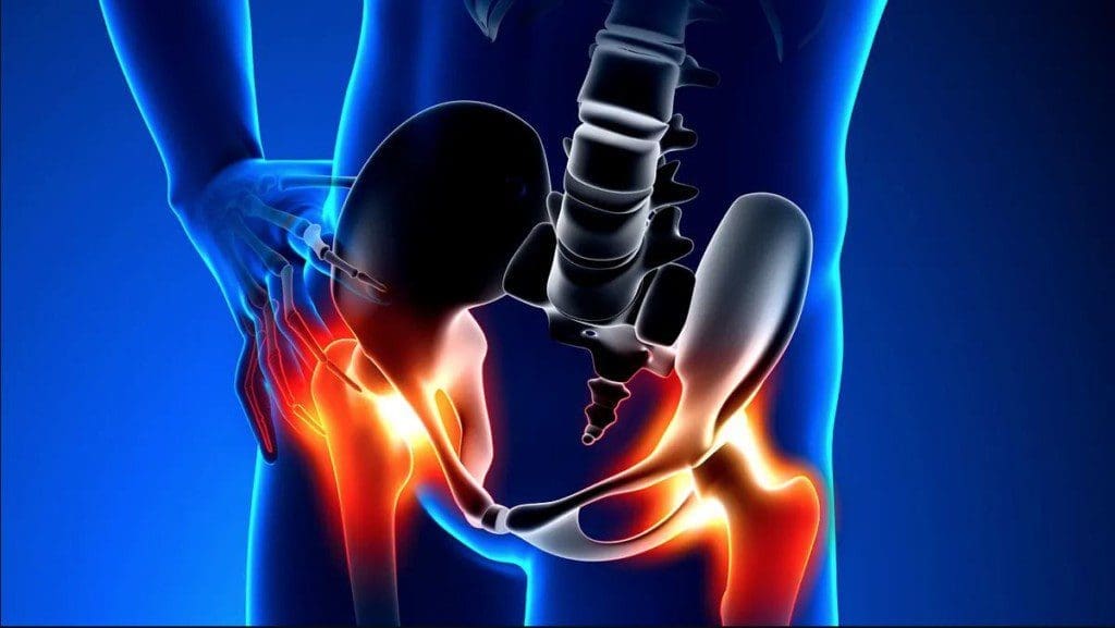
Why the Hip Can Be Injured in a Crash
The hip is a ball-and-socket joint. The “ball” is the femoral head at the top of the thighbone. The “socket” is the acetabulum, which is part of the pelvis. Strong ligaments, muscles, cartilage, and the labrum help keep the joint stable.
Because the hip is built to be strong, it usually takes a major force to dislocate or fracture it. This is why high-energy crashes are a major concern. Research on frontal motor vehicle crashes has found that hip fractures and dislocations are important injury patterns for both doctors and vehicle safety experts (Rupp et al., 2004).
The exact injury often depends on the body’s position during impact. For example:
- A bent knee striking the dashboard can drive force backward through the thighbone.
- A side impact can compress the pelvis and hip socket.
- A locked seatbelt can protect life, but still create force across the pelvis and soft tissues.
- Bracing the foot against the floor can strain the hip flexors, hamstrings, and ligaments.
- A twisting motion can irritate or tear the labrum.
Hip Dislocation: A True Emergency
A traumatic hip dislocation happens when the femoral head is forced out of the socket. This is often linked to dashboard impact, especially when the knee is bent, and the thighbone is driven backward (Masiewicz & Johnson, 2023). The American Academy of Orthopaedic Surgeons explains that motor vehicle collisions are the most common cause of traumatic hip dislocations, and the knee hitting the dashboard is a common mechanism (American Academy of Orthopaedic Surgeons, n.d.-b).
Symptoms may include:
- Severe hip or groin pain
- Inability to stand or walk
- A leg that looks rotated or shortened
- Numbness, tingling, or weakness
- Pain that does not improve with rest
A hip dislocation needs urgent medical care. The joint must be placed back into position by trained medical professionals. Waiting too long can increase the risk of complications, including damage to the blood supply, nerve injury, cartilage injury, and later arthritis.
Acetabular Fractures: When the Hip Socket Breaks
An acetabular fracture is a break in the socket part of the hip joint. This can happen when force drives the femoral head into the acetabulum. AAOS notes that this force can be transmitted through the knee, such as when the knee hits the dashboard in a head-on collision (American Academy of Orthopedic Surgeons, n.d.-a).
This injury is serious because the socket must stay smooth and stable for the hip to move well. If the joint surface is disrupted, the patient may develop long-term pain, stiffness, instability, or arthritis.
Common signs include:
- Deep hip or groin pain
- Pain with movement
- Inability to bear weight
- Swelling or bruising
- Pain after a high-force crash, even if the person can still move
Some acetabular fractures may be treated without surgery, but many require orthopedic evaluation. Surgery may be needed when the joint surface is displaced or unstable.
Femoral Head Fractures: Damage to the Ball of the Joint
A femoral head fracture means the ball at the top of the thighbone is cracked or broken. These injuries often happen with a hip dislocation. When the femoral head hits the socket with force, pieces of bone or cartilage may break.
Femoral head injuries are important because this part of the bone carries weight and helps the hip glide. A rough or damaged joint surface can create pain, catching, stiffness, or early arthritis.
These injuries usually require imaging such as X-rays and CT scans. A patient may also need an MRI if soft tissue damage is suspected.
Hip Labral Tears After a Crash
The labrum is a ring of cartilage around the hip socket. It helps deepen the socket and improves joint stability. Mayo Clinic explains that trauma, including injury or dislocation from a car accident, can cause a hip labral tear (Mayo Clinic, 2024).
A labral tear does not always feel like a simple bruise. It may feel like deep groin pain, catching, clicking, locking, or pinching in the hip.
Common symptoms include:
- Groin or front hip pain
- Clicking or catching
- Stiffness
- Pain with sitting, squatting, or turning
- Pain that returns with activity
- Feeling like the hip is unstable
Labral tears can be difficult to diagnose because symptoms may resemble a strain, a low back problem, a sports hernia, or a pelvic injury. Clinical exam, orthopedic testing, imaging, and sometimes diagnostic injections may be used to identify the source of the pain.
Muscle Strains, Sprains, and Soft Tissue Damage
Not every hip injury after a crash is a fracture or dislocation. Many patients develop soft tissue injuries. These may still be painful and disabling.
Soft tissue injuries may involve:
- Hip flexor strains
- Hamstring injuries
- Gluteal muscle strain
- Ligament sprains
- Trochanteric bursitis
- Tendon irritation
- Deep bruising from seatbelt trauma
- Pelvic and sacroiliac joint irritation
These injuries can affect walking, bending, sleep, work, and exercise. They may also cause the body to compensate, leading to low back pain, knee pain, or an altered gait.
Why Hip Pain May Show Up Late
After a crash, adrenaline can hide pain. Some people feel “okay” at the scene but develop pain hours or days later. Swelling, inflammation, muscle guarding, and joint irritation can build over time.
Delayed hip pain should not be ignored, especially when it follows a high-force crash. Pain that worsens, limits walking, causes numbness, or feels deep in the groin should be evaluated.
Red flags include:
- Inability to bear weight
- Severe pain
- Visible deformity
- Numbness or weakness
- Fever after injury
- Increasing swelling
- Hip pain with abdominal or pelvic pain
- Pain after a dashboard impact
The Role of Imaging and Medical Oversight
A serious hip injury cannot be safely diagnosed by symptoms alone. X-rays may help identify fractures or dislocations. CT scans can show complex bone injuries. MRI may help evaluate the labrum, cartilage, muscles, tendons, and bone marrow swelling.
This is where medical direction matters. In an integrative injury care setting, Dr. Maria Guadalupe Cardenas, MD, provides internal medicine oversight as Medical Director and Collaborative Physician. This helps support safe screening, referral decisions, medical documentation, and coordination when advanced imaging or orthopedic evaluation is needed.
Dr. Jimenez’s clinical approach emphasizes that personal injury patients often need more than pain relief alone. They may need structural evaluation, neurological screening, rehabilitation planning, metabolic support, and clear documentation of injury patterns.
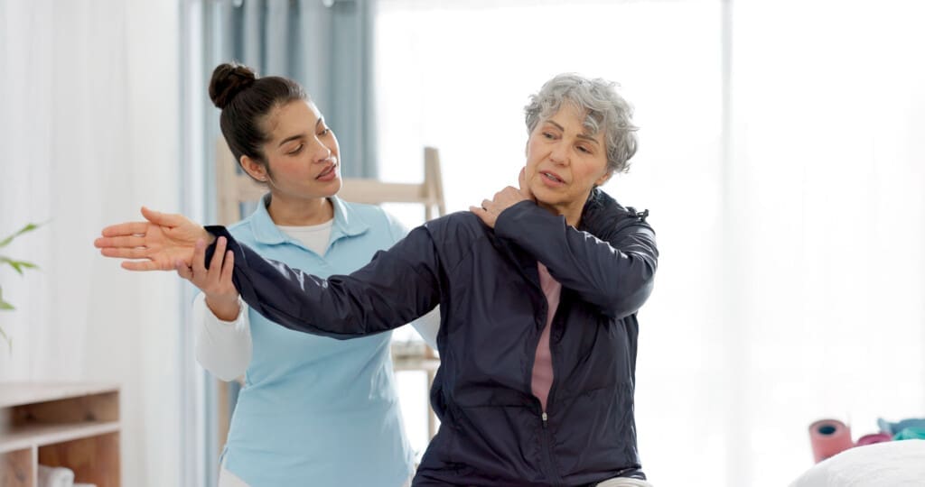
Chiropractic Care for Hip and Pelvic Mechanics
Chiropractic care may help patients with joint restriction, pelvic imbalance, low back compensation, and movement problems after an accident. When the hip is injured, the pelvis, lumbar spine, sacroiliac joints, knees, and ankles may all change the way they move.
Dr. Alex Jimenez, DC, evaluates how the body moves as a connected system. In personal injury care, this can include:
- Posture and gait assessment
- Lumbar spine and pelvic evaluation
- Hip range of motion testing
- Muscle strength testing
- Neurological screening
- Functional movement review
- Referral for imaging when needed
- Rehabilitation planning
Chiropractic care is not a replacement for emergency care in cases of fracture or dislocation. However, after serious injuries are ruled out or medically managed, chiropractic and rehabilitation care may help restore mobility, reduce compensation, and improve function.
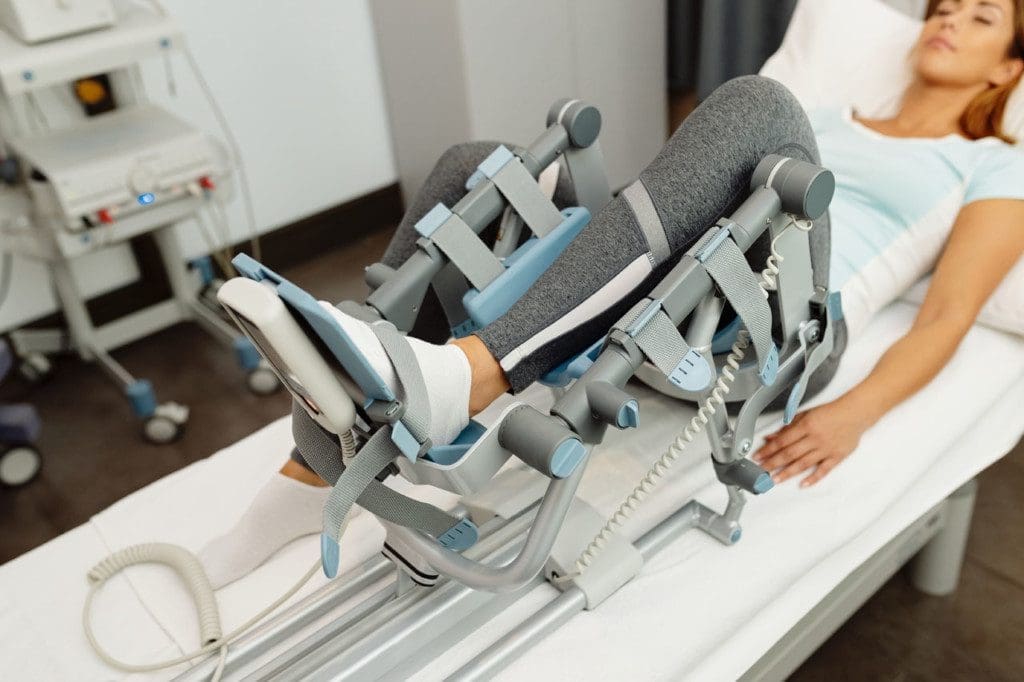
Rehabilitation: Rebuilding Motion and Strength
Rehabilitation is a key part of hip recovery. Pain can cause the body to move poorly. Over time, this may lead to stiffness, weakness, and fear of movement.
A hip rehabilitation plan may include:
- Gentle mobility work
- Hip and core strengthening
- Glute activation
- Balance training
- Gait retraining
- Stretching tight muscles
- Stability work for the pelvis and low back
- Gradual return to work, walking, and exercise
The goal is not just to reduce pain. The goal is to help the hip move better, carry weight safely, and work with the rest of the body.
Functional Medicine Support After Injury
Functional medicine looks at the whole person. After a crash, recovery may be affected by sleep, inflammation, nutrition, blood sugar balance, stress, hydration, and previous health problems.
Dr. Jimenez’s integrative model includes functional medicine principles to support healing. This may include reviewing:
- Nutrition quality
- Protein intake
- Vitamin D status
- Inflammation markers
- Blood sugar control
- Sleep recovery
- Stress load
- Hydration
- Medication history
- Prior injuries
This whole-person view can be helpful because tissue repair requires more than rest. The body needs the right internal environment to heal.
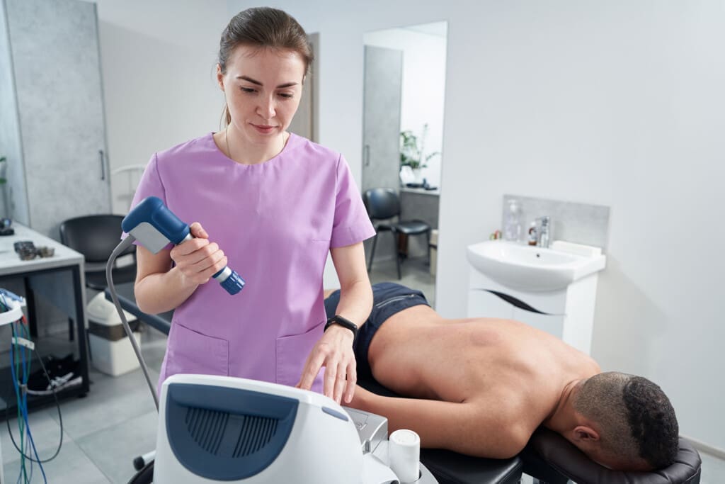
Regenerative Therapies: PRP, PFP, and MFAT
Some hip injuries involve soft tissue irritation, tendon injury, cartilage stress, labral-related pain, or early degenerative changes. In selected cases, regenerative therapies may be considered as part of a broader care plan.
Common regenerative options include:
- PRP: Platelet-rich plasma, made from the patient’s own blood
- PFP: Platelet-focused plasma or platelet-rich fibrin-style preparations, depending on the protocol used
- MFAT: Micro-fragmented adipose tissue, processed from the patient’s own fat tissue
These treatments are designed to support the body’s natural repair signaling. Research on PRP for hip osteoarthritis suggests it may improve pain and function in some patients, especially in mild to moderate cases, though results vary and protocols differ (Berney et al., 2021; Singh et al., 2019). A study on MFAT with PRP reported positive findings for hip osteoarthritis, but further research is needed to identify the optimal candidates and long-term outcomes (Heidari et al., 2022).
It is important to say this clearly: regenerative therapy is not a magic cure, and it does not replace emergency care, fracture treatment, surgery when required, or proper rehabilitation. It is best used as part of a medically guided plan.
When Surgery May Be Needed
Some hip injuries cannot be treated with conservative care alone. Surgery may be needed for displaced acetabular fractures, unstable joints, certain femoral head fractures, loose bone fragments, or severe labral injuries that do not respond to non-surgical care.
An integrative clinic should recognize when a referral is needed. Good care means knowing when chiropractic, rehabilitation, injections, or functional medicine are appropriate and when orthopedic or emergency care is required.
A Multidisciplinary Path for El Paso Injury Patients
At Injury Medical Clinic PA in El Paso, the team approach combines:
- Chiropractic care with Dr. Alex Jimenez
- Medical oversight with Dr. Maria Guadalupe Cardenas, MD
- Personal injury care
- Functional medicine
- Rehabilitation
- Diagnostic coordination
- Regenerative therapy consideration
- Documentation for injury cases when appropriate
This model helps patients move from pain and confusion toward a structured recovery plan. After an accident, the most important first step is a careful evaluation. The second step is matching the treatment plan to the actual injury.
Conclusion: Hip Pain After a Crash Deserves Careful Attention
Hip injuries after motor vehicle accidents can be serious. A dashboard impact, side collision, seatbelt force, or sudden twisting motion can damage the joint, socket, labrum, muscles, tendons, and ligaments.
Some injuries need emergency care. Others need imaging, rehabilitation, chiropractic support, medical oversight, or regenerative options. The best plan depends on the exact diagnosis.
For patients in El Paso, an integrative injury care model can help connect the pieces. With Dr. Alexander Jimenez, DC, APRN, FNP-BC, and Dr. Maria Guadalupe Cardenas, MD, working in a multidisciplinary setting, patients can receive structured evaluation, medically guided care planning, rehabilitation support, and whole-person recovery strategies.
References
American Academy of Orthopaedic Surgeons. (n.d.-a). Acetabular fractures. OrthoInfo.
American Academy of Orthopaedic Surgeons. (n.d.-b). Hip dislocation. OrthoInfo.
Berney, M., McCarroll, P., Glynn, L., Lenehan, B., & Coady, C. (2021). Platelet-rich plasma injections for hip osteoarthritis. Journal of Hip Preservation Surgery.
Heidari, N., et al. (2022). Comparison of the effect of MFAT and MFAT + PRP on osteoarthritis of the hip. Journal of Orthopaedic Surgery and Research.
Masiewicz, S., & Johnson, J. (2023). Posterior hip dislocation. In StatPearls. StatPearls Publishing.
Mayo Clinic. (2024). Hip labral tear: Symptoms and causes.
Rupp, J. D., Flannagan, C. A. C., Kuppa, S. M., & Schneider, L. W. (2004). Injuries to the hip joint in frontal motor-vehicle crashes. Accident Analysis & Prevention, 36(5), 903–911.
Singh, J. R., Haffey, P., Valimahomed, A., & Simunovic, N. (2019). The effectiveness of autologous platelet-rich plasma for osteoarthritis of the hip. Orthopaedic Journal of Sports Medicine.
Dr. Alex Jimenez. (n.d.). El Paso, TX doctor of chiropractic.
Dr. Alexander Jimenez. (n.d.). LinkedIn profile.