Uncover the connection between chiropractic care and bone health in the musculoskeletal system for improved physical well-being.
Chiropractic Care: Your Path to Less Pain and Stronger Bones
Imagine waking up one morning, ready to conquer the day, only to be greeted by a nagging pain in your lower back that feels like a grumpy troll decided to camp out there overnight. Or maybe your neck feels stiffer than a board after a long day hunched over your laptop, making you wonder if you’re secretly turning into a robot. These aches and pains aren’t just annoying—they’re your body’s way of waving a red flag, saying, “Hey, something’s not right with my musculoskeletal system!” Now, add in the worry about keeping your bones strong as you age, especially if you’ve heard one too many stories about osteoporosis turning bones into fragile twigs. Sound familiar? Don’t worry, you’re not alone, and there’s a hero in this story: chiropractic care.
Chiropractic care isn’t just about cracking backs and sending you on your way. It’s a science-backed, non-invasive approach to reducing musculoskeletal pain and supporting bone health, helping you move better, feel better, and maybe even dance like nobody’s watching (or at least without wincing). In this blog post, we’ll dive deep into why your musculoskeletal system is the unsung hero of your daily life, how bone health keeps you standing tall, and why chiropractic care—especially from experts like Dr. Alexander Jimenez, DC, APRN, FNP-BC in El Paso, Texas—can be your ticket to a pain-free, stronger you. We’ll sprinkle in some humor to keep things light, back it all up with clinical insights, and highlight Dr. Jimenez’s unique role in personal injury cases. Ready? Let’s get cracking—er, adjusting!
The Musculoskeletal System: Your Body’s Framework
Think of your musculoskeletal system as the scaffolding of a building: it holds everything together, keeps you upright, and lets you move without collapsing into a puddle of jelly. This system includes your bones, muscles, tendons, ligaments, and joints, all working in harmony like a well-rehearsed band. When one part goes out of tune—like a strained muscle or a misaligned spine—the whole performance suffers, leading to pain, stiffness, or even injury.
Why the Musculoskeletal System Matters
Your musculoskeletal system is the MVP of daily life. It lets you:
- Walk, run, and jump: Whether you’re chasing your dog or sprinting to catch the bus, your bones and muscles make it happen.
- Lift and carry: From groceries to toddlers, your joints and ligaments keep you strong.
- Sit and stand: Your spine and core muscles keep you balanced, even when you’re binge-watching your favorite show.
- Protect vital organs: Your ribcage and skull are like bodyguards for your heart and brain.
But when something goes wrong—like a herniated disc, a sprained ankle, or just plain old bad posture—musculoskeletal pain can turn your day into a groan-fest. According to research, spine pain alone affects over 11% of the world’s population, making it a leading cause of disability (Beltran-Alacreu et al., 2021). Low back pain, neck pain, and joint issues are common culprits, often triggered by poor ergonomics, injuries, or even stress that tightens your muscles like a grumpy cat.
Environmental Factors That Mess with Your Musculoskeletal Mojo
Your environment can be a sneaky saboteur of your musculoskeletal health. Here’s how:
- Sedentary Lifestyle: Sitting all day at a desk or on the couch can weaken muscles and stiffen joints, making your spine crankier than a toddler without a nap.
- Poor Posture: Slouching over your phone or laptop can misalign your spine, leading to pain that feels like a permanent knot in your back.
- Stress: Tense muscles from stress can pull your joints out of whack, creating a vicious cycle of pain and more stress.
- Injuries: Car accidents, falls, or sports mishaps can damage muscles, ligaments, or bones, leaving you sore and sidelined.
- Nutrition: A diet low in calcium or vitamin D can weaken bones, making them more prone to fractures or conditions like osteoporosis.
These factors don’t just cause pain—they can also weaken your bones over time, increasing the risk of osteoporosis, where bones become so brittle they might snap like a dry spaghetti noodle. That’s where bone health comes in, and trust us, it’s more important than you might think.
References
Beltran-Alacreu, H., López-de-Uralde-Villanueva, I., Fernández-Carnero, J., & La Touche, R. (2021). Clinical effectiveness and efficacy of chiropractic spinal manipulation for spine pain. Frontiers in Pain Research, 2, 765921. https://doi.org/10.3389/fpain.2021.765921
Bone Health: The Foundation of a Strong You
If your musculoskeletal system is the scaffolding, your bones are the steel beams that keep it sturdy. Bone health isn’t just about avoiding fractures—it’s about maintaining a strong, resilient skeleton that supports your body through every twist, turn, and TikTok dance challenge.
Why Bone Health Is a Big Deal
Bones are living tissues that constantly remodel themselves, breaking down old bone and building new ones like a never-ending home renovation project. Healthy bones:
- Support your body: They keep you upright and moving, no matter how many Zoom meetings you endure.
- Store minerals: Bones are like a bank for calcium and phosphorus, releasing them when your body needs a boost.
- Protect organs: Your skull, spine, and ribs shield your brain, spinal cord, and heart from life’s bumps and bruises.
- Produce blood cells: Bone marrow is a factory for red and white blood cells, keeping your body oxygenated and infection-free.
But when bone health declines, trouble brews. Osteoporosis, a condition where bones lose density and become fragile, affects millions worldwide, especially older adults and postmenopausal women. A simple fall can lead to a hip fracture, which can be a game-changer, limiting mobility and independence (Sözen et al., 2017). Even younger folks aren’t immune—poor diet, lack of exercise, or chronic stress can weaken bones over time, setting the stage for future problems.
Environmental Factors and Bone Health
Your bones aren’t fans of a lazy or stressful lifestyle. Here’s what can weaken them:
- Low Calcium and Vitamin D: Without these nutrients, your bones can’t rebuild effectively, like trying to bake a cake without flour.
- Inactivity: Weight-bearing exercises like walking or lifting weights stimulate bone growth. Sitting all day? Your bones might start slacking off too.
- Smoking and Alcohol: These habits can interfere with bone remodeling, making your skeleton less sturdy.
- Medications: Long-term use of corticosteroids or other drugs can thin bones, increasing fracture risk.
- Environmental Toxins: Exposure to heavy metals or pollutants can disrupt bone metabolism, especially over time.
The good news? You can fight back with a bone-friendly lifestyle and, yes, chiropractic care. Chiropractors like Dr. Alexander Jimenez don’t just focus on pain relief—they help optimize your musculoskeletal system to support bone health, keeping you strong and mobile.
References
Sözen, T., Özışık, L., & Başaran, N. Ç. (2017). Osteoporosis prevention, screening, and treatment: A review. Journal of Osteoporosis, 2017, 1659707. https://doi.org/10.1155/2017/1659707
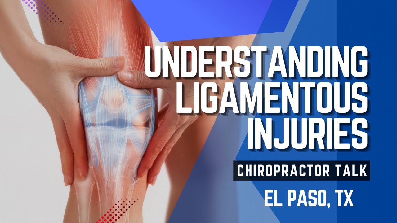
Understanding Ligamentous Injuries- Video
Chiropractic Care: The Spine’s Best Friend
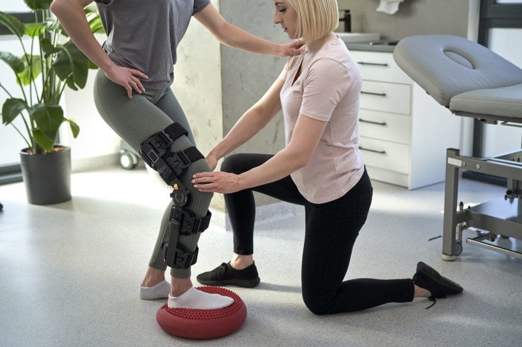
Now, let’s talk about the star of the show: chiropractic care. If your spine were a diva, chiropractors would be its personal trainers, keeping it aligned, flexible, and ready to shine. Chiropractic care focuses on the spine and nervous system, using hands-on techniques to correct misalignments (called subluxations) that can cause pain, stiffness, and even health issues beyond your back.
The Clinical Rationale for Chiropractic Care
Chiropractic care isn’t just a feel-good massage—it’s grounded in biomechanics and neurology. Here’s why it works for musculoskeletal pain:
- Relieves Nerve Pressure: Subluxations can pinch nerves, causing pain that radiates to your arms, legs, or even head. Spinal adjustments realign vertebrae, taking the pressure off nerves and reducing pain (Beltran-Alacreu et al., 2021).
- Reduces Inflammation: Misaligned joints can trigger inflammation, making your muscles and tissues grumpy. Adjustments restore proper movement, calming the inflammation storm.
- Improves Mobility: Stiff joints and tight muscles limit your range of motion. Chiropractic techniques like manipulation and mobilization loosen things up, helping you move like you’re starring in a dance movie (Rubinstein et al., 2019).
- Supports Muscle Balance: Weak or tight muscles can pull your spine out of alignment. Chiropractors often prescribe exercises to strengthen your core and stabilize your spine, like giving your body a tune-up.
For low back pain, chiropractic care is especially effective. A systematic review found that spinal manipulative therapy (SMT) significantly reduces pain and disability in chronic low back pain, often outperforming standard medical care (Rubinstein et al., 2019). Another study showed SMT is just as effective for acute low back pain, with benefits lasting up to six weeks (Paige et al., 2017). Even the Canadian Chiropractic Guideline Initiative recommends SMT as a first-line treatment for low back pain, alongside exercise and patient education (Bussières et al., 2018).
Chiropractic and Bone Health
Chiropractic care isn’t just about pain—it can also support bone health. Here’s how:
- Stimulates Bone Remodeling: Spinal adjustments improve joint function, which can stimulate weight-bearing activity in bones, encouraging them to stay strong (Health Coach Clinic, 2023).
- Enhances Circulation: Better alignment means better blood flow, delivering nutrients like calcium and vitamin D to your bones.
- Prevents Falls: By improving balance and coordination, chiropractic care reduces the risk of falls, a major cause of fractures in older adults (Hawk et al., 2020).
- Complements Bone Healing: Some chiropractors use low-force techniques or recommend devices like electrical stimulators, which research shows can speed up bone healing after fractures (Griffin et al., 2016).
Dr. Alexander Jimenez, a chiropractor and nurse practitioner in El Paso, takes this a step further. His dual expertise allows him to combine chiropractic adjustments with functional medicine, addressing not just pain but also nutritional and metabolic factors that impact bone health. For example, he might recommend a diet rich in calcium and vitamin D or supplements to support bone density, alongside spinal adjustments to keep your musculoskeletal system in top shape (Jimenez, 2025).
References
Beltran-Alacreu, H., López-de-Uralde-Villanueva, I., Fernández-Carnero, J., & La Touche, R. (2021). Clinical effectiveness and efficacy of chiropractic spinal manipulation for spine pain. Frontiers in Pain Research, 2, 765921. https://doi.org/10.3389/fpain.2021.765921
Bussières, A. E., Stewart, G., Al-Zoubi, F., Decina, P., Descarreaux, M., Hayden, J., & Stuber, K. (2018). Spinal manipulative therapy and other conservative treatments for low back pain: A guideline from the Canadian Chiropractic Guideline Initiative. Journal of Manipulative and Physiological Therapeutics, 41(4), 265–293. https://doi.org/10.1016/j.jmpt.2017.12.004
Griffin, X. L., Warner, F., & Costa, M. L. (2016). Efficacy of electrical stimulators for bone healing: A meta-analysis of randomized sham-controlled trials. Scientific Reports, 6, 31724. https://doi.org/10.1038/srep31724
Hawk, C., Schneider, M. J., Haas, M., Katz, P., Dougherty, P., Gleberzon, B., & Killinger, L. Z. (2020). Effects of chiropractic care on strength, balance, and endurance in active-duty U.S. military personnel with low back pain: A randomized controlled trial. Journal of Alternative and Complementary Medicine, 26(7), 592–601. https://doi.org/10.1089/acm.2020.0107
Health Coach Clinic. (2023). Health & wellness: Bone health. https://healthcoach.clinic/health-wellness-bone-health/
Jimenez, A. (2025). Dr. Alexander Jimenez, DC, APRN, FNP-BC, IFMCP, CFMP, ATN. LinkedIn. https://www.linkedin.com/in/dralexjimenez/
Paige, N. M., Miake-Lye, I. M., Booth, M. S., Beroes, J. M., Mardian, A. S., Dougherty, P., & Shekelle, P. G. (2017). Spinal manipulative therapy for acute low-back pain. Cochrane Database of Systematic Reviews, 2017(2), CD008880. https://doi.org/10.1002/14651858.CD008880.pub3
Rubinstein, S. M., de Zoete, A., van Middelkoop, M., Assendelft, W. J. J., de Boer, M. R., & van Tulder, M. W. (2019). Manipulation and mobilization for treating chronic low back pain: A systematic review and meta-analysis. The Spine Journal, 19(5), 866–879. https://doi.org/10.1016/j.spinee.2018.12.013
Dr. Alexander Jimenez: El Paso’s Chiropractic Superhero
If chiropractic care were a comic book, Dr. Alexander Jimenez would be the caped crusader swooping in to save the day. Based in El Paso, Texas, Dr. Jimenez isn’t just a chiropractor—he’s also a board-certified nurse practitioner with over 25 years of experience. His dual licensure makes him a rare gem, blending the hands-on healing of chiropractic with the diagnostic precision of functional medicine.
Why Dr. Jimenez Stands Out
Dr. Jimenez’s practice at Injury Medical & Chiropractic Clinic is like a one-stop shop for musculoskeletal health. Here’s what makes him special:
- Advanced Imaging and Diagnostics: He uses MRI, CT scans, and X-rays to pinpoint injuries like herniated discs or nerve compression, ensuring treatments are as precise as a laser-guided missile (Jimenez, 2025).
- Dual-Scope Procedures: As both a chiropractor and nurse practitioner, he can address biomechanical issues (like spinal misalignments) and systemic ones (like hormonal imbalances or nutritional deficiencies), giving you a 360-degree approach to healing.
- Holistic Care: Dr. Jimenez doesn’t just adjust your spine and call it a day. He incorporates nutritional counseling, physical therapy, and lifestyle advice to keep you pain-free and thriving.
- Personal Injury Expertise: In El Paso, where car accidents and workplace injuries are common, Dr. Jimenez is a trusted ally for personal injury victims. He uses his skills to document injuries thoroughly, supporting legal claims while helping patients recover.
Personal Injury Cases in El Paso
El Paso’s busy roads and active workforce mean personal injuries—like whiplash from car accidents or back strains from heavy lifting—are all too common. These injuries can leave you feeling like you’ve been hit by a runaway train, with pain, stiffness, and even emotional stress piling up. Chiropractic care is a lifeline for these cases, offering non-invasive relief that gets you back on your feet without surgery or heavy meds.
Dr. Jimenez shines in this arena. His detailed reports, backed by advanced imaging, help lawyers and insurance companies understand the extent of your injuries, ensuring you get fair compensation. Meanwhile, his treatments—like spinal adjustments, soft tissue mobilization, and rehab exercises—target the root cause of your pain, not just the symptoms. A 2025 article highlights his ability to bridge medical and legal needs, making him a go-to practitioner for El Pasoans recovering from motor vehicle accidents or workplace injuries (Sciatica Clinic, 2025).
A Day in the Life of a Jimenez Patient
Picture this: You’ve been in a fender-bender, and your neck feels like it’s auditioning for a role in a horror movie. You visit Dr. Jimenez, who starts with a thorough exam, including an MRI to check for hidden damage. He explains your injury in plain English (no medical jargon here!), then uses gentle spinal adjustments to ease the pressure on your nerves. He might throw in some massage therapy to loosen tight muscles and prescribe exercises to strengthen your core. Oh, and he’ll probably remind you to eat more leafy greens for bone health—because he’s that thorough. By the end of your visit, you’re not just feeling better; you’re armed with a plan to stay that way.
References
Jimenez, A. (2025). Dr. Alexander Jimenez DC, APRN, FNP-BC, IFMCP, CFMP, ATN. LinkedIn. https://www.linkedin.com/in/dralexjimenez/
Sciatica Clinic. (2025). Chiropractic care techniques for five musculoskeletal issues. https://sciatica.clinic/
How Chiropractic Care Tackles Overlapping Risk Profiles
Musculoskeletal pain and poor bone health often share the same troublemakers: sedentary habits, poor nutrition, stress, and injuries. These overlapping risk profiles can create a perfect storm, where a weak spine leads to pain, and weak bones increase injury risk, which then worsens pain—it’s like a bad sitcom that keeps getting renewed. Chiropractic care breaks this cycle by addressing both issues at once.
Common Risk Profiles and Chiropractic Solutions
Here’s how chiropractic care tackles the culprits behind musculoskeletal pain and bone health woes:
- Sedentary Lifestyle: Sitting too much weakens muscles and bones. Chiropractors prescribe weight-bearing exercises and adjustments to improve posture and joint function, keeping your spine and skeleton strong (Hawk et al., 2020).
- Poor Nutrition: Low levels of calcium or vitamin D can harm bones and muscles. Dr. Jimenez often pairs adjustments with nutritional counseling, recommending foods like dairy, fish, or fortified cereals to boost bone health (Health Coach Clinic, 2023).
- Stress: Tense muscles pull your spine out of alignment, causing pain. Chiropractic adjustments and relaxation techniques (like acupuncture) reduce stress and muscle tension, calming your nervous system (Jimenez, 2025).
- Injuries: Trauma from accidents can misalign joints and weaken bones. Chiropractors use spinal manipulation and rehab to restore alignment and mobility, while devices like electrical stimulators may speed bone healing (Griffin et al., 2016).
Evidence-Backed Benefits
Research backs chiropractic care’s ability to tackle these risks. A randomized controlled trial found that chiropractic care improved strength, balance, and endurance in active-duty military personnel with low back pain, suggesting it can counteract the effects of inactivity (Hawk et al., 2020). Another study showed that chiropractic care is cost-effective for low back pain, reducing healthcare costs compared to standard medical care (Goertz et al., 2016). For bone health, chiropractic’s focus on alignment and movement complements osteoporosis prevention strategies, like exercise and nutrition, outlined in clinical reviews (Sözen et al., 2017).
Dr. Jimenez’s integrative approach takes this to the next level. By combining chiropractic adjustments with functional medicine, he addresses the whole person—not just the pain. For example, a patient with chronic back pain might get spinal adjustments, a diet plan to boost bone density, and stress management tips to keep muscles relaxed. It’s like getting a full-body makeover, minus the reality TV cameras.
References
Goertz, C. M., Long, C. R., Vining, R. D., Pohlman, K. A., Walter, J., & Coulter, I. (2016). Effectiveness and economic evaluation of chiropractic care for the treatment of low back pain: A systematic review of pragmatic studies. PLoS ONE, 11(8), e0160037. https://doi.org/10.1371/journal.pone.0160037
Griffin, X. L., Warner, F., & Costa, M. L. (2016). Efficacy of electrical stimulators for bone healing: A meta-analysis of randomized sham-controlled trials. Scientific Reports, 6, 31724. https://doi.org/10.1038/srep31724
Hawk, C., Schneider, M. J., Haas, M., Katz, P., Dougherty, P., Gleberzon, B., & Killinger, L. Z. (2020). Effects of chiropractic care on strength, balance, and endurance in active-duty U.S. military personnel with low back pain: A randomized controlled trial. Journal of Alternative and Complementary Medicine, 26(7), 592–601. https://doi.org/10.1089/acm.2020.0107
Health Coach Clinic. (2023). Health & wellness: Bone health. https://healthcoach.clinic/health-wellness-bone-health/
Jimenez, A. (2025). Dr. Alexander Jimenez DC, APRN, FNP-BC, IFMCP, CFMP, ATN. LinkedIn. https://www.linkedin.com/in/dralexjimenez/
Sözen, T., Özışık, L., & Başaran, N. Ç. (2017). Osteoporosis prevention, screening, and treatment: A review. Journal of Osteoporosis, 2017, 1659707. https://doi.org/10.1155/2017/1659707
The Chiropractic Identity: Spine Care and Beyond
Chiropractic isn’t just a treatment—it’s a philosophy centered on the spine as the key to overall health. The spine houses your nervous system, which controls everything from your heartbeat to your ability to touch your toes. When your spine’s out of whack, it’s like a bad Wi-Fi signal: everything slows down, and you’re left frustrated.
Spine Care as the Core of Chiropractic
A 2016 study defines spine care as the heart of chiropractic identity, emphasizing non-invasive techniques to restore spinal alignment and nervous system function (Gliedt et al., 2016). Chiropractors believe that a healthy spine supports a healthy body, reducing pain, improving mobility, and even boosting your mood (because who’s happy when their back hurts?). Techniques like spinal manipulation, mobilization, and soft tissue therapy are the tools of the trade, each tailored to your unique needs.
Dr. Jimenez embodies this philosophy, using spine care to address not just pain but also systemic issues like fatigue or digestive problems. His functional medicine approach digs deeper, looking at how spinal health interacts with nutrition, stress, and lifestyle to keep you in tip-top shape.
Beyond Pain Relief
Chiropractic care goes beyond fixing aches and pains. It’s about prevention and wellness, helping you avoid injuries and maintain bone health. Regular adjustments can:
- Improve Posture: Say goodbye to that hunchback vibe from too much screen time.
- Boost Athletic Performance: Better alignment means better movement, whether you’re a weekend warrior or a couch potato dreaming of 5Ks.
- Enhance Bone Health: By promoting movement and balance, chiropractic care supports strong bones and reduces fracture risk.
In El Paso, where personal injuries are common, this preventive approach is a game-changer. Dr. Jimenez’s patients often report not just pain relief but also more energy, better sleep, and a renewed zest for life—kind of like finding the perfect coffee blend, but for your body.
References
Gliedt, J. A., Hawk, C., Anderson, M., Ahmad, K., Bunn, D., Cambron, J., & Schneider, M. J. (2016). Spine care as a framework for the chiropractic identity. Journal of Chiropractic Humanities, 23(1), 14–21. https://doi.org/10.1016/j.echu.2016.09.004
A Closer Look at Chiropractic Techniques
Chiropractors have a toolbox full of techniques to restore your body to harmony. Here’s a peek at some of the most common ones and how they help:
- Spinal Manipulation: This is the classic “crack” you hear during an adjustment. It realigns vertebrae, reduces nerve pressure, and feels like a mini-vacation for your spine (Bussières et al., 2018).
- Mobilization: Gentle movements stretch and loosen joints, perfect for those who prefer a softer touch.
- Soft Tissue Therapy: Massage or myofascial release targets tight muscles, easing tension and improving blood flow.
- Rehabilitation Exercises: Core-strengthening moves and stretches keep your spine stable and your bones strong.
- Electrical Stimulation: Low-level currents can reduce pain and speed bone healing, like giving your body a tiny pep talk (Griffin et al., 2016).
Dr. Jimenez mixes and matches these techniques based on your needs. Got a herniated disc from a car accident? He might use spinal decompression to ease the pressure, followed by exercises to rebuild strength. Worried about osteoporosis? He’ll focus on adjustments and nutrition to keep your bones rock-solid.
References
Bussières, A. E., Stewart, G., Al-Zoubi, F., Decina, P., Descarreaux, M., Hayden, J., & Stuber, K. (2018). Spinal manipulative therapy and other conservative treatments for low back pain: A guideline from the Canadian Chiropractic Guideline Initiative. Journal of Manipulative and Physiological Therapeutics, 41(4), 265–293. https://doi.org/10.1016/j.jmpt.2017.12.004
Griffin, X. L., Warner, F., & Costa, M. L. (2016). Efficacy of electrical stimulators for bone healing: A meta-analysis of randomized sham-controlled trials. Scientific Reports, 6, 31724. https://doi.org/10.1038/srep31724
Conclusion: A Serious Note on Chiropractic Care
Chiropractic care is more than a quick fix for back pain—it’s a powerful, evidence-based approach to reducing musculoskeletal pain, supporting bone health, and improving your overall quality of life. By addressing spinal alignment, nerve function, and lifestyle factors, chiropractors like Dr. Alexander Jimenez help you break free from the cycle of pain and weakness, empowering you to live stronger and healthier. In El Paso, where personal injuries can disrupt lives, Dr. Jimenez’s expertise as a chiropractor and nurse practitioner makes him a beacon of hope, guiding patients through recovery with precision and compassion.
Disclaimer: This blog post is for informational purposes only and should not be taken as medical advice. Always consult a qualified healthcare professional, such as a licensed chiropractor or physician, before starting any treatment. Individual results may vary, and chiropractic care may not be suitable for all conditions. If you’re experiencing pain or have concerns about your bone health, contact a provider like Dr. Jimenez at Injury Medical & Chiropractic Clinic (915-850-0900) to discuss your options. Your health is worth it—take the first step today.
References
Beltran-Alacreu, H., López-de-Uralde-Villanueva, I., Fernández-Carnero, J., & La Touche, R. (2021). Clinical effectiveness and efficacy of chiropractic spinal manipulation for spine pain. Frontiers in Pain Research, 2, 765921. https://doi.org/10.3389/fpain.2021.765921
Bussières, A. E., Stewart, G., Al-Zoubi, F., Decina, P., Descarreaux, M., Hayden, J., & Stuber, K. (2018). Spinal manipulative therapy and other conservative treatments for low back pain: A guideline from the Canadian Chiropractic Guideline Initiative. Journal of Manipulative and Physiological Therapeutics, 41(4), 265–293. https://doi.org/10.1016/j.jmpt.2017.12.004
Gliedt, J. A., Hawk, C., Anderson, M., Ahmad, K., Bunn, D., Cambron, J., & Schneider, M. J. (2016). Spine care as a framework for the chiropractic identity. Journal of Chiropractic Humanities, 23(1), 14–21. https://doi.org/10.1016/j.echu.2016.09.004
Goertz, C. M., Long, C. R., Vining, R. D., Pohlman, K. A., Walter, J., & Coulter, I. (2016). Effectiveness and economic evaluation of chiropractic care for the treatment of low back pain: A systematic review of pragmatic studies. PLoS ONE, 11(8), e0160037. https://doi.org/10.1371/journal.pone.0160037
Griffin, X. L., Warner, F., & Costa, M. L. (2016). Efficacy of electrical stimulators for bone healing: A meta-analysis of randomized sham-controlled trials. Scientific Reports, 6, 31724. https://doi.org/10.1038/srep31724
Hawk, C., Schneider, M. J., Haas, M., Katz, P., Dougherty, P., Gleberzon, B., & Killinger, L. Z. (2020). Effects of chiropractic care on strength, balance, and endurance in active-duty U.S. military personnel with low back pain: A randomized controlled trial. Journal of Alternative and Complementary Medicine, 26(7), 592–601. https://doi.org/10.1089/acm.2020.0107
Health Coach Clinic. (2023). Health & wellness: Bone health. https://healthcoach.clinic/health-wellness-bone-health/
Jimenez, A. (2025). Dr. Alexander Jimenez, DC, APRN, FNP-BC, IFMCP, CFMP, ATN. LinkedIn. https://www.linkedin.com/in/dralexjimenez/
Paige, N. M., Miake-Lye, I. M., Booth, M. S., Beroes, J. M., Mardian, A. S., Dougherty, P., & Shekelle, P. G. (2017). Spinal manipulative therapy for acute low-back pain. Cochrane Database of Systematic Reviews, 2017(2), CD008880. https://doi.org/10.1002/14651858.CD008880.pub3
Rubinstein, S. M., de Zoete, A., van Middelkoop, M., Assendelft, W. J. J., de Boer, M. R., & van Tulder, M. W. (2019). Manipulation and mobilization for treating chronic low back pain: A systematic review and meta-analysis. The Spine Journal, 19(5), 866–879. https://doi.org/10.1016/j.spinee.2018.12.013
Sciatica Clinic. (2025). Chiropractic care techniques for five musculoskeletal issues. https://sciatica.clinic/
Sözen, T., Özışık, L., & Başaran, N. Ç. (2017). Osteoporosis prevention, screening, and treatment: A review. Journal of Osteoporosis, 2017, 1659707. https://doi.org/10.1155/2017/1659707