Table of Contents
When an EMG and MRI Tell Different Stories: Understanding Nerve Injury Evidence
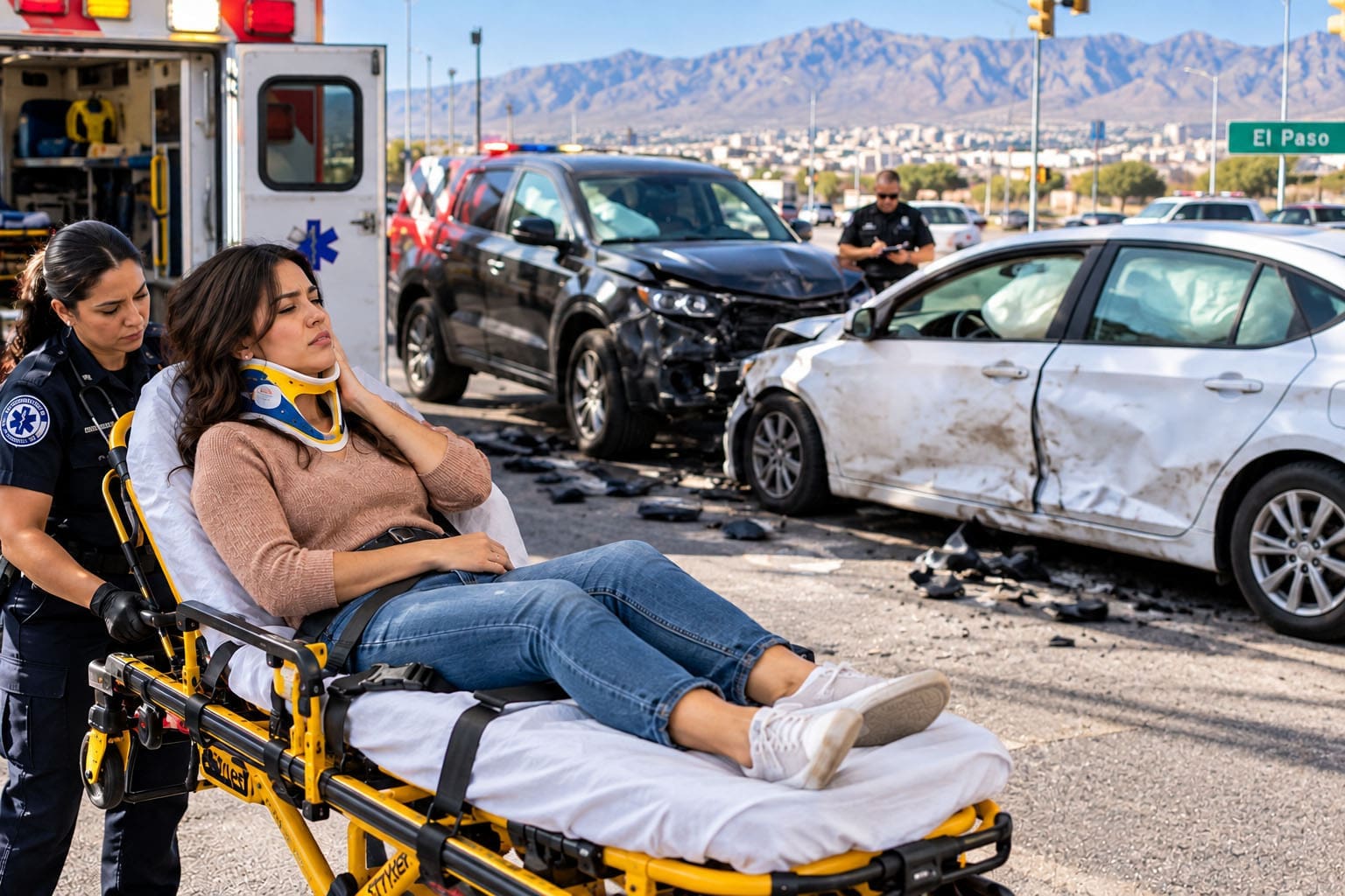
After a collision on I-10 or Loop 375, many people leave the scene thinking their low back will settle in a few days. Weeks later, the pain still shoots down one leg, sitting is difficult, and sleep is broken. An MRI shows a disc herniation pressing near a nerve root. An electromyography (EMG) study comes back “normal” or points to a different level. The patient wonders if the pain is real. The attorney wonders whether the records will hold up. Both tests are useful. Neither one, by itself, tells the full story of nerve injury. Understanding what each measures, what it misses, and when it is performed helps clarify conflicting results and leads to a clearer clinical picture.

What MRI Actually Shows
Magnetic resonance imaging creates detailed pictures of soft tissues. It can show a disc that has moved out of place, narrowed openings where nerves exit the spine, or swelling around a nerve root. These findings are anatomical. They describe structure. A disc extrusion visible on MRI can explain pain, numbness, or weakness in a particular part of the leg. Yet MRI cannot measure whether the nerve is still carrying signals normally. Many people over age 40 have disc changes on MRI that never cause symptoms. The image alone does not prove that the visible abnormality caused the current problem or that it began on the day of the collision. Clinical correlation—matching the picture to the person’s history, exam, and timing—is required. More information on how imaging fits into the evaluation of radiculopathy can be found in the clinic’s guide to radiculopathy and sciatica.
What Electrodiagnostic Testing Measures
EMG and nerve conduction studies (NCS) measure physiology—how nerves and the muscles they supply are working. Needle EMG looks for abnormal electrical activity in muscles. After a nerve root is injured, the muscle fibers it supplies can show spontaneous activity (fibrillations or positive sharp waves) once Wallerian degeneration has occurred. Nerve conduction studies measure the speed and size of signals traveling along nerves. In true single-root radiculopathy, the distal sensory responses are often normal because the sensory cell bodies sit outside the spine; the abnormality is more often found in the motor root and the muscles it reaches. These tests can confirm a dysfunctional nerve root and help separate a root problem from a more peripheral nerve problem, such as peroneal neuropathy or carpal tunnel. Details on how these studies assess nerve health are covered on the nerve conduction velocity page.
Timing Changes: What the Tests Can Show
Timing is one of the most common reasons EMG and MRI appear to disagree. After an acute nerve-root injury, certain EMG abnormalities take time to develop. Fibrillation potentials may appear in the paraspinal muscles within about one to two weeks and in the limb muscles several weeks later. An EMG performed in the first ten to fourteen days after a collision can look normal even when a significant root injury is present. Later studies are more likely to capture the changes. MRI, by contrast, can show structural change almost immediately, yet that change may not have produced enough axonal loss for the EMG to detect. Performing the studies at the right window and repeating them when needed reduces the chance of false reassurance or false alarm.
Limitations of Each Test
MRI has high sensitivity for disc herniations and foraminal narrowing but lower specificity for the patient’s current symptoms. Degenerative changes are common in asymptomatic adults; an MRI “positive” for disc bulge does not automatically mean that bulge is responsible for the leg pain that began after the crash. EMG often shows the opposite profile in many studies: it is more specific for true root dysfunction once enough time has passed, yet it is not fully sensitive. Purely sensory root irritation, a mild demyelinating lesion, or limited axonal loss that spares most sampled muscle can leave the EMG normal. Sampling only a few muscles, skipping the paraspinals, or testing too early further lowers the chance of detecting an abnormality. Neither test is perfect. Concordance between them is often only modest when clinicians don’t also consider clinical findings.
Why the Stories Differ and What to Do Next
Apparent contradictions usually have ordinary explanations. An MRI may show an L5 disc extrusion while the EMG is normal because the compression mainly irritates sensory fibers, the study was performed too early, or the motor axons reaching the tested muscles have not yet degenerated enough to produce abnormal potentials. Conversely, an EMG may show chronic denervation in an L4–L5 pattern while the current MRI looks unremarkable if the acute structural change has resolved, if the injury is chemical (inflammatory) rather than purely compressive, or if the imaging cuts missed the exact level of irritation.
In either case, the next step is not to discard one result. Instead, integrate both with the history, physical examination (reflexes, strength, sensory map, and tension signs), and timeline relative to the trauma. Additional clinical evaluation—sometimes a carefully timed repeat EMG, sometimes advanced imaging interpretation focused on acute versus chronic features, sometimes functional testing of range and strength—clarifies which findings are relevant to the present injury. Attorneys evaluating neurological evidence can obtain practical guidance on building causation opinions from the clinic’s resource on medical-legal causation opinions.
Building a Coherent Record for Patients and Attorneys
In personal-injury cases, the goal is to show that the collision mechanics (rapid deceleration, flexion-compression, or rotational forces) produced the documented nerve-root dysfunction rather than merely uncovering an unrelated age-related change. Timed clinical notes, digital range-of-motion measurements, and consistent symptom tracking help link the objective findings to the event. When EMG and MRI appear mismatched, the integrated record explains the discrepancy instead of leaving it for a defense expert to exploit. Patients gain clarity about why one test looks “normal” while they still cannot sit through a meeting or sleep through the night. Attorneys gain objective material that survives scrutiny under the Texas Eggshell Plaintiff principle: the defendant takes the plaintiff as found, and documented traumatic aggravation of a nerve root is compensable even when some degeneration already existed.
What Makes This Clinic Distinct
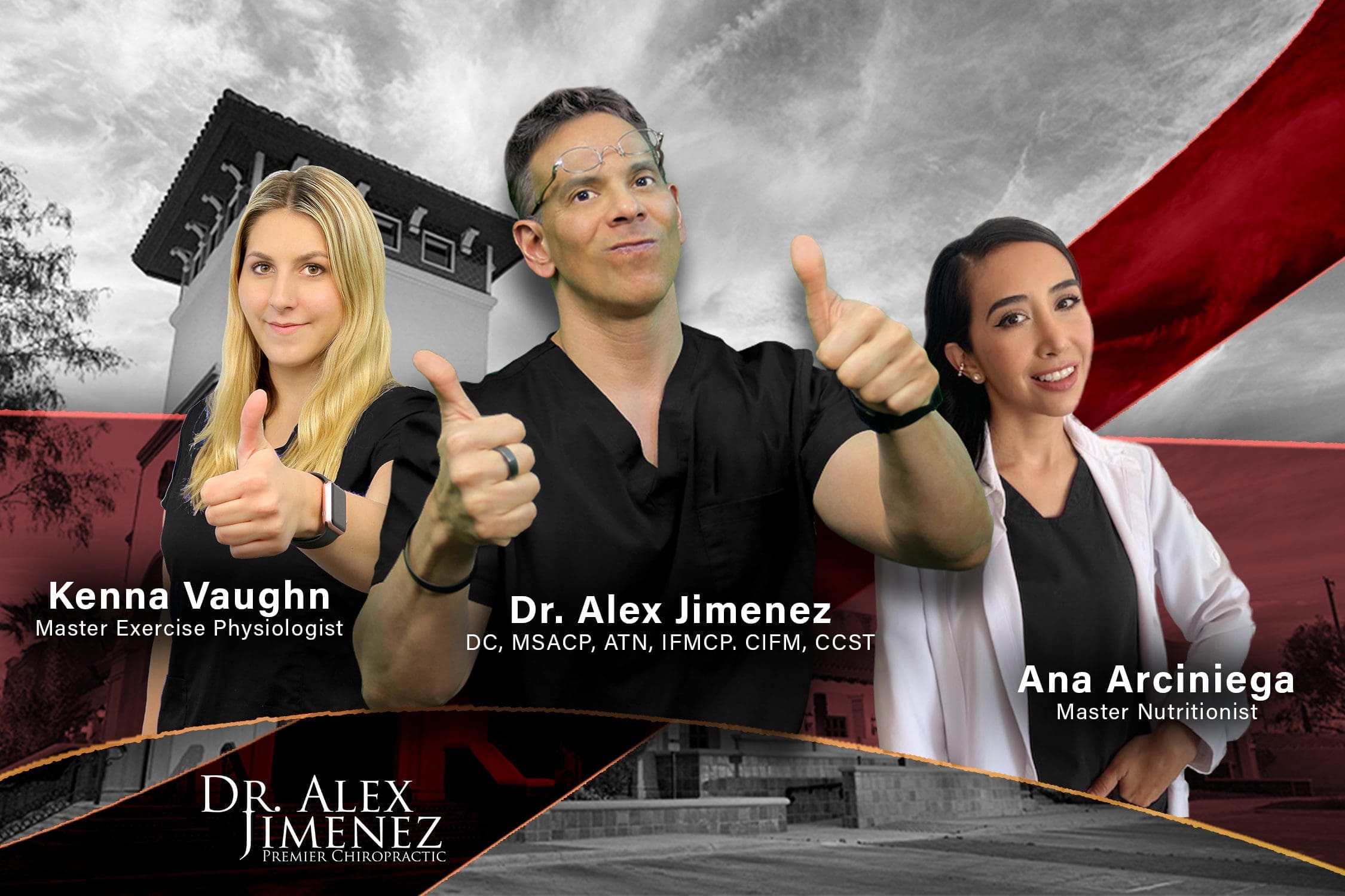
Injury Medical Clinic PA is the only clinic in El Paso, Texas, that combines a medical director with a nurse practitioner specializing in soft-tissue injury. Dr. Maria Guadalupe Cardenas, MD, Board Certified in Internal Medicine, serves as Medical Director and Collaborative Physician. She directs advanced laboratory panels, risk stratification, and coordination of internal medicine issues that often accompany traumatic nerve injury. Dr. Alex Jimenez, DC, APRN, FNP-BC, CFMP, IFMCP, is dual-licensed and focuses on soft-tissue, structural, and neurofunctional care. Together, they provide clinical correlation, timely electrodiagnostic interpretation, and multidisciplinary documentation that single-specialty settings rarely bring together. This structure supports both root-cause recovery and the clear records needed when causation is later examined.
Reclaim Your Mobility: Chiropractic Care for Sciatica Recovery | El Paso, TX
Practical Benefits of Coordinated Care
With a clearer clinical picture, treatment can target the real sources of limitation. Chiropractic adjustments restore segmental motion and reduce mechanical irritation at the root. Neurofunctional acupuncture and electroacupuncture modulate pain signals and support nerve recovery without relying on long-term medication. Patients typically report less radiating pain, improved ability to sit and walk, better sleep, and a faster return of confidence in movement. These gains are tracked with objective measures, so both the individual and any reviewing attorney can see progress. The approach rests on three bioethical pillars: beneficence (combining structural, neurological, and medical care solely for root-cause improvement), non-maleficence (favoring non-invasive, drug-free options that lower the risk of unnecessary procedures or medication dependence), and autonomy (educating the patient so they remain the informed decision-maker and can coordinate with any existing medical team).
A coherent explanation of why the EMG and MRI differ does more than resolve confusion. It turns two incomplete pictures into a single, defensible account of nerve injury that serves both the person living with the symptoms and the attorney responsible for presenting the case. Further resources on causation and diagnostic strategy are available through the master guide at dralexjimenez.com.
References
American Association of Neuromuscular & Electrodiagnostic Medicine. (2010). Utility of electrodiagnostic testing in evaluating patients with lumbosacral radiculopathy: An evidence-based review. Muscle & Nerve, 42(2), 276–282. https://doi.org/10.1002/mus.21759
Cho, S. C., Ferrante, M. A., Levin, K. H., Harmon, R. L., & So, Y. T. (2010). Utility of electrodiagnostic testing in evaluating patients with lumbosacral radiculopathy: An evidence-based review. Muscle & Nerve, 42(2), 276–282. https://doi.org/10.1002/mus.21759
Dillingham, T. R., Annaswamy, T. M., & Plastaras, C. T. (2020). Evaluation of persons with suspected lumbosacral and cervical radiculopathy: Electrodiagnostic assessment and implications for treatment and outcomes (Part II). Muscle & Nerve, 62(4), 474–484. https://doi.org/10.1002/mus.27008
Murtaza, F., Haider, S., Amin, A., Zahid, A., & Rehman, A. U. (2023). Correlation between electrodiagnostic study and magnetic resonance imaging in lumbar radiculopathy patients in a tertiary care hospital. Case Reports in Clinical Medicine, 12(10), 363–370. https://doi.org/10.4236/crcm.2023.1210051
Nardin, R. A., Patel, M. R., Gudas, T. F., Rutkove, S. B., & Raynor, E. M. (1999). Electromyography and magnetic resonance imaging in the evaluation of radiculopathy. Muscle & Nerve, 22(2), 151–155. https://doi.org/10.1002/(SICI)1097-4598(199902)22:2<151::AID-MUS3>3.0.CO;2-B
Wilbourn, A. J., & Aminoff, M. J. (1998). AAEM minimonograph 32: The electrodiagnostic examination in patients with radiculopathies. Muscle & Nerve, 21(12), 1612–1631. https://doi.org/10.1002/(SICI)1097-4598(199812)21:12<1612::AID-MUS2>3.0.CO;2-0
Professional Scope of Practice *
The information herein on "EMG MRI Nerve Evidence for Injury Cases Explained" is not intended to replace a one-on-one relationship with a qualified health care professional or licensed physician and is not medical advice. We encourage you to make healthcare decisions based on your research and partnership with a qualified healthcare professional.
Blog Information & Scope Discussions
Welcome to El Paso's Premier Wellness and Injury Care Clinic & Wellness Blog, where Dr. Alex Jimenez, DC, FNP-C, a Multi-State board-certified Family Practice Nurse Practitioner (FNP-BC) and Chiropractor (DC), presents insights on how our multidisciplinary team is dedicated to holistic healing and personalized care. Our practice aligns with evidence-based treatment protocols inspired by integrative medicine principles, similar to those found on this site and our family practice-based chiromed.com site, focusing on restoring health naturally for patients of all ages.
Our areas of multidisciplinary practice include Wellness & Nutrition, Chronic Pain, Personal Injury, Auto Accident Care, Work Injuries, Back Injury, Low Back Pain, Neck Pain, Migraine Headaches, Sports Injuries, Severe Sciatica, Scoliosis, Complex Herniated Discs, Fibromyalgia, Chronic Pain, Complex Injuries, Stress Management, Functional Medicine Treatments, and in-scope care protocols.
Our information scope is multidisciplinary, focusing on musculoskeletal and physical medicine, wellness, contributing etiological viscerosomatic disturbances within clinical presentations, associated somato-visceral reflex clinical dynamics, subluxation complexes, sensitive health issues, and functional medicine articles, topics, and discussions.
We provide and present clinical collaboration with specialists from various disciplines. Each specialist is governed by their professional scope of practice and their jurisdiction of licensure. We use functional health & wellness protocols to treat and support care for musculoskeletal injuries or disorders.
Our videos, posts, topics, and insights address clinical matters and issues that are directly or indirectly related to our clinical scope of practice.
Our office has made a reasonable effort to provide supportive citations and has identified relevant research studies that support our posts. We provide copies of supporting research studies upon request to regulatory boards and the public.
We understand that we cover matters that require an additional explanation of how they may assist in a particular care plan or treatment protocol; therefore, to discuss the subject matter above further, please feel free to ask Dr. Alex Jimenez, DC, APRN, FNP-BC, or contact us at 915-850-0900.
We are here to help you and your family.
Blessings
Dr. Alex Jimenez DC, MSACP, APRN, FNP-BC*, CCST, IFMCP, CFMP, ATN
Email: [email protected]
Multidisciplinary Licensing & Board Certifications:
Licensed as a Doctor of Chiropractic (DC) in Texas & New Mexico*
Chiropractic Licenses:
Texas DC License #: TX5807, Verified: TX5807
New Mexico DC License #: NM-DC2182, Verified: NM-DC2182
Nurse Practitioner Licenses:
Texas APRN License #: 1191402, Verified: 1191402 *
New Mexico CNP License #: 90560, Verified 90560
Florida APRN License #: 11043890, Verified: APRN11043890 *
Colorado License #: C-APN.0105610-C-NP, Verified: C-APN.0105610-C-NP
New York License #: N25929, Verified N25929
Georgia APRN License #: GAA-NP005701
Multi-State Advanced Practice Registered Nurse (APRN*) Texas & Multi-States
Multi-state Compact APRN License by Endorsement (43 States)
Compact Status: Multi-State License: Authorized to Practice in 43 States*
Nursing Licensure Compact: Updated Here
DEA Registration: (Drug Enforcement Agency Registered)
All medical (MDs) and family practice providers (FNP-APRN) are registered and licensed to offer various levels of medication.
Verify Providers Here
License Verification Link: Nursys License Verifier
* Prescriptive Authority Authorized (DEA Registered Providers). Call if Required
Board Certification:
ANCC FNP-BC: Board Certified Nurse Practitioner*
Education:
Graduate with Honors: ICHS: MSN-FNP (Family Nurse Practitioner Program)
Degree Granted. Master's in Family Practice, MSN Diploma (Cum Laude)
Dr. Alex Jimenez, DC, APRN, FNP-BC*, CFMP, IFMCP, ATN, CCST
(Board Certified: Family Practice Nurse Practitioner—Multistate)*
(Licensed Nurse Practitioner & Chiropractor - Multistate)*
Clinical Director
DC & FNP License (Review Above)
Digital Business Card
NPI: 1205907805
Dr. Maria Cardenas, MD
(Board Certified: Internal Medicine)
(Licensed Medical Doctor)
Medical Director, Clinical Director & Collaborative Physician
NPI # 1164426748
MD License #: J2933
Licenses and Board Certifications:
MD: Medical Doctor
DC: Doctor of Chiropractic
APRNP: Advanced Practice Registered Nurse
FNP-BC: Family Practice Specialization (Multi-State Board Certified)
FNP-BC: Family Practice Across Life Span (Neonatal to Geriatrics)
RN: Registered Nurse (Multi-State Compact License)
CFMP: Certified Functional Medicine Provider
MSN-FNP: Master of Science in Family Practice Medicine
MSACP: Master of Science in Advanced Clinical Practice
IFMCP: Institute of Functional Medicine
CCST: Certified Chiropractic Spinal Trauma
ATN: Advanced Translational Neutrogenomics
Family with Primary Care Focus (Family Nurse Practitioner or FNP)
- The Family Nurse Practitioner (FNP) promotes, maintains, and restores health for individuals and families across the lifespan. FNPs also identify health risks, promote wellness, and diagnose and manage acute and chronic illness.
- The FNP focuses on comprehensive primary care, promoting healthy lifestyles for patients across the lifespan in settings such as private practice, physician offices, and community health centers.
Memberships & Associations:
TCA: Texas Chiropractic Association: Member ID: 104311
TNA: Texas Nurse Association: Member ID: 06458222
TNP: Texas Nurse Practitioner Association ID: 2025091511
AANP: American Association of Nurse Practitioners: Member ID: 2198960
ANA: American Nurses Association: Member ID: 06458222 (District TX01)
| Primary Taxonomy | Selected Taxonomy | State | License Number |
|---|---|---|---|
| No | 111N00000X - Chiropractor | NM | DC2182 |
| Yes | 111N00000X - Chiropractor | TX | DC5807 |
| Yes | 363LF0000X - Nurse Practitioner - Family | TX | 1191402 |
| Yes | 363LF0000X - Nurse Practitioner - Family | FL | 11043890 |
| Yes | 363LF0000X - Nurse Practitioner - Family | CO | C-APN.0105610-C-NP |
| Yes | 363LF0000X - Nurse Practitioner - Family | NY | N25929 |
| Yes | 363LF0000X - Nurse Practitioner - Family | NM |
90560 |
| Yes | 363LF0000X - Nurse Practitioner - Family | GA | GAA-NP005701 |
Dr. Alex Jimenez, DC, APRN, FNP-BC*, CFMP, IFMCP, ATN, CCST
(Board Certified: Family Practice Nurse Practitioner—Multistate)*
(Primary Care Across Lifespan—Neonatal / Pediatric / Adult / Geriatrics)
(Licensed Nurse Practitioner & Chiropractor - Multistate)*
Clinical Director
Digital Business Card
NPI: 1205907805
Dr. Maria Cardenas, MD
(Board Certified: Internal Medicine)*
(Licensed Medical Doctor)*
Medical Director, Clinical Director & Collaborative Physician
NPI # 1164426748
MD License #: J2933
📆 Schedule Appointment: Schedule 24/7 (Click Here)